
Abstract
Amylase-resistant, periodic acid-Schiff (PAS)–positive inclusions were identified in the skeletal muscle of four of twenty-four purpose-bred beagle dogs from a routine toxicology study. Affected myofibers contained amorphous material filling up to 20% of the sarcoplasm that stained lightly basophilic with hematoxylin and eosin and was strongly PAS–positive with amylase resistance. Transmission electron micrographic examination of the inclusions revealed granular, non-membrane–bound, electron-dense material, consistent with polysaccharide. Although skeletal muscle inclusions with similar features have been reported in dogs in conjunction with systemic metabolic disorders and less often in muscle adjacent to nonmyogenic sarcomas, all four of these dogs lacked clinical or pathological findings diagnostic of a concurrent systemic metabolic or localized skeletal muscle disorder. Furthermore, these skeletal muscle inclusions were present in both vehicle- and test article–treated dogs and were considered an incidental finding that may occur spontaneously in clinically normal beagle dogs; as such, their presence in drug-treated animals should be interpreted with caution.
Amylase-resistant, periodic acid-Schiff (PAS)–positive inclusions in skeletal muscle are reported in multiple species in a variety of disease conditions, most often in disorders of carbohydrate metabolism (Cabello et al. 1981; Firshman et al. 2006; Harvey et al. 1990; Tsujino et al. 2000; Valentine et al. 2000). Equine polysaccharide storage myopathy (EPSSM) is the most commonly described disorder in the veterinary literature in which these inclusions are a diagnostic feature (Valentine et al. 2000). In dogs, the majority of reports of PAS–positive and amylase-resistant material in skeletal muscle are in cases of phosphofructokinase deficiency (PFK), a condition analogous to human inherited type IV glycogen storage disease (glycogenosis type IV) that disrupts normal anaerobic glycolysis (Drash and Field, 1971; Harvey et al. 1990; Tsujino et al. 2000). In addition to PFK and other metabolic disorders, complex polysaccharide inclusions were recently reported in skeletal muscle adjacent to osseous and synovial cell sarcomas in dogs with no identified systemic metabolic dysfunction (Valentine et al. 2002). In contrast to previous cases, this report describes the presence of complex polysaccharide inclusions in clinically normal beagle dogs from a routine toxicology study.
These were naïve sixteen- to eighteen-month-old purpose-bred beagle dogs from Marshall Farms that were part of a one-month oral toxicity study that included twelve males and twelve females. The dogs were housed within the Pfizer facility at Amboise (France) in individual kennels and fed a pelleted commercial laboratory animal food supplied by Scientific Animal Food and Engineering, with free access to water. Clinical examinations, body weights, electrocardiograms, clinical hematology/biochemistry, and urinalysis were periodically performed throughout the conduct of the study. The procedures used in this study were consistent with the guidelines of the Pfizer Institutional Animal Care and Use Committee.
At study termination, the animals were fasted overnight prior to sacrifice by exsanguination under intravenous barbiturate anesthesia and were then necropsied. Tissue samples, including the semimembranosus muscle, were collected, fixed in 10% buffered formalin, and routinely processed to hematoxylin and eosin (H&E) slides. To further characterize light microscopic findings, a histochemical stain for PAS, with and without prior amylase digestion, was applied to sections of skeletal muscle from one female control (vehicle) animal and one female high-dose animal. Sections of liver were used as the positive controls for PAS and PAS-amylase stains. For transmission electron microscopic (TEM) examination, a formalin-fixed and paraffin-embedded sample of skeletal muscle from one female high-dose animal was deparaffinized and processed for TEM. The sample was washed in 0.15M phosphate Sörensen buffer (pH 7.2 to 7.4) with 0.2% sodium chloride. The tissue was then postfixed in 2% osmium tetroxide in phosphate Sörensen buffer and then washed in sterile water. The sample was routinely processed and embedded in epoxy resin. Ultrathin sections were stained with uranyl acetate and lead citrate. Observations were made using a Jeol 1200 EX II electron microscope, and digital pictures were taken.
Morphologic changes in skeletal muscle were similar for all animals and were detected in two control females, as well as one low-dose female and one high-dose female. Approximately 5% to 10% of myofibers contained peripherally to less frequently centrally located, irregularly shaped, amorphous material within the sarcoplasm that lacked a distinct membrane structure and filled up to 10% to 20% of the total myofiber cross-sectional area (Figure 1 ). All inclusion material stained lightly basophilic in hematoxylin and eosin (H&E) sections and was magenta pink (positive) in both PAS (Figure 2B ) and PAS-amylase stained sections (Figure 2D); in contrast, the control liver slide was PAS positive (Figure 2A), but amylase sensitive (Figure 2C). Affected and unaffected myofibers were of similar size, and there was no associated inflammation. Transmission electron microscopy revealed granular, non-membrane–bound, minimally electron-dense material disrupting the sarcoplasmic reticulum (Figures 3A and 3B ).
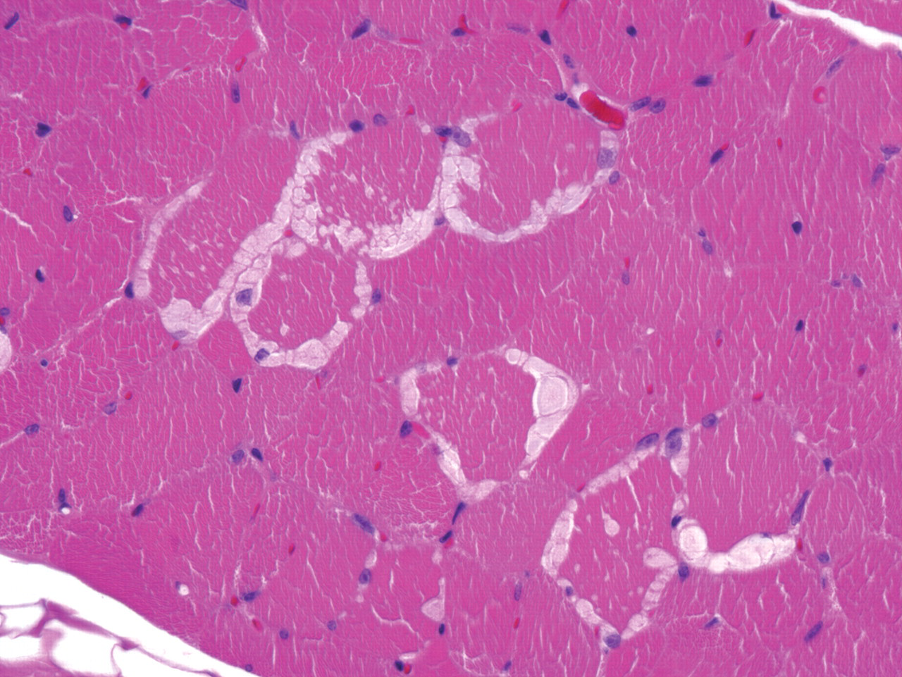
Photomicrograph of section of canine semimembranosus muscle containing well-defined, irregularly shaped lightly basophilic inclusions. (H&E, 40×).
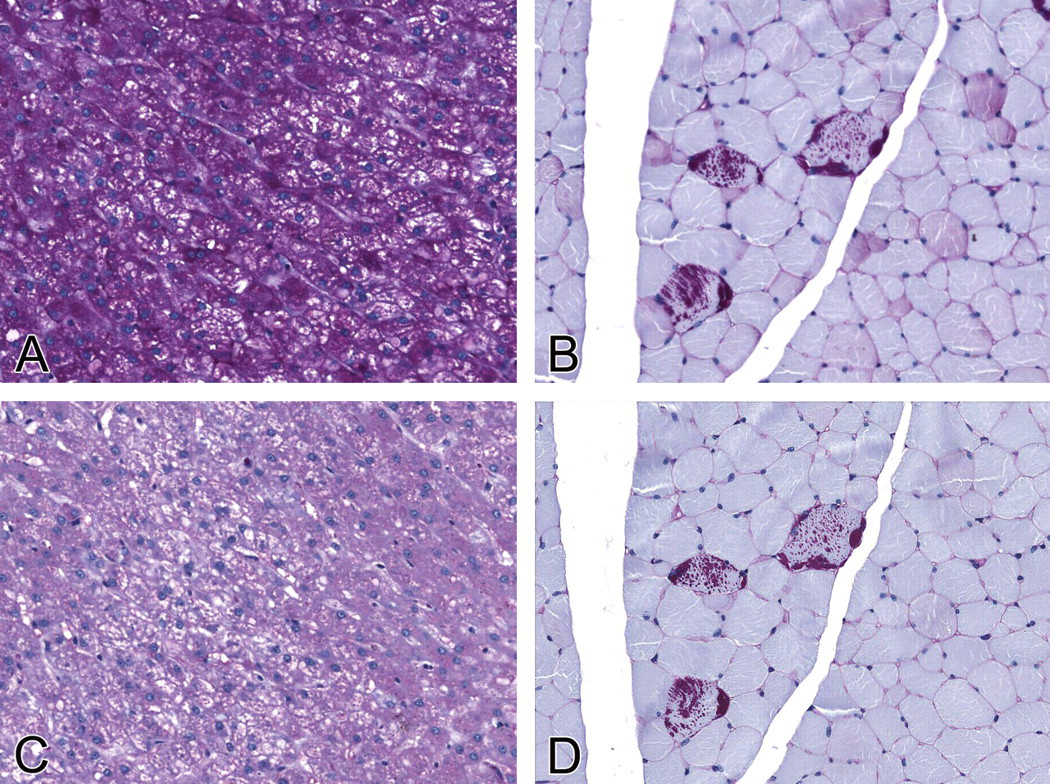
(A) Photomicrograph of a section of liver with strong positive staining of hepatocyte cytoplasm (PAS, 20×). (B) Photomicrograph of a section of canine semimembranosus muscle with strong positive staining of the muscular inclusions (PAS, 20×). (C) Photomicrograph of a section of liver with amylase sensitivity (PAS-amylase, 20×). (D) Strong positive staining of the muscular inclusions after digestion with amylase, indicating the presence of complex polysaccharides (PAS-amylase, 20×).
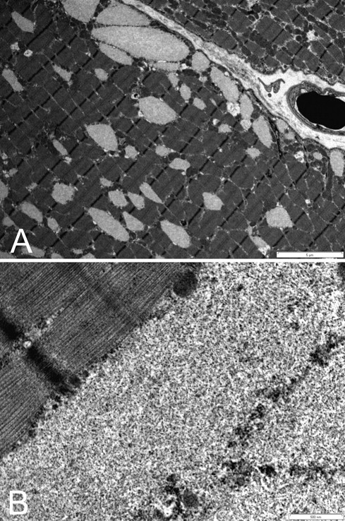
(A) TEM photomicrographs of canine semimembranosus muscle containing non-membrane–bound, moderately electron-dense intrasarcoplasmic inclusions distorting individual sarcomere units (Bar =5 µM). (B) Inclusions consist of moderately electron-dense granular material (Bar = 500 nM).
The amylase-resistant, PAS-positive material in these cases is consistent with previous descriptions of skeletal muscle inclusions, in multiple species, seen in association with a variety of disease conditions (Cabello et al. 1981; Firshman et al. 2006; Harvey et al. 1990; Tsujino et al. 2000; Valentine et al. 2000). The prominent granular material seen with TEM has been described in earlier canine reports (Harvey et al. 1990; Valentine et al. 2002). Those studies also reported filamentous structures admixed with the granular material; such structures were not identified in this case, and their absence may be because of the limited sample selection (Harvey et al. 1990; Valentine 2002). The histological appearance of the inclusion material is similar to the descriptions in previous canine reports, both in staining properties and variation in size and distribution of the myofiber inclusions (Harvey et al. 1990; Valentine 2002). Formalin-fixed tissues are considered reliable to identify these inclusions; a recent review of fourteen histological criteria used to diagnose EPSSM found that PAS–positive, amylase-resistant material was the most consistent diagnostic feature including those observed in formalin-fixed samples from affected horses (Firshman et al. 2006).
Although the histological and TEM findings indicate that these inclusions are comparable with those previously reported, these canine cases differ greatly in that no underlying systemic metabolic or localized skeletal muscular disorder was apparent in any of the affected animals. Although no direct testing was performed for PFK or other systemic causes of complex polysaccharide inclusions such as glycogen or branched-chain amino acid disorders and familial myoclonic epilepsy (Valentine et al. 2002), none of the animals exhibited any clinical signs or clinical pathology characteristic of these conditions. Phosphofructokinase deficiency is seen primarily in English Springer and American Cocker Spaniel breeds and characterized by hemolytic disease and minimal signs of neuromuscular dysfunction (Harvey et al. 1990). Similar to the dogs presented in this case report, in some cases of EPPSM, affected horses have no apparent clinical signs (Valentine et al. 2000).
The complex polysaccharide inclusions in the skeletal muscle of these dogs were considered incidental, as they occurred with a similar incidence in vehicle- and test article–treated animals. Reports of complex polysaccharide inclusions in the skeletal muscle of otherwise clinically normal laboratory beagle dogs were not found in the literature. Although skeletal muscle is a tissue routinely examined in Good Laboratory Practice (GLP) toxicology studies, routine sectioning may easily miss the inclusions, given their low total number and multifocal distribution. Although these inclusions were present only in female animals in this study, the influence of sex on their incidence cannot be ascertained from this small sample size. Genetics may play a role, but this possibility would need to be determined with further research.
In conclusion, the presence of PAS–positive and amylase-resistant material in canine skeletal muscle may occur as a spontaneous finding in routine toxicity studies, and their presence in compound-treated animals should be interpreted with caution.
Footnotes
Acknowledgments
The writing of this article, by Lyn M. Wancket, was partially supported by a fellowship from Pfizer organized by the American College of Veterinary Pathologists and Society of Toxicologic Pathology Coalition for Veterinary Pathology Fellows. We thank Marie-Thérèse Masson for performing ultrastructural investigations and illustrations, Walt Bobrowski for assistance with figures, and Drs. Laura Rush and Katy Gropp for their critical review of the manuscript.