
Abstract
To compare the susceptibility to toxicity of semicarbazide hydrochloride (SEM-HCl) between young and adult rats, 3- and 20-week-old female SD rats were given a diet containing SEM-HCl at 0, 500, or 1,000 ppm and 0 or 1,000 ppm, respectively, for 4 weeks. Half of the animals were then maintained on basal diet for a further 2 weeks as recovery groups. Only in young rats was deformation of the knee joints as well as thorax and tail observed at 500 and 1,000 ppm. Histopathologically, severe osteochondral lesions, such as disarrangement and thickening of the epiphyseal cartilage and deformation of articular cartilage, were observed, but the severity of these lesions became reduced during the recovery period. In adult rats, osteochondral lesions were relatively mild. Fissures in the cartilage matrix of the tibia were characteristic of adult rats, and in these, reduction of severity was not obvious in the recovery group. In the thoracic aorta, the appearance of elastic laminae was altered only in young rats in both the 4-week treatment and recovery groups. These results suggest that growing animals are more susceptible to toxicity of SEM-HCl than adults are. Effects and the induced lesions link to the developing stage of the target organs.
Introduction
Semicarbazide (SEM), a metabolite of the banned veterinary antibiotic nitrofurazone, has been proposed as a marker for detection of nitrofurazone abuse (Effkemann and Feldhusen 2004; de la Calle and Anklam 2005). However, SEM has in fact been found in food in glass jars sealed with plastic gaskets manufactured using azodicarbonamide as a blowing agent, such as baby foods, fruit juices, jams, and conserves (European Food Safety Authority 2003; Stadler et al. 2004). Other sources of contamination, such as from food processing by hypochlorite treatment, have further been suggested (Hoenicke et al. 2004; Saari and Peltonen 2004). The possibility of exposure through various foods independent of nitrofurazone usage has thus raised concerns about the health risk of SEM.
SEM is also known to inhibit enzymes, such as lysyl oxidase, semicarbazide-sensitive amine oxidase (SSAO), and glutamic acid decarbocylase (Magyar, Mészáros, and Mátyus 2001; Dawson, Rinaldi, and Pöch 2002; Macedo et al. 2007). It thereby acts as an osteolathyrogen, inducing osteochondral and vascular lesions in young rats due to impaired cross-linking reactions of collagen and elastin through inhibition of lysyl oxidase or SSAO (Ramamurti and Taylor 1959; Langford et al. 1999; Dawson et al. 2002; Mercier et al. 2007). In addition, teratogenic effects such as induction of cleft palate and aortic aneurysms have been reported (Steffek, Verrusio, and Watkins 1972; de la Fuente del Rey 1986; Gong et al. 2006). SEM has weak genotoxicity in vitro but not in vivo (Parodi et al. 1981; Abramsson-Zetterberg and Svensson 2005; European Food Safety Authority 2005, Food Safety Commission 2007). In mice, SEM hydrochloride (SEM-HCl) at a high dose in the drinking water increased incidences of lung and blood vessel tumors that were commonly observed in untreated mice, suggesting that SEM-HCl might be only a weak carcinogen (Toth, Shumizu, and Erickson 1975). In rats, although no carcinogenic effects were observed after feeding a diet containing SEM-HCl at 500 and 1,000 ppm for 78 or 32 weeks (Weisburger et al. 1981), data for carcinogenicity remain insufficient for evaluation.
So far, the risk of adverse effects of SEM exposure to human beings has been considered low because there is a sufficient margin of exposure (Nestmann et al. 2005). However, intake of SEM for infants is estimated to be much higher than for adults because of high consumption of baby food in glass jars and the infants' small body weight (European Food Safety Authority 2005; Nestmann et al. 2005). Generally, developing bones and cartilage of infants and children have a different susceptibility from that of their adult counterparts (Schwenk et al. 2003), and previous studies of effects of other osteolathyrogens such as beta-aminopropionitrile (BAPN) and aminoacetonitrile hydrochloride indicated young animals to be more susceptible than adults (Morgan and Bellamy 1976; Davies and Schofield 1980). In man, although there has been no report of osteolathyrism caused by SEM, Haque et al. (1997) reported lesions induced by BAPN. Accordingly, because infants or children might be more vulnerable to SEM than adults, evaluation of toxic effects of SEM with the focus on age-related susceptibility is important for risk assessment in human health. In the present study, we therefore compared histopathological osteochondral and vascular lesions in young and adult female rats given SEM-HCl for 4 weeks. Reversibility of the induced lesions was also assessed.
Materials and Methods
Chemicals
SEM-HCl (CAS no. 563-41-7) was purchased from Hayashi Pure Chemical Ind., Ltd. (Osaka, Japan), as a white powder with a purity of 99.3% and well mixed at concentrations of 0, 500, or 1,000 ppm into a powdered basal diet (CRF-1; Oriental Yeast Co., Tokyo, Japan). More than 89% stability of the test compound was confirmed for up to 4 weeks of storage at 4°C and room temperature. Test diets were prepared every 2 weeks and stored at 4°C before use.
Animals and Treatments
Thirteen pregnant Crl:CD (SD) rats were obtained from Charles River Japan Inc. (Kanagawa, Japan) at gestational day 10. They were housed individually in polycarbonate cages with wood chip bedding and maintained in an air-conditioned animal room (temperature 24°C ± 1°C, relative humidity 55% ± 5%, 12-hour light-dark cycle) with powdered basal diet (CRF-1) and tap water available ad libitum. At weaning, 30 female pups (210 or 22-day-old) were allocated to 3 groups, each consisting of 10 animals from different dams, and given diet containing 0, 500, or 1,000 ppm SEM-HCl for 4 weeks (young group). The dietary dose levels were determined as toxic levels in a 2-week dose-finding study conducted on the basis of a previous report (Weisburger et al. 1981). Dams were kept untreated for 5 weeks after weaning, and 12 animals showing no abnormalities at 20 weeks of age were divided into 2 groups and fed diet containing 0 or 1,000 ppm SEM-HCl for 4 weeks (adult group). Observations for mortality and clinical signs, including posture and gait abnormalities and deformation of 4 limbs, thorax, and tail, were conducted daily. Body weight and food consumption were recorded every week. After 4-weeks treatment, half of the animals from each group were euthanized by exsanguination from the abdominal aorta under deep anesthesia with ether. The remaining animals were maintained on basal diet for another 2 weeks as recovery groups and then subjected to necropsy in the same manner. The experimental protocol using animals was reviewed and approved by the Animal Care and Use Committee of the National Institute of Health Sciences, Japan.
Histopathological Examination
After macroscopic examination, all organs were removed and fixed in 10% neutral buffered formalin. For examination of osteochondral lesions, the nasal cavity, sternum, right femur, right tibia, left knee joint, right and left ankles, spine (cervical, thoracic, lumbar, and caudal vertebrae with corresponding spinal cord) were fixed in 10% neutral buffered formalin and then decalcified in EDTA solution at room temperature for a month. The tissues were then routinely processed for paraffin embedding, sectioned, and stained with hematoxylin and eosin. Victoria blue staining was applied to three transverse sections of the descending thoracic aorta cut at 5-mm intervals to demonstrate elastic fibers. The number of elastic laminae of each section was counted.
Statistical Analysis
Variance in data for body weights and food consumption of young rats was checked for homogeneity by Bartlett’s procedure. If the variance was homogeneous, the data were assessed by one-way analysis of variance. If not, the Kruskal-Wallis test was applied. When statistically significant differences were detected, the Dunnett’s multiple test was employed for comparison between the 0-ppm and treatment groups. In the adult group, the data for body weights and food consumption were analyzed by the Student’s or Welch’s t-test following a test for equal variance.
Results
In-Life Parameters
No deaths occurred during the experiment. Body weights in young groups increased constantly, and body weight gain at 1,000 ppm was significantly suppressed from week 1 (Fig. 1) throughout the study. In contrast, body weights of adult animals in both the 0- and 1,000-ppm groups stayed flat and were comparable between the two groups throughout the study. Data for food consumption and SEM-HCl intake are summarized in Table 1 . Although the mean values for food consumption per animal in young and adult rats were similar, the mean value for food consumption per kilogram of body weight in young animals was nearly doubled that in adults. Accordingly, intake of SEM-HCl in young animals was also twice as much as that in adult rats.
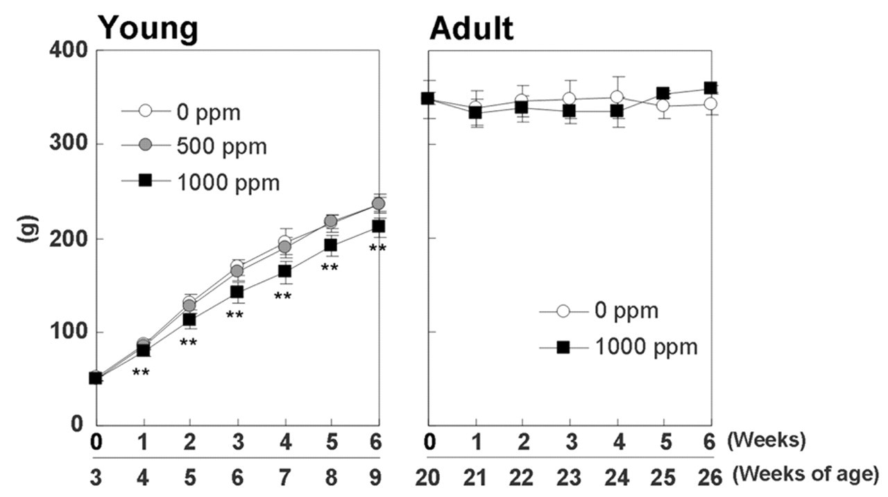
Growth curves for young and adult rats given semicarbazide hydrochloride in diet for 4 weeks and then maintained on basal diet for 2 weeks as a recovery period. **Significantly different from the 0-ppm group at p < 0.01.
Food consumption and intake of SEM-HCl by young and adult rats.
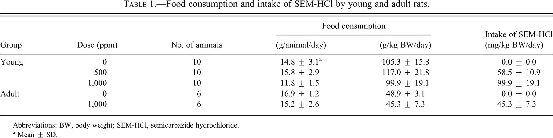
Abbreviations: BW, body weight; SEM-HCl, semicarbazide hydrochloride.
a Mean ± SD.
Clinical Findings and Necropsy
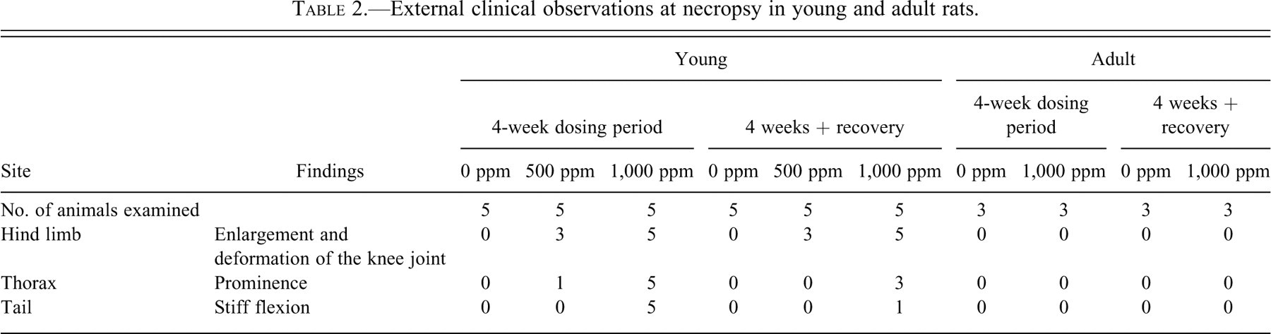
The external abnormalities observed during the study are summarized in Table 2 . Enlargement and deformation of the knee joints was apparent in young rats given 500 and 1,000 ppm from week 1. Tails in these groups exhibited stiff flexion from week 2, and prominence of the thorax was also found at week 4. Such changes remained after the 2-week withdrawal period, although the number of animals with the abnormalities and their severities were somewhat decreased. In adults, no external findings were found in either the 4-week treatment or recovery groups.
External clinical observations at necropsy in young and adult rats.
Histopathological Examination
Both in young and adult groups, histopathological lesions were mainly observed in the bones, cartilages, and aorta, and no treatment-associated changes were found in any other organs. The findings for histopathological examination of the bones and joints are summarized in Table 3 .
Histopathological findings for the bones and joints in young and adult rats.
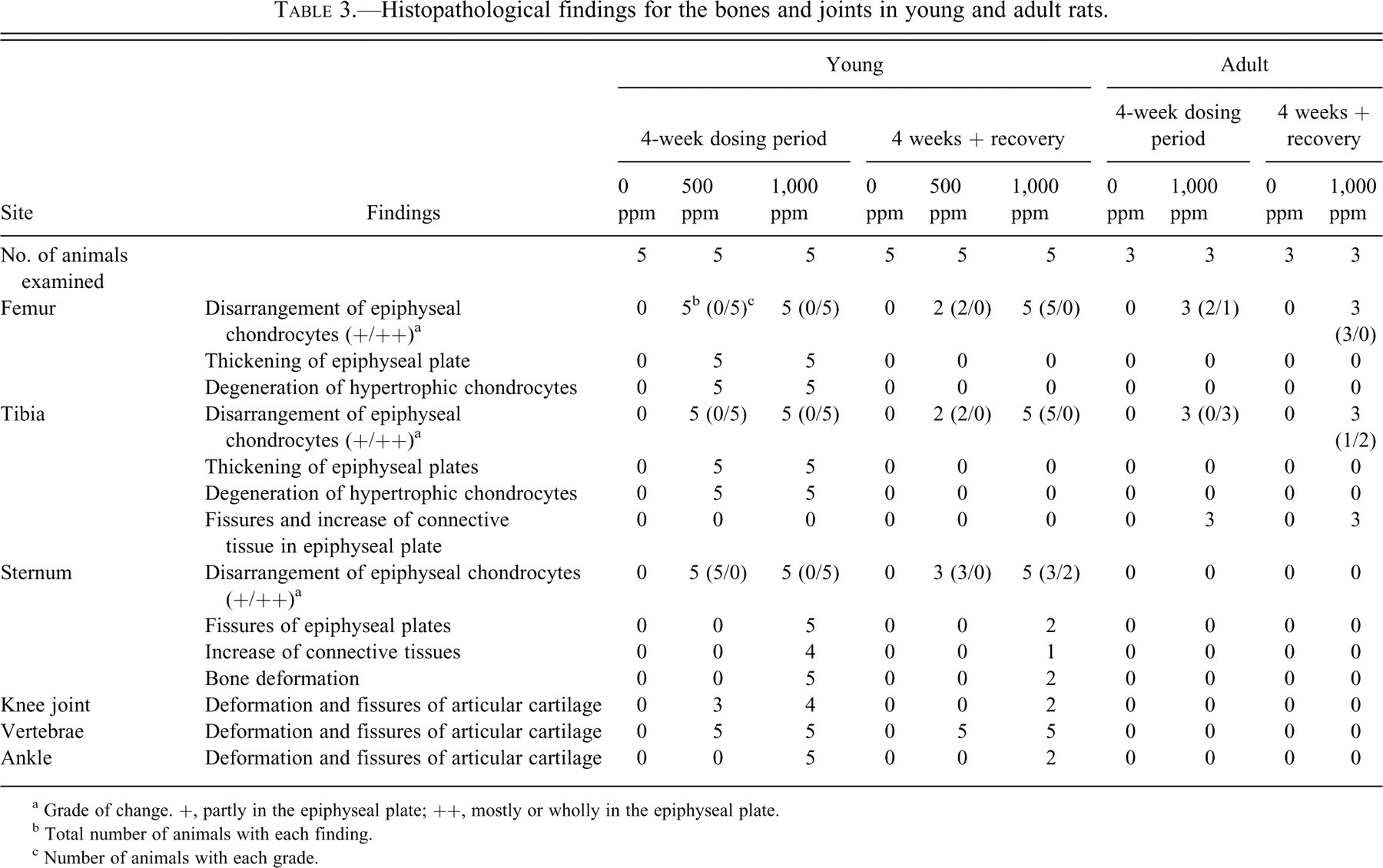
a Grade of change. +, partly in the epiphyseal plate; ++, mostly or wholly in the epiphyseal plate.
b Total number of animals with each finding.
c Number of animals with each grade.
In young rats, severe osteochondral lesions were observed in various sites in the body at both 500 and 1,000 ppm. As compared with the 0-ppm group (Fig. 2A and B), the epiphyseal plates at the proximal ends of the tibia were thickened in animals given the two doses at similar severities, and disarrangement of epiphyseal chondrocytes and degeneration of hypertrophic chondrocytes was also observed in the thickened cartilage plates (Fig. 2C and D). Staining intensity of the cartilage matrix in these groups was reduced. After the recovery period, thickening of the epiphyseal plates and the degeneration of hypertrophic chondrocytes had disappeared, and the severity of disarrangement of epiphyseal chondrocytes was reduced (Fig. 2E and F). The staining of the cartilage matrix had completely recovered to that seen in the control animals. The femurs exhibited similar histological lesions to the tibias with the same severity. In the sternum, disarrangement of epiphyseal chondrocytes was observed in young animals at 500 and 1,000 ppm in a dose-dependent manner, and fissures with increased connective tissue and bone deformation were evident at 1,000 ppm. After the recovery period, the severities of these lesions were reduced as in the case in the tibia. In addition, in young animals only, deformation and fissures of articular cartilage were observed in the knee joints, intervertebral joints (from cervical to caudal), and ankles at 500 and 1,000 ppm (Fig. 3). Although some lesions in the articular cartilage were decreased after the recovery period, such reduction was not apparent in the intervertebral joints.
Histopathology of the tibia of young rats. Normal proximal end of a tibia (A) and high magnification of an epiphyseal plate from the 0-ppm group (B). In a rat given 1,000 ppm for 4 weeks, thickening of the epiphyseal plate and reduction of staining intensity of the cartilage matrix are apparent (C). At high power, disarrangement and degeneration of hypertrophic chondrocytes are evident in the thickened cartilage plate (D). After the recovery period, the thickness and staining intensity of the epiphyseal plate are normal (E), and the severity of disarrangement of epiphyseal chondrocytes is reduced (F). Hematoxylin and eosin stain. Bars = 500 μm (A, C, E), 100 μm (B, D, F).
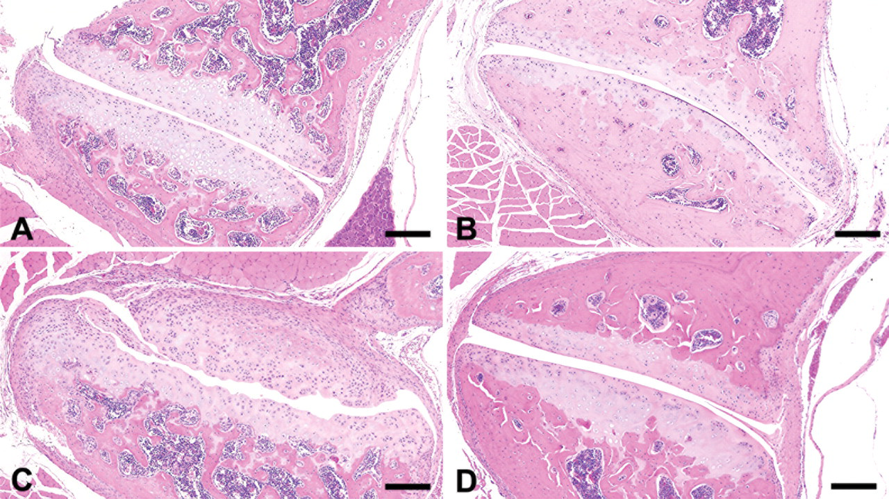
Histopathology of the cervical vertebrae of young and adult rats. Normal intervertebral joints of young (A) and adult (B) rats of the 0-ppm group. In a young animal receiving 1,000 ppm for 4 weeks, deformation and fissures of articular cartilage is apparent (C), while there are no significant lesions in an adult case (D). Hematoxylin and eosin stain. All bars = 200 μm.
In adult rats, the osteochondral lesions were relatively mild and limited to the femur and tibia. Disarrangement of epiphyseal chondrocytes was found in the femur and tibia but without thickening of the epiphyseal plate and degeneration of hypertrophic chondrocytes. Fissures in the cartilage matrix in the anterior areas of the tibias were characteristic in adult rats of the 1,000-ppm group, accompanied by an increase of connective tissues (Fig. 4C and D), and these persisted in the recovery group. There were no significant lesions in the sternum or articular cartilage in adult rats. In both young and adult groups, no treatment-related changes in cartilage of the nasal cavity, trachea and bronchi, or Achilles tendons were found. Most lesions in young animals at 500 ppm were clearly more severe than those in adult animals at 1,000 ppm.
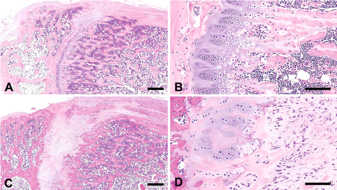
Histopathology of the tibia of adult rats. Normal proximal end of a tibia (A) and high magnification of its epiphyseal plate from the 0-ppm group (B). In the 1,000-ppm group, disarrangement of epiphyseal chondrocytes and the fissures with increased connective tissues are found in the anterior area (C, D). Hematoxylin and eosin stain. Bars = 500 μm (A, C), 100 μm (B, D).
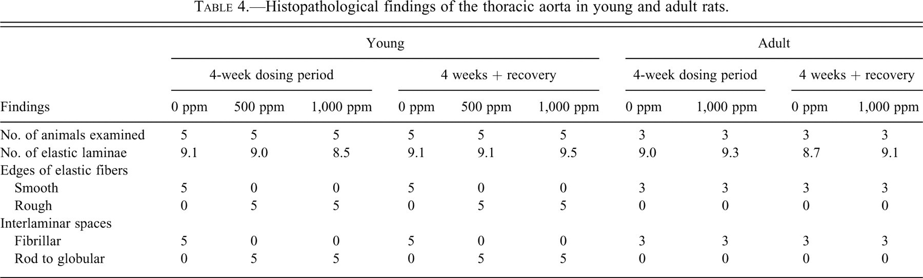
Histopathological findings for the thoracic aorta are summarized in Table 4. Although the numbers of elastic laminae were unchanged, their edges became roughened only in young rats at 500 and 1,000 ppm, and the interlaminar spaces in treated groups had a rod or globular appearance, in contrast to the fibrillar appearance in the 0-ppm group (Fig. 5). These lesions were similarly found after the recovery period. In adult cases, histology of elastic laminae did not differ between the 0- and 1,000-ppm groups, after both the 4-week treatment and the recovery period.
Histopathological findings of the thoracic aorta in young and adult rats.
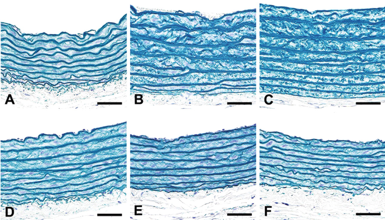
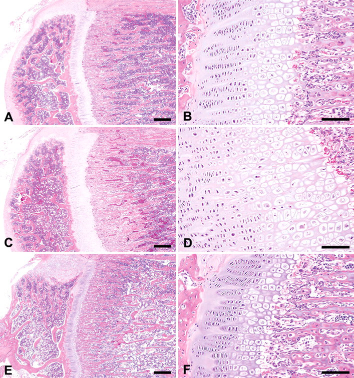
Transverse sections of the thoracic aorta of young and adult rats. As compared with the 0-ppm group (A), elastic laminae in the 1,000-ppm group show rough edges in the young group (B), and the interlaminar spaces in treated groups have a rod or globular appearance, in contrast to the fibrillar appearance in the 0-ppm group. These changes were similarly found after the recovery period (C). In adult cases, the histology of elastic laminae was unchanged between 0 ppm (D) and 1,000 ppm in both the 4-week treatment (E) and recovery groups (F). Victoria blue stain. All bars = 50 μm.
Discussion
In the present study, toxicologic effects of SEM-HCl were mainly observed in the bones, cartilage, and aorta, and histopathologic changes observed in young groups were basically consistent with the previous reports using rats during the growing period (Ramamurti and Taylor 1959; Langford et al. 1999; Mercier et al. 2007). While the target sites of SEM-HCl were similar in both young and adult groups, the lesions induced were much more diverse and serious in young animals. Young animals generally take more food per body weight during the growing period than their adult counterparts do, and during the present study, the intake of SEM-HCl per kilogram of body weight in young animals was about twice that in adult rats. However, even when comparing young animals at 500 ppm and adults at 1,000 ppm, the lesions in young animals were clearly more severe. Therefore, young rats are more susceptible to SEM-HCl than adults. This is in line with the fact that SEM impairs cross-linking reactions of collagen and elastin, which are essential for maturation of connective tissues, including bones and blood vessels (Ramamurti and Taylor 1959; Langford et al. 1999; Dawson, Rinaldi, and Pöch 2002; Mercier et al. 2007).
After the recovery period, the severities of osteochondral lesions were markedly reduced in young animals, especially in the long bones growing up rapidly, such as the femur and tibia. On the other hand, histological changes of the aorta were similarly observed in both 4-week treatment and recovery groups. In SD rats, the growth of bones is reported to reach equilibrium by approximately 20 weeks of age (Horton et al. 2008). It is known that in the aorta, elastic laminae are formed by 4 weeks of age and mature by 8 weeks of age, and turnover of elastin of the aortic wall is extremely slow in adult animals (Davis 1995; Katsuda et al. 2002). Therefore, the present results suggest that the induced lesions in the bones and cartilages are reversible during the period when they grow vigorously, even if these effects of SEM-HCl are more severe. In contrast, while the aorta is affected by SEM-HCl during the period of formation of elastic laminae, the lesions are irreparable after completing maturation of elastic laminae.
Reduction of staining intensity of the cartilage matrix is considered to reflect the decreased polymerization of cartilage matrix or absence of mineralization (Ramamurti and Taylor 1959; Maranghi et al. 2009), indicating alteration of matrix properties caused by inhibition of cross-linking reactions of collagen. Because fissures specifically occurred in the sites subject to body weight loading such as the anterior area of the tibia, alteration of matrix properties might result in reduction in the strength of cartilage.
Taking the present results and the literature together, SEM-HCl induces characteristic histopathological lesions of bones, cartilage, and the elastic laminae of arteries. Although the intake of SEM-HCl per kilogram of body weight in young animals was about twice that in adult rats, the lesions in young animals at 500 ppm were clearly of greater intensity than in adult animals at 1,000 ppm. Therefore, animals with growing processes are considered to be more susceptible than adults, and the toxicologic effects and induced lesions appear to depend on the growing stage of the target organs. For risk assessment of SEM exposure in humans, it is thus important to take into account the development stage of the bones and blood vessels.
Footnotes
Acknowledgment
We thank Miss Ayako Kaneko for technical assistance in conducting the animal study.