
Abstract

Dose Coefficients for External Exposures to Environmental Sources
ICRP PUBLICATION 144
Approved by the Commission in May 2019
Abstract–This publication presents radionuclide-specific organ and effective dose-rate coefficients for members of the public resulting from environmental external exposures to radionuclide emissions of both photons and electrons, calculated using computational phantoms representing the International Commission on Radiological Protection's (ICRP) reference newborn, 1-year-old, 5-year-old, 10-year-old, 15-year-old, and adult males and females. Environmental radiation fields of monoenergetic photon and electron sources were first computed using the Monte Carlo radiation transport code PHITS for source geometries representing environmental radionuclide exposures including planar sources on and within the ground at different depths (representing radionuclide ground contamination from fallout or naturally occurring terrestrial sources), volumetric sources in air (representing a radioactive cloud), and uniformly distributed sources in simulated contaminated water. For the above geometries, the exposed reference individual is considered to be completely within the radiation field. Organ equivalent dose-rate coefficients for monoenergetic photons and electrons were next computed employing the PHITS code, thus simulating photon and electron interactions within the tissues and organs of the exposed reference individual. For quality assurance purposes, further cross-check calculations were performed using GEANT4, EGSnrc, MCNPX, MCNP6, and the Visible Monte Carlo radiation transport codes. From the monoenergetic values, nuclide-specific effective and organ equivalent dose-rate coefficients were computed for 1252 radionuclides of 97 elements for the above environmental exposures using the nuclear decay data from ICRP Publication 107. The coefficients are given as dose-rates normalised to radionuclide concentrations in environmental media, such as radioactivity concentration (nSv h−1 Bq−1 m2 or nSv h−1 Bq−1 m3), and can be renormalised to ambient dose equivalent (Sv Sv−1) or air kerma free in air (Sv Gy−1). The main text provides effective dose-rate coefficients for selected radionuclides; details including age- and sex-dependent organ dose-rate coefficients are provided as an electronic supplement to be downloaded from the ICRP and SAGE websites. The data show that, in general, the smaller the body mass of the phantom, the higher the organ and effective dose due to: (1) closer proximity to the source (in the case of ground contamination); and (2) the smaller amount of body shielding of internal organs in the younger and smaller reference phantoms. The difference in effective dose between an adult and an infant is 60–140% at a photon energy of 0.05 MeV, while it is less than 70% above a photon energy of 0.10 MeV, where smaller differences are observed for air submersion and the largest differences are observed for soil contamination on the surface of the ground. For realistic exposure situations of radionuclide environmental contamination, the difference is found to be more moderate. For example, for radioactive caesium (134Cs, 136Cs, 137Cs/137mBa) deposited on and in the ground, the difference in effective dose between an adult and an infant is in the range of 30–60%, depending on the radioactivity deposition depth within the soil.
© 2020 ICRP. Published by SAGE.
Keywords: External radiation; Environmental; Effective dose; Organ equivalent dose; Dose coefficients; Ambient dose equivalent; Soil contamination; Air submersion; Water immersion
MAIN POINTS
EXECUTIVE SUMMARY
External irradiation from environmental sources of radionuclides is an important pathway of exposure to members of the public which may result from both routine discharges and major accidental releases from nuclear facilities, regions of high naturally occurring radionuclide soil concentrations, or environmental contamination following radiological terrorist events envolving radioactive materials. Age-dependent dose coefficients for internal exposures have been evaluated comprehensively by the International Commission on Radiological Protection (ICRP) in Publications 56, 67, 69, 71, and 72 (ICRP, 1990, 1993, 1995a,c,d), with updates published for the reference adults in the Occupational Intakes of Radionuclides series (ICRP, 2015, 2016a,b, 2017a, 2019). However, age-dependent dose coefficients for external environmental exposures have not been evaluated previously by ICRP. These data are especially important for dose evaluation in the environment where individuals across a wide range of age groups can be potentially exposed. The purpose of this publication is, therefore, to provide reference age-dependent dose-rate coefficients for external environmental exposures for members of the general public. Dose-rate coefficients are needed to evaluate effective dose from measured or evaluated data on environmental radioactivity concentrations, air kerma rates, absorbed dose-rates in air, or ambient dose equivalent rates. Calculation of dose-rate coefficients requires evaluation of the environmental field (such as exposure geometry, density and composition of soil, and radionuclide concentration distribution in the environmental media), information on the emitted radiations, anatomic computational models of the human body (such as reference voxel phantoms representing exposed members of the general public), and transport simulations of emitted radiations within both the environmental media and anatomy of the exposed individuals. Organ equivalent doses depend on body size as, in external photon exposures, increasing amounts of overlying tissue (skeletal muscle and subcutaneous fats in particular) enhance the shielding of deeper radiosensitive organs (ICRP, 2010). Resultantly, this publication considers the full range of ICRP reference individuals (newborn to adult) in these calculations. The most probable exposure scenarios were identified: exposure to contamination on or below the ground surface and at different depths (ground exposure); submersion in a contaminated atmospheric cloud (air submersion); and immersion in contaminated water (water immersion). In the first two scenarios, air-over-ground geometry and a human body standing upright above the ground surface were assumed. Organ and effective dose-rate coefficients for environmental exposures were computed for the ICRP voxel-based adult male and female reference computational phantoms in Publication 110 (ICRP, 2009a), as well as for the 10 ICRP Reference Male and Female paediatric phantoms (ICRP, 2020). These phantoms have been formally adopted by ICRP for use by Committee 2 in the development of age-dependent dose coefficients following the 2007 Recommendations (ICRP, 2007). ICRP establishes, for the first time, reference dose-rate coefficients for exposure to radionuclides in the environment in ground, air, and water. Radiations considered include direct photons from radionuclide decays, scattered photons in the environment, beta particles and electrons, and bremsstrahlung x rays from beta particles and from conversion and Auger electrons. For contaminated ground and air, computations were performed in three steps. In Step 1, radiation transport of monoenergetic particles (photons and electrons) from the contaminated environment was conducted and the resulting radiation field (particle type, energy, and direction) was recorded on the surface of a virtual cylinder surrounding the exposed individual (a so-called ‘coupling cylinder’). In Step 2, the recorded particles on the surface of the coupling cylinder were transported, in turn, within the body of each of the 12 reference phantoms and monoenergetic source particles. In Step 3, values of organ equivalent dose rate for monoenergetic particles were spectrum-weighted to yield radionuclide-specific dose-rate coefficients. Additional simulations under Step 2 included the placement of an air sphere for tallying ambient dose equivalent rate and air kerma rate at a height of 1 m from the surface of the ground in order to report organ and effective dose-rate coefficients normalised to either the environmental radionuclide concentration, or measured values of ambient dose equivalent rate or air kerma rate, where the latter might be obtained from radiation environmental monitoring data. Section 1 provides an introduction and Section 2 describes the schema for dose assessment from environmental exposures. Section 3 gives a brief description of the quantities used currently in radiation protection for external environmental dosimetry. Section 4 is a brief summary of the ICRP adult and paediatric voxel phantoms employed in the calculations. Section 5 illustrates the characteristics of the environmental fields simulated, as well as the main aspects of their simulation (Step 1). Section 6 highlights the organ dose-rate simulations in the computational phantoms (Step 2). Section 7 depicts the estimation of dose-rate coefficients for radionuclides (Step 3), and Section 8 depicts the estimation of nuclide-specific dose-rate coefficients for planar sources in specific depths and volumetric sources. Section 9 gives some concluding remarks on the use and limitations of the given dose-rate coefficients. Annex A gives the reference coefficient rates for effective dose for all ages considered, ambient dose equivalent, and air kerma for selected radionuclides for soil contamination at a depth of 0.5 g cm−2, air submersion, and water immersion. Tables for all radionuclides as well as for further planar sources and exponential volumetric sources can be found in the electronic supplement which may be downloaded from the ICRP and SAGE websites. Annexes B and C discuss the special considerations for skeletal and skin dosimetry, respectively, and Annex D gives some examples of calculations of the dose-rate coefficients tabulated in this publication. The electronic supplement presents reference dose-rate coefficients for effective dose and organ equivalent doses for those organs for which tissue weighting factors are assigned in Publication 103 (ICRP, 2007) (red bone marrow, colon, lungs, stomach, breast, stomach wall, gonads, bladder wall, liver, oesophagus, thyroid, endosteum, brain, salivary glands, and skin). The organ equivalent dose-rate coefficients are given separately for the adult male and female models. In addition, dose-rate coefficients are given for ambient dose equivalent and air kerma for soil contamination and submersion in contaminated air. For description of the content of the electronic supplement, please see Annex E. A data viewer code is provided which allows interactive, comfortable viewing and downloading of the dose-rate coefficient data.
1. INTRODUCTION
(1) Members of the public are exposed to various external radiation sources, including naturally occurring radionuclides in soil and other environmental media, as well as cosmic radiation originating from solar particle events and galactic cosmic rays. Moreover, small quantities of radionuclides could be discharged in the environment under routine operations, for example, from nuclear or other facilities that process radioactive materials, leading to small but continuing exposures to the public. In the case of a major nuclear facility accident, potentially large quantities of radionuclides could be released into the environment, resulting in wide geographic regions of contamination. Such was the case following the nuclear power plant (NPP) accidents in Chernobyl, Ukraine in 1986 and in Fukushima Prefecture, Japan in 2011. Both resulted in exposures to members of the general public, with the former being substantially more significant than the latter. In such cases, accurate evaluation of radiation doses to the exposed public is important for evaluation of the impact of the accident and to guide decisions on appropriate radiological protection countermeasures. (2) External exposure to environmental sources is an important pathway of exposure of the public after major releases of radionuclides to the environment. In the early stage after a nuclear accident, internal exposures due to inhalation and ingestion of radionuclides are likely to contribute significantly to organ equivalent and effective dose, together with external dose contributions from submersion within the radioactive cloud or plume, depending on many factors, such as the regional weather conditions. However, sometime after an accidental release, and if appropriate restrictions of foodstuffs are implemented, external exposures from deposited radionuclides on and in the ground become the dominant contributor to the radiation dose to members of the public. As shown in Fig. 1.1, this was specifically the case following the accident at Fukushima NPP in 2011. (3) Age-dependent dose coefficients for internal exposures to members of the public have been evaluated comprehensively by the International Commission on Radiological Protection (ICRP) in Publications 56, 67, 69, 71, and 72 (ICRP, 1990, 1993, 1995a,c,d), and revisions are in progress. However, to date, reference values of age-dependent dose coefficients for external environmental exposures have not been evaluated by ICRP. In this publication, environmental radionuclide external exposures to the full range of ICRP reference individuals are addressed to provide age-dependent dose-rate coefficients. (4) Doses are calculated using the range of reference paediatric phantoms (ICRP, 2020) as well as the reference adult phantoms (ICRP, 2009a); that is, male and female models for newborn, 1-year-old, 5-year-old, 10-year-old, and 15-year-old children as well as the adult. However, it should be noted that the current ICRP system of radiological protection uses a simplified set of tissue weighting factors in the calculation of effective dose, based on sex- and age-averaged relative detriment values, and specifies only two nominal detriment values: 5.7 × 10⁻² Sv−1 for the whole population and 4.2 × 10⁻² Sv−1 for adults (ICRP, 2007). Thus, recognised differences in detriment and relative detriment (the contribution of the various organs and tissues to total detriment) as a function of age at exposure are not taken into account other than in the differences between the two nominal detriment values (ICRP, 1991, 2007). This approach is adopted principally because application of the effective dose coefficients is for the protection of both the public or workers, and dose limits, constraints, and reference levels are set to apply to each of these groups. Thus, differences in effective dose-rate coefficients as a function of age shown in this publication relate only to differences in physical size and organ masses, and do not address age differences in detriment per Sv. Similarly, differences in organ absorbed dose-rate coefficients do not inform on differences in stochastic risk per Gy as a function of age at exposure. (5) For external exposures to environmental sources, the dosimetric quantities of interest are the radiation doses received by the radiosensitive organs and tissues of the body due to photons and electrons emitted by radionuclides distributed in soil, air, or water. The types of radiation considered are those of importance for external exposure by radionuclides: photons including bremsstrahlung and electrons including beta particles. The neutron dose from radionuclides released to the environment after a nuclear accident is considered to be negligible. Neutrons as well as muons from cosmic radiation are not dealt with in this publication. If there is a need to estimate doses from cosmic radiation, the reader is referred to the study by Sato (2016). (6) The geographic pattern of radionuclide distribution in air or soil after an accident is dependent on the time and duration of release, deposition pathways, the chemical form of released radionuclides, and prevailing meteorological conditions at the time of the release. The latter can include wind direction and any rainfall or snowfall occurring during the passage of the plume. For a routine or extended release, wind direction can be expected to vary over time. In the longer term, rainfall, snowfall, and weathering will allow penetration of deposited radionuclides into soil, and some migration via water pathways or through resuspension. The deposition densities of released radionuclides are often quite heterogeneous. Generally, in the longer term, one or a few radionuclides will dominate as the principal contributors to human exposure (such as 137Cs and 134Cs in the case of the Fukushima NPP accident) (UNSCEAR, 2008, 2013; ICRP, 2009b; Saito et al., 2015). (7) Ground contamination is the most important source in large-scale accidents as deposited radionuclides continue to expose members of the general public over extensive geographical regions for long time periods (UNSCEAR, 2008, 2013; Mikami et al., 2015; Saito et al., 2015). Deposited radionuclides in the ground with respect to depth (via migration) may be represented sometime after the accident by an exponentially decreasing concentration profile from the soil surface; moreover, the so-called ‘relaxation mass per unit area’ (in g cm−2) is an indicator of radionuclide migration into the ground, and is observed to increase with elapsed time since initial soil deposition (ICRU, 1994; Matsuda et al., 2015). Further, the deposited radionuclides could have various concentration profiles – mostly exponential or profiles exhibiting a peak at a certain soil depth that can be approximated by a hyperbolic secant function (Matsuda et al., 2015) or a time-dependent migration factor, both likely to be found on undisturbed soils. However, mechanical processing may result in different activity distributions or profiles. Ploughing of agricultural lands could lead to non-exponential profiles, such as inverted or uniform to the depth of ploughing. Decontamination activities may also change the depth distributions, for example when the contaminated upper soil is removed or covered by a non-contaminated layer. As it is not possible to simulate all possible ground types and soil depth distributions, simulations for planar sources at fixed depths below the ground surface can provide basic data to enable the reconstruction of diverse and complex radionuclide sources with different depth profiles. Therefore, this publication provides dose-rate coefficients for plane sources on the surface and at various depths in the soil. Although the respective data for concrete and asphalt, which are surfaces typically found in urban areas, are not directly provided, the data in this publication could be used for estimation of dose coefficients relevant to urban areas. This would require appropriate adjustment of the mass per unit area of the contamination depth profiles by considering the density of the respective medium, as per the discussion in Sections 8.1.1 and 8.1.2. A similar approach was employed for the US Environmental Protection Agency Federal Guidance Reports Nos 12 (Eckerman and Ryman, 1993) and 15 (Bellamy et al., 2019), and by the International Commission on Radiation Units and Measurements (ICRU, 1994), where dose-rate coefficients for planar sources were convoluted to approximate any specific or desired radionuclide concentration soil depth profile. (8) Similarly, the source conditions following a radioactive release in air could change in various ways according to the prevailing and time-dependent meteorological conditions. Near the release point, the radionuclide concentrations in air are often modelled by Gaussian distributions perpendicular to the wind axis (Gaussian plume model), and typical meteorological conditions are classified into several categories due to atmospheric turbulence conditions and temperature–altitude profiles. The degree of radionuclide dispersion could be entirely different according to these meteorological conditions; therefore, the relation of dose-rates attributed to radionuclide concentrations and their distributions in air can vary greatly. Consequently, it is not practical to evaluate dose-rate coefficients to cover all possible diverse conditions. At locations sufficiently far from the release point, the radionuclide distributions in air could be approximated to be uniform, and the hemispherical submersion model is considered to be a good approximation at all exposure locations due to the rapid dispersal of radioactive material in air resulting in homogenous distribution. (9) Water immersion is a rare pathway of environmental exposure; however, radioactive releases to the oceans and seas, or the contamination of surface waters have been observed following major radiological accidents. In a large accident, aquatic systems such as rivers, ponds, and seas might be contaminated, and inhabitants might be immersed in water containing radionuclides. Generally, it is anticipated that exposure from water immersion is not significant in most cases, but to enable the evaluation of such exposures, dose-rate coefficients for water immersion are also provided. (10) A number of scientific articles have reported dose-rate coefficients for external irradiation of the body for monoenergetic sources or for radionuclides distributed in the environment (Dillman, 1974; Poston and Snyder, 1974; O'Brien and Sanna, 1976; DOE, 1988; Petoussi et al., 1989, 1991; Jacob et al., 1990; Saito et al., 1990, 1991, 1998; Eckerman and Ryman, 1993; Zankl et al., 2002; Petoussi-Henss and Saito, 2009). Most of the above articles are based on mathematical computational phantoms, mainly of adults. Data on organ equivalent doses for external exposures to the newborn and children are scarce. The first calculated data based on voxel computational phantoms stemmed from work published by Jacob et al. (1990), Saito et al. (1990), and Petoussi et al. (1991), who computed the dose-rate coefficients for an 8-week-old baby and a 7-year-old child. (11) After 2011, many research studies revisited these calculations using current and more state-of-the-art Monte Carlo methods and anatomic phantoms. An update of the work of Jacob et al. (1990) and Saito et al. (1990) can be found in Petoussi-Henss et al. (2012). Saito et al. (2012) estimated effective dose-rate coefficients, assuming an exponential distribution of radioactivity in the ground and over a wide range of depths, for both adults and the newborn. Yoo et al. (2013a,b) presented nuclide-specific dose-rate coefficients for air submersion, ground surface contamination, and water immersion exposure situations for the ICRP adult reference phantoms. Satoh et al. (2015) presented dose-rate coefficients for exposure to both 134Cs and 137Cs for different age groups using both the ICRP adult reference phantoms and the University of Florida paediatric non-uniform rational B-spline (NURBS)-based computational phantoms. Bellamy et al. (2016) employed age-specific mathematical phantoms for calculations of effective dose-rates for submersion in radioactive air and for water immersion. Veinot et al. (2017) computed these values for the same phantoms following exposure to contaminated soil. Recently, the US Environmental Protection Agency published Federal Guidance Report No. 15 (Bellamy et al., 2019), which replaces Federal Guidance Report No. 12 (Eckerman and Ryman, 1993). This publication tabulates age-specific organ and effective dose-rate coefficients for Reference Persons obtained employing the Oak Ridge National Laboratory stylised phantoms (Cristy and Eckerman, 1987; Han et al., 2006). (12) The purpose of the present publication is to provide ICRP reference age-dependent dose-rate coefficients for external exposures to radionuclides for use in both prospective and retrospective radiological protection assessment to exposed populations. Experience from post-accident situations suggests that there is broad public concern that children are at higher risk from radiation exposure than adults, and that the protection of children, in particular, is of high importance to the population and, consequently, for radiological protection. The variability of organ equivalent dose with sex, body size, and age has been demonstrated by investigations covering various types of external exposures (Zankl et al., 2002; Johnson et al., 2009; Cassola et al., 2011; Petoussi-Henss et al., 2012; Lv et al., 2017). (13) Today the main method for assessment of absorbed doses in the human body from external radiation fields is by the application of Monte Carlo radiation transport methods. The simulation results are then expressed in terms of organ equivalent dose-rate coefficients giving the organ equivalent dose-rate per unit of environmental activity concentration or external dose-rate measurement. Hereafter in this publication, they will be referred to as ‘dose-rate coefficients’ or simply as ‘coefficients’. (14) For simulating the exposure to fields of environmental radiation, the following three typical cases of environmental sources have been addressed in this publication: (1) soil (ground) contamination, simulated as fully infinite planar sources on the ground surface and at selected depths below the ground surface; (2) air submersion, simulated as a semi-infinite volume source of radionuclides in air; and (3) water immersion, simulated as a fully infinite volume source of radionuclides in water. Considering the characterisation of the geometries of the sources, for ground contamination, the size of the source is considered to be infinite whereas the irradiation geometry is considered to be semi-infinite. Thus, the geometry is termed as ‘infinite plane source in the ground’ or ‘infinite plane source in horizontal directions’. During air submersion, the body is irradiated by an infinite air source on the surface of the ground and the geometry is therefore ‘semi-infinite’. During water immersion, the body is surrounded completely by water and the geometry is therefore considered to be ‘infinite’. (15) The dose-rate coefficients have been computed for the ICRP voxel-based adult male and female reference computational phantoms (ICRP, 2009b), as well as for the 10 ICRP paediatric NURBS-based voxelised phantoms (ICRP, 2020). Computations performed for soil contamination and submersion in contaminated air were carried out in three distinct steps. Step 1 involves radiation transport of monoenergetic particles from the contaminated environment (soil or air) to a virtual cylinder surrounding the exposed individual, subsequently referred to as the ‘coupling cylinder’. Step 2 involves transport of the primary and secondary radiation particles recorded on the surface of the coupling cylinder into the phantom to provide dose-rate coefficients as a function of energy of the initially monoenergetic particles emitted from the contaminated environmental media. Step 3 entails spectrum weighting of the resultant organ equivalent doses to yield radionuclide-specific dose-rate coefficients. Additional simulations under Step 2 include the placing of an air sphere for tallying air kerma and ambient dose equivalent rates at 1 m above the ground surface. This additional step is needed in order to report organ and effective dose-rate coefficients in terms of environmental radionuclide concentration and also in terms of these measured quantities. Separation of Steps 1 and 2 significantly improves the calculation efficiency and statistical accuracy of the computed results because the same radiation fields recorded at the coupling cylinder can be used repeatedly for different exposed computational phantoms. For water immersion, the organ equivalent dose-rate coefficients for monoenergetic particles were computed directly, without use of the coupling cylinder. (16) The expected applications of the dose-rate coefficients are: (1) pre-accidental evaluations in order to predict the possible impacts on the public by postulated radiological accidents; (2) post-accidental evaluations to estimate doses in order to develop a radiological protection strategy for the exposed population; (3) evaluations following discharge of radionuclides from nuclear and radioisotope facilities during routine operations; and (4) evaluations of naturally occurring radionuclides in the environment. The pre-/post-accident analyses are typically performed by software packages (e.g. codes for severe accidents). The software predicts the dispersion, migration, and distribution of radionuclides in the environment. The dose-rate coefficients of the present publication can thus be implemented in these codes. (17) It should be noted that dose-rate coefficients are calculated for idealised and hypothetical source geometries, such as semi-infinite and uniform distributions, for reference phantoms wearing no clothing and for an idealised, upright posture, even for the exposed newborn. As a result, they do not fully reflect actual exposures for a particular situation and exposed individual. Estimated district-averaged effective doses to adults, children, and infants living in Fukushima City (UNSCEAR, 2013). (Reproduced with permission from United Nations Scientific Committee on the Effects of Atomic Radiation). Note: the contribution of the external plume is too small to be visible on the graph.

2. SCHEMA FOR DOSE ASSESSMENT FROM ENVIRONMENTAL EXPOSURE
(18) Dose-rate coefficients are needed to evaluate effective dose from measurable quantities such as radioactivity concentrations (i.e. surface activity density and air activity density), air kerma rate, absorbed dose-rates in air, or ambient dose equivalent rates. These quantities are mostly obtained from environmental measurements, but also from evaluation using computational models or computer simulations. Calculation of dose-rate coefficients requires evaluation of the environmental field (i.e. exposure geometry, density and composition of soil and air, and radionuclide concentration depth profiles), anatomic models of the human body (i.e. reference phantoms for various members of the general public), and simulation of radiation transport through the environment and into the body of the exposed individual. Organ equivalent doses depend on body size as, in external exposures, increasing amounts of overlying muscle and adipose tissue enhance the shielding of deeper-seated radiosensitive organs (ICRP, 2010). Furthermore, the characteristics of radiation fields change with height above ground soon after the deposition, especially for sources on and in the ground, and thus body height and – by extension – the differing locations of radiosensitive internal organs can impact the magnitude of assessed organ equivalent dose. For example, in the early stages after the accident at Fukushima NPP, it was reported that the dose-rate in air at 0.5 m above ground was higher than that at 1 m above ground, which caused many concerns regarding the reliable evaluation of exposures to children (UNSCEAR, 2013). (19) Fig. 2.1 shows a schematic representation of the evaluation of organ equivalent and effective dose-rates in the environment. The measurable quantities used predominantly for the evaluation of exposures in the environment are the radionuclide concentrations in soil, air, or water, and the dose-rates in air at 1 m above ground. To evaluate organ equivalent dose-rates or effective dose-rates from these quantities, dose (rate) coefficients are necessary. Generally, there are three methods for dose assessment for external environmental exposures, as shown in Fig. 2.1. (20) The first method (DC1 in Fig. 2.1) is the direct conversion from radionuclide concentration in the environmental media such as soil, air, and water. The radionuclide concentrations, expressed in Bq kg−1 or Bq m−3, are usually determined by collection and analyses of environmental samples of these environmental media. In the case of soil contamination, deposition density per unit area (Bq m−2) is often used because this quantity indicates the contamination level of a location regardless of radionuclide depth profile. Alternatively, in-situ measurements using portable germanium semiconductor detectors are sometimes performed (Mikami et al., 2015). Furthermore, computer modelling could be used to determine radionuclide concentrations in the environment. For example, simulations of air dispersion enable analysis of the movement of radionuclides within the environment, and thus provide predicted estimates of radionuclide concentrations in air and on the ground. For evaluating exposure, these data need to be related to the effective dose-rates or organ equivalent dose-rates experienced by exposed individuals located within the vicinity of where the modelled or measured radionuclide environmental concentration is present. (21) The second method (DC2 in Fig. 2.2) employs conversions based upon measurement of dose-rates in air. Historically, dose-rates in air have been measured in terms of air kerma rate or air absorbed dose-rate (both in Gy h−1). After introduction of the operational quantity ‘ambient dose equivalent’, the ambient dose equivalent rate (in Sv h−1) was also applied to environmental radiation monitoring and has been widely used. An enormous amount of air dose-rate data has been accumulated in terms of Gy h−1 and Sv h−1, and these data are converted to effective dose and equivalent dose-rate with dose-rate coefficients expressed in Sv Gy−1 or Sv Sv−1. United Nations Scientific Committee on the Effects of Atomic Radiation (UNSCEAR) (2013) has used the value of 0.7 for conversion from air absorbed dose (Gy) to effective dose (Sv). This is considered to be a representative value for adults; however, this value could change according to the source distribution, energy spectrum, and age of the exposed individual. For example, this value is obviously lower for low-energy photon sources. It must be noted that ambient dose equivalent also needs to be converted to effective dose for appropriate dose evaluations in the environment, despite the fact that the units are the same (i.e. Sv h−1). After the radiological accident at Fukushima NPP, ambient dose equivalent was often erroneously regarded to be equal to effective dose without the application of any dose-rate coefficients, and this led to overestimation of exposure doses to members of the public. Ambient dose equivalent is useful as a conservative estimate for initial dose estimates in emergency response and planning, when detailed information on the conversion from ambient dose equivalent to effective dose is not available. However, this conversion is required for medium and long term decision-making and planning of remediation activities. (22) The third method (DC3 and then DC2) demonstrated in Fig. 2.1 is employed when the estimation of dose-rates in air (i.e. absorbed dose-rates in Gy h−1 and ambient equivalent dose-rates in Sv h−1) is necessary in addition to the estimation of effective dose and equivalent dose-rate (Sv h−1). From the viewpoint of environmental radiation monitoring, the measured dose-rate in air at 1 m above ground is a very important quantity and can be compared with the value calculated using DC3. To calculate the effective or equivalent dose, a two-step approach – DC3 followed by DC2 – can be used. First, the radionuclide concentration in the environment (Bq m−2 or Bq m−3) is converted to dose-rate in air at 1 m above ground using DC3, and then the dose-rate in air can be converted to effective dose and equivalent dose-rate with DC2. In principle, the effective dose and equivalent dose-rate obtained by the two-step method provide similar values to those which would have been obtained directly using DC1 given the same initial conditions. A similar procedure has been employed in Publication 136 (ICRP, 2017b) for the estimation of dose coefficients for non-human biota environmentally exposed to radiation. (23) If the source conditions are not typical and DC3 cannot provide reliable estimation of dose-rates in air, and if direct measurements are difficult, a modified two-step approach could be applied. First, dose-rates are evaluated, taking into account the specific conditions of the contamination situation, and then the evaluated air dose-rates are converted to effective dose and equivalent dose-rate using DC2. This approach was used after the Fukushima NPP accident for cases where the deposition density per unit area and depth profile of radionuclides deposited on the ground varied significantly with location, especially if decontamination was performed. In such complex contamination conditions, dose-rates in air in terms of ambient dose equivalent rate are evaluated, taking into account the precise horizontal and vertical distribution of radioactive caesium, as reported by Malins et al. (2016) who aimed to investigate the efficiency of decontamination work postulating different decontamination methods and extents. The dose-rate in air obtained in this way could be further converted into effective dose or equivalent dose-rates by applying DC2; this approach is less sensitive to source distribution. Schematic representation of evaluation of effective and organ equivalent dose rates in the environment. DC1, DC2, and DC3 indicate the different methods of dose evaluation, as explained in Section 2.

3. DOSIMETRIC QUANTITIES USED IN RADIOLOGICAL PROTECTION
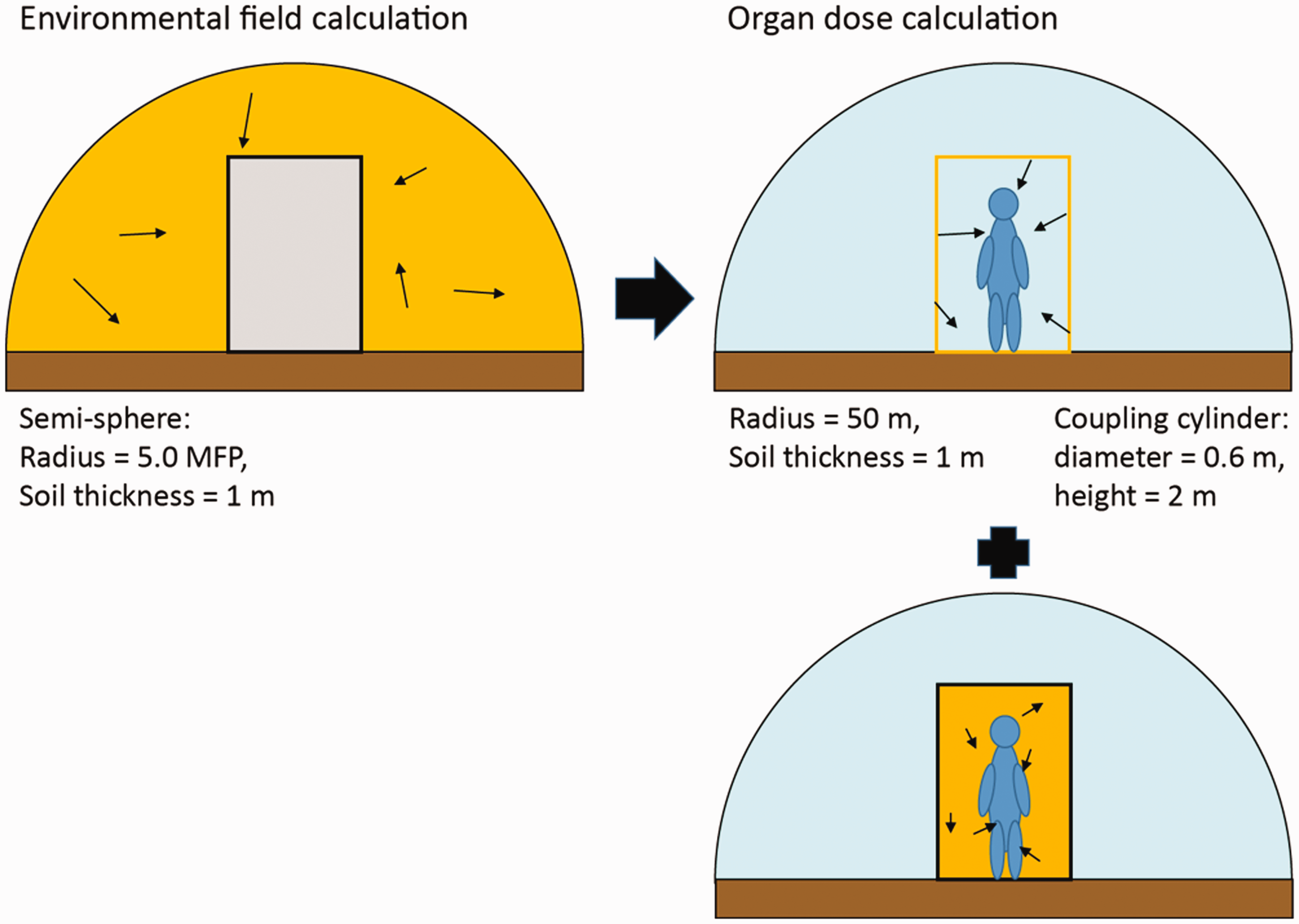
3.1. Organ absorbed dose and equivalent dose
(24) The mean absorbed dose averaged over the volume of organs and tissues is the primary scientific quantity from which effective dose is calculated. Absorbed dose, D, is defined as the quotient of mean energy, (25) The SI unit of absorbed dose is J kg−1 and its special name is gray (Gy). Absorbed dose is derived from the mean value of the stochastic quantity of energy imparted, ɛ, and does not reflect the random fluctuations of the interaction events in tissue. While it is defined at any point in matter, its value is obtained as an average over a mass element, dm, and hence over many atoms or molecules of matter. (26) When using the quantity ‘absorbed dose’ in radiological protection, doses are averaged over tissue volumes. It is assumed that for low doses, the mean value of absorbed dose averaged over a specific organ or tissue can be correlated with radiation detriment for stochastic effects in that tissue with an accuracy sufficient for the purposes of radiological protection. The averaging of absorbed dose is carried out over the volume of a specified organ (e.g. liver) or tissue (e.g. active bone marrow), or the sensitive region of a tissue (e.g. endosteal surfaces of the skeleton). (27) Equivalent dose, HT, to a tissue or organ is defined as:
Radiation weighting factors of the International Commission on Radiological Protection.


3.2. Effective dose
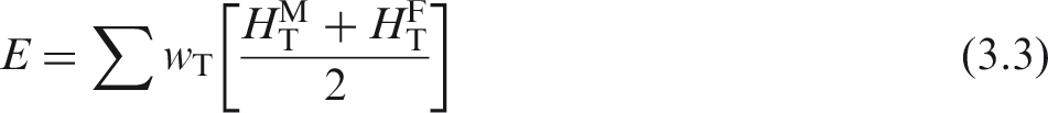
(28) Effective dose, E, introduced in Publication 60 (ICRP, 1991) is the risk-related quantity in radiation protection and is defined as a weighted average of organ equivalent doses. In accordance with the definition of effective dose in Publication 103 (ICRP, 2007), the effective dose is computed as:
(29) Effective dose was originally introduced for the control of occupational exposures to external and internal sources of radiation. While the concept has remained essentially unchanged through Publication 60 (ICRP, 1991) to Publication 103 (ICRP, 2007), its use has been extended to members of the public of all ages, including in-utero exposures of the fetus (ICRP, 2001, 2004, 2006). ICRP provides effective dose coefficients for situations of external and internal exposures of workers and members of the public, and for radiopharmaceutical administrations to patients as reference values for use in prospective and retrospective dose assessments. (30) The tissue weighting factors of Table 3.2 are sex- and age-averaged values for all organs and tissues, including the male and female breast, testes, and ovaries (i.e. gonads, related to possible carcinogenic and heritable effects). This averaging implies that the application of this approach is restricted to the determination of effective dose in radiological protection (ICRP, 2007). (31) Effective dose is calculated for sex-averaged Reference Persons at specified ages as defined in Publication 89 (ICRP, 2002). The Publication 103 (ICRP, 2007) definition includes the specification of Reference Male and Female anatomical models for radiation transport calculations. While exposures may relate to individuals or population groups, effective dose is calculated for Reference Persons exposed in the same way. (32) Effective dose, in sieverts (Sv), is accepted internationally as the central radiological protection quantity and is used for regulatory purposes worldwide, providing a risk-adjusted measure of total body dose from both external and internal sources in relation to stochastic risks of cancer and hereditary effects, expressed in terms of detriment. It has proved to be a valuable and robust quantity for use in the optimisation of protection and for setting dose criteria such as dose limits, dose constraints, and reference levels for the protection of workers or members of the public. Tissue weighting factors of the International Commission on Radiological Protection (ICRP, 2007). Remainder tissues: adrenals, extrathoracic regions of the respiratory tract, gall bladder, heart, kidneys, lymphatic nodes, muscle, oral mucosa, pancreas, prostate (male), small intestine, spleen, thymus, and uterus/cervix (female).

3.3. Air kerma
(33) For measuring external radiation, basic physical quantities that relate the radioactivity in the environment with protection and operational quantities are required. National and international standards laboratories maintain standards and reference radiation fields that are specified and described in terms of these quantities for calibration of instruments and dosimeters. Air kerma free-in-air, (34) Air kerma, K, for ionising uncharged particles, is given by:

3.4. Operational quantities
(35) The protection quantities ‘organ equivalent dose’ and ‘effective dose’ are not measurable, and therefore cannot be used directly as quantities in radiation monitoring. Operational quantities are thus used for assessment of the protection quantities (effective dose or equivalent dose in tissues or organs). The operational quantities aim to provide a reasonable estimate of the values of protection quantities relevant to the exposure of humans to external radiations under most irradiation conditions (ICRU, 1985, 1988, 1993), and they are often used in practical regulations or guidance. (36) The operational quantities are defined using the quantity ‘dose equivalent’ (H) (ICRU, 1985). H is the product of Q and D at a point in tissue; thus, (37) For area monitoring, two quantities – ambient dose equivalent, (38) For individual monitoring, the personal dose equivalent, (39) The recommended values of d are chosen for the assessment of various doses:
4. THE ICRP REFErENCE PHANTOMS
4.1. Adult reference computational phantoms
(40) Computational phantoms of the human body – together with radiation transport codes – have been employed for many years in the evaluation of organ equivalent dose-rate coefficients in environmental radiation protection. Over the last two decades, voxel phantoms have been introduced that are derived mostly from (whole-body) medical image data of real persons instead of the older stylised computational body models. A voxel model (or phantom) is a three-dimensional representation of the human body in the form of an array of identification numbers, arranged in slices, rows, and columns. Each entry in this array represents a tissue voxel; organs are then represented by those voxels having the same identification number, and are spatially arranged to represent the organ volume. More information on voxel phantoms, their development, and their use can be found elsewhere (Xu and Eckerman, 2010). (41) For the computation of organ absorbed doses, the adult male and female reference computational phantoms, representing ICRP Reference Adult Male and Reference Adult Female (ICRP, 2007), were used in this publication. These phantoms were adopted by ICRP and ICRU as the phantoms for computation of the ICRP reference dose coefficients, and are described extensively in Publication 110 (ICRP, 2009a). The reference computational phantoms are based on human computed tomographic (CT) data and were constructed by modifying the voxel models (Zankl and Wittmann, 2001; Zankl et al., 2005) of two individuals (Golem and Laura) whose body height and mass closely resembled the reference data. The organ masses of both phantoms were adjusted to the ICRP data given in Publication 89 (ICRP, 2002) on the Reference Male and Reference Female with high precision, without significantly altering their realistic anatomy. The phantoms contain all target regions relevant to the assessment of human exposure to ionising radiation for radiological protection purposes, including all tissues and organs that contribute to the protection quantity ‘effective dose’ (ICRP, 2007). (42) The male reference computational phantom consists of approximately 1.95 million tissue voxels (excluding voxels representing the surrounding vacuum), each with a slice thickness (corresponding to the voxel height) of 8.0 mm and an in-plane resolution (i.e. voxel width and depth) of 2.137 mm, corresponding to a voxel volume of 36.54 mm3. The number of slices is 220, resulting in body height of 1.76 m and total body mass of 73 kg. The female reference computational phantom consists of approximately 3.89 million tissue voxels, each with a slice thickness of 4.84 mm and an in-plane resolution of 1.775 mm, corresponding to a voxel volume of 15.25 mm3. The number of slices is 346, resulting in body height of 1.63 m and total body mass of 60 kg. The number of individually segmented structures is 136 in each phantom, and 53 different tissue compositions have been assigned to them. The various tissue compositions reflect both the elemental composition of the tissue parenchyma (ICRU, 1992) and each organ’s blood content (ICRP, 2002) (i.e. organ composition inclusive of blood). Fig. 4.1 shows frontal (coronal) views of the male (right) and female (left) computational phantom, respectively. (43) Due to the limited resolution of the source tomographic data upon which these phantoms were constructed, and the very small dimensions of some of the ICRP defined source and target regions, it was not possible to represent all tissues explicitly. In the skeleton, for example, the target tissues of interest are the haematopoietically active bone marrow located within the marrow cavities of spongiosa, as well as the endosteal layer lining the surfaces of the bone trabeculae and the inner surfaces of the medullary cavities of the long bones (presently assumed to be 50 µm in thickness). Due to their small dimensions, these two target tissues had to be incorporated as homogeneous constituents of spongiosa within the reference phantoms. At lower energies of photons and neutrons, secondary charged-particle equilibrium is not fully established in these tissue regions over certain energy ranges. Consequently, more refined techniques for accounting for these effects in skeletal dosimetry were used in this publication, and are discussed in more detail in Annex B. Images of the adult male (right) and adult female (left) computational phantoms (ICRP, 2009a). The following organs can be identified by different surface colours: breast, colon, eyes, lungs, liver, pancreas, salivary glands, small intestine, stomach, thyroid and urinary bladder, testes, and teeth. Muscle and adipose tissue are semi-transparent.

4.2. Paediatric phantoms
(44) The 10 ICRP paediatric computational phantoms, described in Publication 143 (ICRP, 2020), are as follows:
newborn – male and female; 1-year-old – male and female; 5-year-old – male and female; 10-year-old – male and female; and 15-year-old – male and female. (45) These phantoms were derived from a series of computational phantoms developed originally at the University of Florida (UF) and later in collaboration with the National Cancer Institute (NCI). Consequently, the original phantoms from which the ICRP paediatric phantoms were derived are presently referred to as the ‘UF/NCI phantom series’ (Lee et al., 2010). The UF/NCI phantoms are a third generation of phantom technology – hybrid phantoms – in which the outer body contour and internal organ surfaces are modelled using the computer animation techniques of either polygon mesh (PM) or non-uniform rational B-spline (NURBS) surfaces, depending on the complexity of anatomical structures. PMs are a cluster of adjacent triangles, while the NURBS surfaces are a cluster of three-dimensional points in space between which a surface is interpolated. In the past few years, it has become possible to use computational phantoms in these two formats directly in some Monte Carlo transport codes. However, most transport codes still utilise a voxel format, composed of tiny cuboidal prisms. A computer script was thus used to convert the UF/NCI hybrid phantoms from their surface format to a voxel format for Monte Carlo simulations conducted in the current publication. These ICRP reference paediatric phantoms in voxel format are thus consistent with the format of the Publication 110 reference adult phantoms (ICRP, 2009a). (46) As noted in Lee et al. (2010), the UF/NCI series of phantoms can be traced directly to real human anatomy. The newborn phantom is based on full-body CT imaging of a 6-day-old female cadaver, while the remainder of the paediatric series (1-year-old to 15-year-old phantoms) are based upon combinations of head CT images, full torso CT images, and rescaled CT-based images of adult arms and legs. The latter approach was necessary as medical imaging of children rarely includes the arms within the imaging field. From the initial series of segmented images, various anatomic sources were used to resize both internal organ anatomy and exterior body size. The most important document used was Publication 89 (ICRP, 2002), providing internal organ masses, total weight, and total height. Additional reference sources were used to target various body circumferential dimensions not given as reference values in Publication 89. The final series of the UF/NCI hybrid phantoms thus fully conforms to reference anatomy specified by ICRP, and is fully traceable to real human CT anatomy. In this manner, the ICRP paediatric phantom series is fully compatible with the process used to develop the Publication 110 (ICRP, 2009a) phantoms, which were also based on segmentation of real human CT anatomy. (47) Another unique feature of the ICRP paediatric phantoms (and of the UF/NCI phantoms) is their explicit coupling to micro-CT-based models of skeletal dosimetry. As noted in Hough et al. (2011) and Johnson et al. (2011), an extensive series of cadaver bone harvests, ex-vivo skeletal CT imaging, and ex-vivo spongiosa core micro-CT imaging were used to construct models of tissue dosimetry in the skeletons of the ICRP reference adult phantoms. This work is described more formally in Annexes D and E of Publication 116 (ICRP, 2010). The paediatric series of ICRP reference phantoms similarly has accompanying models of skeletal anatomy at both macrostructural and microstructural dimensions. Thus, the methods proposed in Publication 116 for external photons and neutrons, and in Publication 133 (ICRP, 2016a) for internal beta and alpha particles, as well as photons, for the Publication 110 (ICRP, 2009a) adult phantoms are available in reporting skeletal tissue dosimetry to paediatric members of the reference series. (48) The following further refinements have been made to the UF/NCI series of paediatric phantoms (Pafundi, 2009; Wayson et al., 2012):
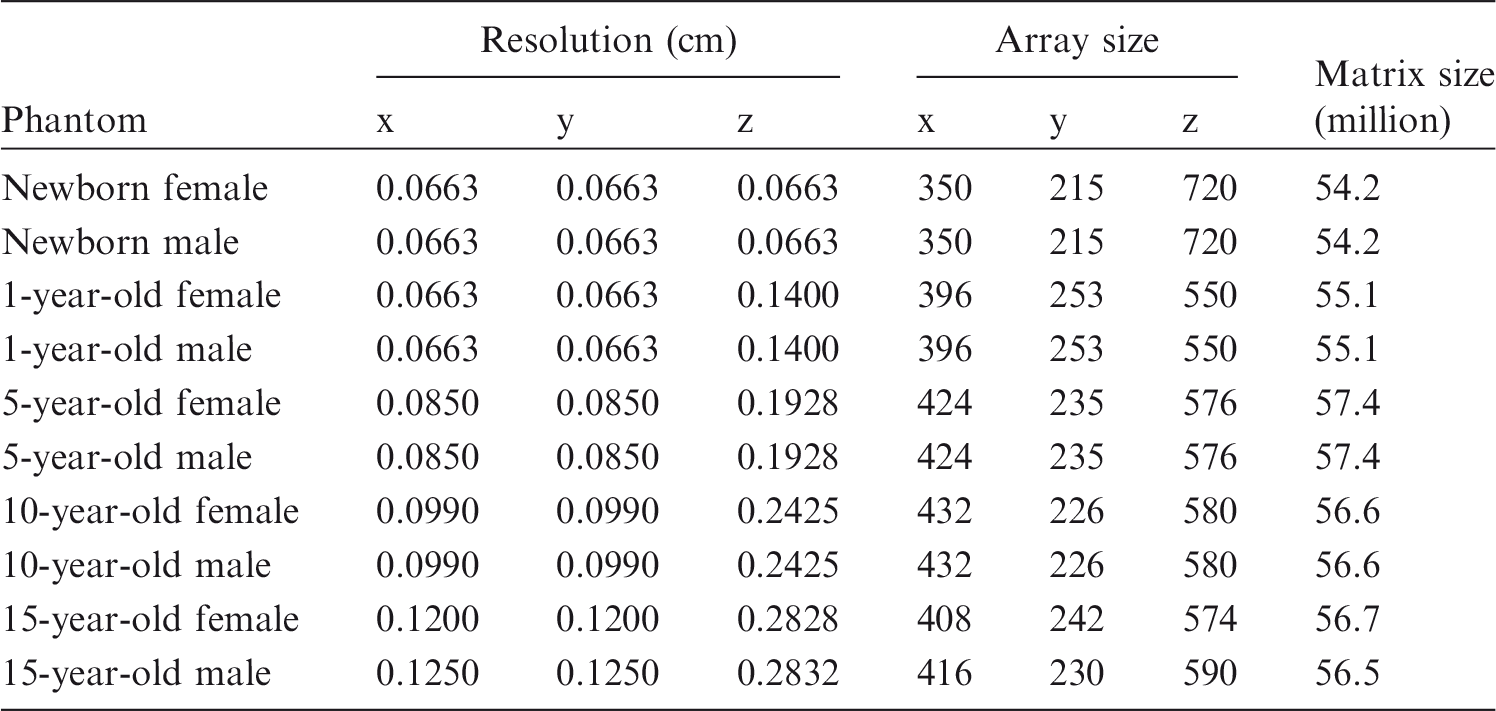
a sub-segmented skeletal model to include regions of cortical bone, spongiosa, and medullary marrow; photon dose–response functions for internal and external photon dosimetry to active marrow and endosteum; a new age-specific regional blood distribution model (Wayson, 2018); a corresponding model of the major blood vessels; separation of subcutaneous fat and skeletal muscle from what was formally residual soft tissues; and inclusion of lymphatic nodes (Lee et al., 2013). (49) The series of ICRP paediatric reference phantoms (ICRP, 2020) is in voxel format, and fully conforms to the framework established in Publication 110 (ICRP, 2009a). All organs and tissue structures modelled in the Publication 110 reference adult male and female phantoms are included with consistent identification numbers (see Annex A of Publication 110). Representative images of the ICRP paediatric series are given in Fig. 4.2. (50) While the ICRP paediatric reference phantoms are identical in format to the Publication 110 (ICRP, 2009a) adult phantoms regarding the identification numbers of the various source and target organs, one important difference is the voxel resolution. One of the main advantages of hybrid phantom technology is that in conversion of the PM/NURBS format of the phantom to the voxel format of that same anatomy, one can select the voxel resolution. Table 4.1 tabulates the voxel resolutions, array size, and total matrix size finally adopted for the ICRP paediatric phantoms. These ensure continuous conformance with the 1% matching of reference masses, and also conform to reference total skin thickness as given by data in Publication 89 (ICRP, 2002). It is noted that for the newborn phantom, the voxels are cubic (i.e. same thickness in x, y, and z directions), while rectangular prisms with larger z dimensions than xy dimensions were adopted for the older phantoms in order to keep the matrix size between 55-58 million voxels in total. In contrast, the Publication 110 adult male and female phantoms have total matrix sizes of 1.9 and 3.9 million voxels, respectively. The need for higher resolution is to preserve organ anatomy in the smaller anatomy of the paediatric reference individuals. Voxel resolution, voxel number, and total matrix size of the International Commission on Radiological Protection paediatric computational phantom series. Series of International Commission on Radiological Protection reference paediatric phantoms (ICRP, 2020). The male and female newborn, 1-, 5-, and 10-year-old phantoms are anatomically identical, except for their gonads.

5. SIMULATION OF THE ENVIRONMENTAL RADIATION FIELD (STEP 1)
(51) Photons emitted from sources distributed in the environment are scattered and/or absorbed in both air and soil, and their energy spectrum and angular distribution in air have specific features dependent on the initial energy and spatial distribution of the emission sites. In the case of volumetric sources in air or ground, the angular distribution of incident photons is nearly uniform for the hemisphere from which the source originates, while small amounts of scattered photons emerge from the opposing semi-sphere (Saito et al., 1998). In the case of deposited sources in the ground, the dominant component of photons is incident along horizontal directions. The Monte Carlo method is a suitable tool able to simulate particle transport and the detailed environmental conditions. (52) For simulating exposure to environmental radiation, the following three typical cases of environmental sources have been addressed in this publication: (1) soil (ground) contamination, simulated as semi-infinite planar sources on the surface and at different depths in the ground; (2) air submersion, simulated as a semi-infinite volume source in air; and (3) water immersion, simulated as a fully infinite source in water. The first source simulates the contamination of radionuclides on and below the ground surface, by assuming an infinite planar source on the surface and in the soil. This geometry is supposed to be semi-infinite in extent as the radiation is emitted from and below the air–ground interface. The second source configuration models the gaseous radioactive release into the atmosphere at locations which are not too near to the release point, by assuming homogeneous contamination of the air in a hemispherical, semi-infinite region above a smooth air–ground interface with a radius that depends on the mean free path (mfp) of the photons of interest. The third source simulates immersion in uniformly contaminated water. For the first and second source configurations, the human body is assumed to be standing upright on the ground, while for water exposures, the human body is assumed to be fully immersed. (53) The transport of radiation particles in the environment was simulated using the Monte Carlo simulation package ‘Particle and Heavy Ion Transport code System’ (PHITS) (Sato et al., 2013). PHITS is a multi-purpose Monte Carlo code that simulates the transport and interaction of hadrons, leptons, and heavy ions in arbitrary three-dimensional geometries. Version 2.66 of the PHITS code was used in this publication (Sato et al., 2013). For simulating photon and electron transport, respectively, the atomic data libraries MCPLIB04 (White, 2003) and EL03 (Adams, 2000) were employed. These libraries provide precise cross-section data and can treat various physical processes of both photons and electrons. (54) PHITS defines the geometry of the calculation model in terms of the combinatorial geometry and the general geometry. In addition, a capability for describing repeated structures and lattice geometries is available to define three-dimensional voxel phantoms. PHITS has a function to draw two-dimensional and three-dimensional figures of the calculation geometries as well as the computed data results using the ANGEL graphic package (Niita et al., 2010). (55) In the environmental radiation transport simulation for photon sources, only photons were transported; secondary electrons generated by photon interactions were not followed. This is because the secondary electrons lose their energies continuously and stop within a short distance in the environmental media, for example at 10 and 400 cm for 0.1 and 1.0 MeV electrons, respectively, in air. However, bremsstrahlung photons generated by secondary electrons have maximum energies comparable to those of the secondary electrons, and are able to propagate across long distances. Production of the bremsstrahlung photons, and their energy and emission angle were sampled at the interaction point based on a thick-target bremsstrahlung approximation model (MCNP, 2003). For electron sources, both primary electrons and their secondary photons were transported in the environment. (56) As mentioned previously, the radiation field computed due to monoenergetic radiation emissions from within the contaminated air and soil was expressed as the position, angle of incidence, and energy of the particles incident on the surface of a virtual cylinder of 2 m height and 0.6 m diameter, which surrounds the exposed individual, and is termed the ‘coupling cylinder’. As the phantom was not present in this first step, the same coupling cylinder source could be applied for all phantoms. Fig. 5.1 shows schematically which particles were recorded on the surface of the coupling cylinder. (57) To cover the wide energy range of radiations that are emitted by many different radionuclides, the monoenergetic photon and electron energies considered varied from 0.01 to 8 MeV. Schematic representation of particle transport during Step 1 and Step 2 of the calculation.

5.1. Soil contamination
(58) Soon after deposition, radionuclides deposited on the ground are assumed to form a planar source at the ground surface. Over time, these radionuclides will migrate or leach into the soil, thereby developing diverse concentration depth profiles in terms of both shape and degree of soil penetration (Matsuda et al., 2015). In many cases, the distribution of radionuclides with respect to soil depth could be approximated as being due to many infinite planar sources within the ground. These functions could have various characteristics, showing peaks at different depths in the soil. As it is not practical to simulate every radionuclide/soil migration function, the simulation of a series of planar radiation sources at different depths can provide basic data enabling one to extrapolate or interpolate these results to model diverse source profiles within the contaminated ground. (59) The air-over-ground geometry was modelled, such as air bounds on the ground with an infinite flat surface. In the real environment, the terrain is not normally flat nor infinite; however, the infinite flat terrain is a conservative representation of the air-soil interface for dose calculations. For example, in the case of an exponentially distributed ground source with a relaxation mass per area of 1 g cm−2, which is a typical depth observed soon after radionuclide ground deposition, approximately half of the measured ambient dose equivalent at 1 m above ground is attributed to photons from sources within a radius of 5 m in the ground (Malins et al., 2015). Thus, a limited series of flat ground surface areas is considered to adequately model the exposure for many real exposure situations. (60) The monoenergetic radioactive sources were defined as planar sources at depths in soil expressed in terms of mfp of photons in soil: 0.0 (i.e. contamination is on the surface), 0.2, 1, 2.5, and 4 mfp. For most exposure situations, the consideration of mfp up to 4 would be sufficient (Eckerman and Ryman, 1993); however, the source depth profile might be changed due to, for example, ploughing. Thus, dose-rate coefficients for a wider range of mfp would be useful, and had been considered to facilitate an accurate integration when determining dose-rate coefficients for continuous source-depth profiles. The air–ground interface (0 mm) is a flat planar source without any soil covering the source. This is an idealised geometry and does not exist in reality as various factors provide shielding from ground surface sources. These include the presence of vegetation, surface roughness, and particle movement due to gravitational forces (Burson and Profio, 1977; Kocher and Sjoreen, 1985; Jacob and Paretzke, 1986). (61) Fig. 5.2 (left) shows schematically the simulation geometry, which consists of a right circular cylinder constructed from a layer of air with a height of 3 mfp and soil with a depth depending on the photon energy: 2 mfp of photons in soil for source depth 0.0 mfp and 0.2 mfp; 3 mfp for source depth of 1.0 mfp; 3.5 mfp for source depth of 2.5 mfp; and 5 mfp for source depth of 4.0 mfp. The additional thickness of at least 1 mfp below the source depth was considered sufficient to account for backscatter events in the deeper layers. The radius of the cylinder corresponds to approximately five times the mfp of the relevant photons in air, and approximates the fully infinite planar geometry of the source. A previous study (Satoh et al., 2014) has shown that this size of simulation geometry is sufficient to properly treat photon transport in the contaminated environment. (62) Table 5.1 lists the density and elemental composition of air and soil adopted in the computations of this publication. The values were obtained from the data for soil (Type 1) provided by ICRU (1994) and dry air from the National Institute of Standards and Technology (NIST) (Berger et al., 2005), respectively. The densities of soil and air were considered to be 1.0 g cm−3 and 1.2 × 10⁻³ g cm−3, respectively. In real environmental exposure situations, the soil densities are mostly higher than 1.0 g cm−3 and could vary according to both location and depth; however, this variation does not affect the relation of source intensity to the radiation field in air, if source depth is expressed in terms of g cm−2. Furthermore, it has been shown that changes in soil composition do not significantly alter the transported photon fields at the phantom coupling surface (Saito and Jacob, 1995). (63) The radiation field was derived for 25 initial photon energies, ranging from 0.01 to 8 MeV, in order to cover the wide energy spectra of naturally occurring and artificially produced radionuclides. The soil was assumed as a planar air–ground interface, and scatter and absorption of the radiation fields in both air and ground were considered in the calculations. (64) Planar sources emitting electrons were also considered for the surface of the soil; for other depths, primary electron sources were not transported as they will not travel sufficiently far to reach the surface. Initial electron energies from 0.01 to 8 MeV were considered, and both electrons and secondary photons were transported. It should be noted that bremsstrahlung x rays were considered in both the soil and air exposure scenarios. (65) From the transport calculations in the environment, individual particles were recorded at the surface of the coupling cylinder. This cylinder is positioned on the ground concentric with the simulation geometry, as depicted in Fig. 5.2 (right). The diameter of the cylinder is 0.6 m and its height is 2 m. The phase–space coordinates are recorded for particles that cross the surface of the cylinder, and consist of the spatial coordinates (x, y, z), momentum (px, py, pz), kinetic energy, and Monte Carlo weight. In order to avoid counting a particle as it exits the cylinder, the space inside the coupling cylinder is treated as an ideal absorber such that the Monte Carlo code terminates the transport of the particle when it enters this region. The data were recorded to an external file in ASCII format to be used for the Step 2 calculations – organ equivalent dose calculations within the phantoms. The small fraction of those photons that could be scattered back into the cylinder from the ground or air is followed in Step 2 calculations (i.e. particles starting from the surface of the coupling cylinder). More details on the method can be found in Satoh et al. (2015). (66) To reduce the variance of the Monte Carlo simulations, the uniform source was reproduced by increasing the number of photons or electrons emitted per unit area and decreasing the Monte Carlo weight of photons or electrons as their emission point approached the coupling cylinder (Satoh et al., 2015). The number of Monte Carlo histories in Step 1 calculations was determined in order to achieve 1% or less statistical uncertainty of simulations of air kerma for the respective environmental radiation. (67) Fig. 5.3 shows an example of the energy and angular distribution of environmental photons from a source of 0.5 MeV at a depth of 0.2 mfp, at heights 0–0.40 m and 1.60–2.00 m, as recorded on the surface of the coupling cylinder. The incident directions of photons are expressed as the sine of a vector parallel to the ground surface and the angles are expressed as elevation angles; for example, (68) Uncollided photons are recorded in the highest energy bin. Overall, approximately 20% of the recorded photons on the coupling cylinder do not interact with the air or soil. It should be noted that the shape of the energy and angular spectra are almost independent of height. (69) The directional distributions of scattered and uncollided photons for a source of 0.1 MeV at 1 and 4 mfp depths in the ground, respectively, are shown in Fig. 5.4. The scattered photons show a small local maximum at shallow directions downwards, which is more pronounced for 1 mfp than for 4 mfp. This is in agreement with the angular dependence of air kerma for sources at 1 and 4 mfp, respectively, as reported by Eckerman and Ryman (1993). The relative number of uncollided photons is considerably decreased from approximately 22% at 1 mfp to approximately 7% at 4 mfp. Density and elemental composition of air (Berger et al., 2005) and soil (ICRU, 1994). Schematic representation of the geometry simulating the environmental field due to soil contamination. mfp, mean free path. Energy (left) and angular (right) distribution of an isotropic infinite source in the soil at a depth of 0.2 mfp, emitting 0.5 MeV monoenergetic photons at different height ranges. Left: the y axis shows the number of photons per energy bin, divided by the total number of photons within the indicated spectrum and the energy bin size (in MeV). Right: The y axis shows the number of photons per sine angle at the two indicated height ranges. The respective distributions are normalised to the total number of photons recorded on the coupling cylinder. To differentiate these distributions from the respective total distribution (Φ), they are marked by the superscript j. Angular distribution of scattered and uncollided photons (summarised over all heights) for an isotropic infinite source in the soil at a depth of 1 mfp (left) and 4 mfp (right) emitting 0.1 MeV monoenergetic photons. The y axis shows the number of photons per sine angle. To differentiate these distributions from the respective total distribution (Φ), they are marked by the superscript j. The distributions are normalised to the total number of photons.
5.2. Submersion in contaminated air
(70) In the air submersion exposure scenario, the contaminated air represents the gaseous radioactive release into the atmosphere at locations which are not too close to the release point, and are assumed to be homogeneous in air activity concentration (i.e. well-mixed air) above a smooth air–ground interface. Near the release point, the air submersion geometry used in this publication may not be appropriate. Photons enter a human body mostly from above, if the human body is positioned below the plume, while incident angles of photons are biased in horizontal directions if the human body is distant from the plume. Evaluating dose-rate coefficients considering these complex situations is not practical, and thus the submersion model of this publication is a conservative approximation of exposures for most cases. Fig. 5.5 shows schematically the air submersion geometry. The geometry is considered to be semi-infinite in extent. The air–ground interface is assumed to be an uncontaminated flat surface of infinite area. The elemental composition of air is shown in Table 5.1 and corresponds to dry air at a density of 1.2 × 10⁻³ g cm−3. Bellamy et al. (2019) estimated air kerma as a function of air density, and Fig. 5.6 shows an example of these results for 1 MeV photons. The authors found that the functional relation between air kerma and air density is virtually independent of photon energy. Using these values, the dose-rate coefficients for air submersion can be scaled to account for different air densities. With increasing humidity, air density increases and, consequently, air kerma decreases (see Fig. 5.6). (71) The number of histories, reduction variance techniques, and scoring of the particles were similar to those mentioned above for soil contamination. (72) The particles originated from the air region are transported and scored on the surface of the coupling cylinder, placed on the air–ground interface. The coupling surface records the position, angle, energy of incident photons, and Monte Carlo weight as discussed in Section 5.1 regarding soil contamination. This method produces energy-dependent fluences. As for the soil contamination exposure scenario, calculations were performed for 25 monoenergetic sources of photons and electrons ranging from 0.01 to 8 MeV. (73) Fig. 5.7 shows the energy spectrum of environmental photons from a source of 0.5 MeV at heights of 0–0.40 m and 1.60–2.00 m. The incident directions of photons are expressed as the sine of the vector parallel to the ground surface. It can be seen that for many photons, no scattering is observed, and most photons come from upper directions with little dependence of their directional distribution on height. Schematic view of the geometry simulating submersion in contaminated air. The region coloured yellow indicates the source region (left). For organ equivalent dose calculations (right), the medium inside the coupling cylinder is air. For electron exposures, the particles start not only from the surface of the cylinder (right top) but also from the inside of the cylinder (right bottom). For photons, this is not necessary as the mean free path of photons in air is long and the source inside the cylinder does not significantly contribute to the results of organ equivalent dose calculations. Air kerma as a function of air density for 1 MeV photons (Bellamy et al., 2019). Energy (left) and angular (right) distribution of a semi-infinite source in the air emitting 0.5 MeV monoenergetic photons. Left: the y axis shows the number of photons per energy bin, divided by the total number of photons within the indicated spectrum and the energy bin size (in MeV). Right: The y axis shows the number of photons per sine angle at the indicated height range. To differentiate these distributions from the respective spectrum for all heights (Φ), they are marked by the superscript j.

5.3. Water immersion
(74) Water immersion might be rare in the pathway of environmental exposure. Nevertheless, many facilities have routine liquid effluent releases, and radioactive releases to the sea or contamination of surface waters have been observed after major radiological accidents. Highly contaminated water from a damaged reactor core and water pools resulting from damaged nuclear fuels can be released through direct or ground water discharge, as occurred as a consequence of the accident at Fukushima NPP in 2011 (Buesseler et al., 2017). Radionuclides such as radioactive iodine and caesium were detected in tap water, and the exposure due to contaminated water used for bathing had to be estimated. Moreover, radionuclides were released into the sea and could have been potentially harmful for people who enter the sea around the NPP following the accident. (75) Fig. 5.8 shows schematically the water immersion geometry. The source geometry is assumed to be infinite in extent. The water density is 1.0 g cm−3 and the composition by mass fraction is 0.112 for H and 0.888 for O, representing pure liquid water. The phantoms are assumed to be completely immersed in the water, and are placed at the centre of a sphere with a radius of 2 m, corresponding to 5 mfp at a photon energy of 8 MeV in water. Monoenergetic sources of photons and electrons are generated uniformly in the contaminated water. The secondary photons and electrons, as well as bremsstrahlung photons, are transported directly by the PHITS Monte Carlo transport code. The organ equivalent dose-rate coefficients for water immersion of the male and female phantoms at the six reference ages have been calculated in a single step, and thus no coupling cylinder was required. Schematic view of water immersion. The sphere is centred on the midpoint of the three axes of the phantom.

5.4. Calculation of air kerma and ambient dose equivalent in the environmental field
(76) Air kerma and ambient dose equivalent have been widely used for the purpose of radiation protection after environmental exposures (ICRU, 1994; IAEA, 2000a). Air kerma is a basic quantity related to photon energy fluence. In natural environments, air kerma is substantially equivalent to the air absorbed dose which has been used by the UNSCEAR to express dose-rates in air (UNSCEAR, 2000, 2008). Absorbed dose in air has been used in environmental monitoring for many years, but it has been gradually replaced by the operational quantity ‘ambient dose equivalent’ as seen in worldwide environmental monitoring data (European Commission Joint Research Centre, 2017). The ambient dose equivalent, H*(10), defined as the dose equivalent for aligned and expanded radiation fields at a depth of 10 mm in the ICRU sphere consisting of ICRU four-element tissue, is aimed to conservatively evaluate effective doses for diverse exposures (ICRU, 1993). Although the operational quantity ‘ambient dose equivalent’ was originally introduced for radiation protection of workers (workplace monitoring), this dosimetric quantity has also been applied to environmental monitoring: the instruments for monitoring of H*(10) generally have an isotropic response with respect to both energy and angular distributions of incident photons. Resultantly, these instruments work well in environmental fields which exhibit complex angular and energy distributions, even if they are calibrated under simple conditions such as for unidirectional irradiation using monoenergetic sources. Consequently, environmental monitoring data worldwide are generally expressed as dose-rates in air reported as the operational quantity ‘ambient dose equivalent’ (European Commission Joint Research Centre, 2017; Cinelli et al., 2019). (77) In order to relate the air contamination and ground contamination densities of a radionuclide to dose-rates in air, coefficients are required for both air kerma and ambient dose equivalent rates. Many authors have published coefficients relating radionuclide concentration in the environment to air kerma rate (Dillman, 1974; O'Brien and Sanna, 1976; ICRU, 1994; Saito and Jacob, 1995) and to ambient equivalent rate (Lemercier et al., 2008; Saito and Petoussi-Henss, 2014), and these data have been used in environmental dose evaluations. In the present publication, these coefficients have been recalculated considering air kerma and ambient dose equivalent rates at 1 m above ground for both the soil contamination and air submersion exposure geometries described above from monoenergetic photon sources. For the simulations, the Monte Carlo code PHITS (see Section 5) was used, and the same environmental conditions were considered as those applied in the calculation of the environmental fields (see Sections 5.1 and 5.2). (78) The calculation was made by simulating a 30 cm-diameter sphere filled with and surrounded by air, 1 m above ground, and scoring the particles entering the sphere. The 30 cm diameter was chosen to represent the size of the torso of adults. The transport simulation was restarted from the surface of the coupling cylinder using the data in the external file created in Step 1 of the calculations. Fig. 5.9 shows a schematic representation of the calculation geometry. The photon fluence scored at the air sphere is converted to an air kerma rate and ambient dose equivalent rate using the dose coefficients given in Publication 74 (ICRP, 1996). The relative uncertainties of these quantities were less than 1%. (79) For soil and air contamination, air kerma depends on distance from the ground; while this dependence is weak for sources in air, it is pronounced for planar sources in the ground. (80) On the basis of these results, ambient dose equivalent rate coefficients, (81) Individual monitoring of effective dose is performed with dosimeters calibrated in terms of personal dose equivalent, Hp(d). ICRP Task Group 90, which developed this publication, decided not to include dose coefficients for Hp(d). Hp(d) is defined as the dose equivalent in soft tissue at an appropriate depth, d, below a specified point on the body. The specified point is usually given by the position where the individual’s dosimeter is worn. Calibration of personal dosimeters is performed by exposure to unidirectional radiation at an incident angle, α, where α is the angle between the direction of incidence of radiation and the reference direction of the personal dosimeter mounted on the front face of the calibration phantom. These calibrations are typically performed for normally incident radiations, i.e. α = 0°. On the other hand, for the environmental fields considered in this publication, photons of wide energy distribution are incident from various angles. Standardising reference radiation fields and calibration procedures simulating environmental radiation have not been recommended by international organisations, such as the International Organization for Standardization and the International Electrotechnical Commission. The coefficients for Hp(10) could be calculated using phantoms and environmental radiation sources by Monte Carlo simulations. However, methods of calibration of personal dosimeters to link dosimeter readings to the dose-rate coefficients of Hp(d) have not yet been established for environmental exposures. (82) Individual monitoring in environmental radiation fields has often been performed in Japan since 2011, with dosimeters calibrated by irradiation of unidirectional radiation (Nuclear Regulation Authority Japan, 2013). It is of significant concern whether the personal dosimeters calibrated in this way provide reasonable values for assessment of effective dose in the environmental fields. To address the question, Satoh et al. (2017) analysed the relation of effective dose, H*(10), and Hp(10) in radiation fields originating from photon emissions from both 134Cs and 137Cs distributed in different depths in soil, where the personal dosimeters were calibrated under the above simplified exposure conditions. A conclusion of their analysis is that both area monitoring and individual monitoring provide reasonably conservative estimates of effective dose for the conditions investigated. Schematic representation of calculation of air kerma and ambient dose equivalent.

6. DETERMINATION OF DOSE-RATE COEFFICIENTS FOR MONOENERGETIC PARTICLES (STEP 2)
6.1. Monte Carlo photon and electron transport calculation in the anthropomorphic phantoms
(83) For the aforementioned exposure geometries of contaminated soil and air, Step 2 calculations involved the computation of organ equivalent dose-rate coefficients in each sex- and age-specific phantom resulting from the simulated radiation fields from Step 1 calculations. The recorded particle histories on the coupling cylinder were used as the source irradiating the phantom, and each phantom was placed inside the air-filled coupling cylinder. (84) Particle transport calculations starting from the surface of the coupling cylinder were performed with PHITS Version 2.66 (Sato et al., 2013). The atomic data libraries MCPLIB04 (White, 2003) and EL03 (Adams, 2000) were used for photon and electron transport, respectively. (85) Step 2 considers photon as well as electron fields as these were recorded on the coupling cylinder. For photon fields, secondary electrons were also transported. The combined relative uncertainty (i.e. one standard deviation) from both Step 1 and Step 2 computations was less than 10% for most organs and tissues, where the dominating contribution stems from the environmental field calculations. (86) The computational methods for determining the equivalent dose-rate to active marrow and skeletal endosteum are described in Annex B. (87) For evaluating the absorbed doses to the sensitive layer of the skin, which is considered to be 50–100 µm below the skin surface, polygon mesh (PM) formats of the reference phantoms were applied, together with the Monte Carlo code GEANT4 (Agostinelli et al., 2003). Further details on estimates of skin dose are given in Annex C. (88) The organ equivalent doses were evaluated in the form of dose-rate coefficients, giving the mean organ equivalent dose-rate normalised to a measurable environmental radioactivity quantity. The doses are then estimated on the basis of the measured ground contamination levels (i.e. surface activity densities) or photon dose rate in the air, normalised to the activity density of each monoenergetic source emitter. As gamma-ray measurements in the environment are performed at 1 m above the ground surface, the normalisation quantity for measurements in air was selected to be air kerma and ambient dose equivalent at 1 m above ground at the position of the body's longitudinal axis. Values of air kerma and ambient dose equivalent at 1 m above ground normalised to source activity are also given (see Sections 5.4 and 6.6). These coefficients are used to facilitate normalisation to source activity (i.e. photon emission per unit area or per unit volume). (89) Organ equivalent dose-rate coefficients for all defined organs/tissues, including all those noted explicitly in the definition of effective dose, are given as equivalent dose-rates per radioactivity concentration. As this publication refers to environmental exposures of photons and electrons, both of which have a radiation weighting factor, wR, equal to unity, the equivalent dose-rate coefficients are numerically equivalent to their corresponding absorbed dose-rate coefficients. (90) In order to avoid fluctuation by statistical uncertainty and obtain smooth curves of the organ equivalent dose-rate coefficients as a function of photon and electron energy, data fitting was applied using the piecewise cubic Hermite function (Fritsch and Carlson, 1980). (91) For monoenergetic photon and electron sources below 0.05 and 0.10 MeV, respectively, it should be noted that the organ equivalent dose-rate coefficients were set to zero if their contribution to effective dose was below 1% and the value of the dose coefficient of the precedent energy was zero. This was done to avoid discontinuities on the curves or to improve their smoothness. (92) Reference values of the equivalent dose-rate coefficients of organs and tissues for which tissue weighting factors are defined (ICRP, 2007), as well as for the remainder tissues, can be found in the electronic supplement to this publication. The data are given separately for the male and female adult and paediatric reference phantoms. The values of the effective dose-rate coefficients are also in the electronic supplement. As these were calculated with the ICRP reference phantoms for reference geometries and following ICRP methodology, they are considered to be the ICRP reference data. (93) The results of these calculations are used to derive radionuclide-specific dose-rate coefficients through energy interpolation to obtain coefficients for the detailed photon and electron decay spectrum of each radionuclide as given in Publication 107 (ICRP, 2008) (see Section 7).
6.2. Dose-rate coefficients for soil contamination
(94) The absorbed dose delivered to internal organs and tissues is calculated by exposing the computational phantoms to the radiation fields obtained previously. Particle transport is restarted based on information on particle history written in the external file during Step 1. Calculation efficiency is improved significantly by using the environmental radiation field obtained in Step 1 which is in common to the Step 2 calculations for the reference phantoms of each age and sex. (95) The right-hand image in Fig. 6.1 illustrates the geometry of the Step 2 calculation. The phantom is placed inside the coupling cylinder, and the remaining space is filled with air. Due to the fact that the simulation geometry is cylindrically symmetrical, the transport calculation is repeated 36 times by rotating the source position in 10° steps at the surface of the cylinder around the central axis to avoid any directional bias. (96) Dose-rate coefficients for soil contamination were evaluated as the effective dose-rate per activity concentration for monoenergetic sources of photons and electrons in soil, whose energy ranges from 0.01 to 8 MeV along 25 energy points. The coefficients are given in nSv h−1 Bq−1 m2. The effective dose rates were evaluated from the data of organ equivalent dose-rates computed using the ICRP adult and paediatric (newborn, 1-, 5-, 10-, and 15-year-old) reference computational phantoms as described in Section 3.2. The data for photons were evaluated for the sources uniformly distributed in the soil over a planar area and at specific depths of 0.0, 0.2, 1.0, 2.5, and 4.0 mfp for the photons emitted in the soil. The dose-rate coefficients for continuous source-depth profiles (i.e. planar and volumetric sources) can be obtained by integration using the above data (see Sections 8.1.1 and 8.1.2). For monoenergetic electron emission, only sources on the air–ground interface were considered (i.e. primary electrons emitted at depth within the soil were not considered). However, photons (bremsstrahlung) arising as electrons slow down within the soil were considered in the manner of US Environmental Protection Agency Federal Guidance Reports 12 (Eckerman and Ryman, 1993) and 15 (Bellamy et al., 2019) using the scaled bremsstrahlung cross-section data of Pratt et al. (1977). (97) For each source depth, 10–20 million photon histories were started, depending on the photon energy, from the recorded distributions on the coupling cylinder; this led to coefficients of variance that were generally approximately 0.5% for large organs and approximately 1% for small organs. For electron irradiations, 20 million to 2 billion particle histories were followed. This led to coefficients of variance that were generally approximately 5% for all organs. Note that these coefficients of variance refer only to Step 2 organ equivalent dose calculations and not to the environmental field calculations. (98) For photon sources, coefficients for air kerma and ambient dose equivalent rates were also evaluated in air at 1 m above ground, as described in Section 5.4. (99) The effective dose-rate coefficients are shown graphically in Figs 6.2–6.6 for photon sources at depths of 0.0, 0.2, 1.0, 2.5, and 4.0 mfp together with data on the corresponding ambient dose equivalent rates. The effective dose-rate coefficients for electron sources on the ground surface are shown in Fig. 6.7. The above data of effective dose are also tabulated in the electronic supplement to this publication. The equivalent dose-rate coefficients for all organs contributing to the effective dose as well as of the remainder tissues are tabulated in regard to both age and sex, and are also compiled in the electronic supplement. (100) The figures demonstrate the age dependence of the effective dose-rate for environmental photon and electron exposures. For most energies and all geometries, the smaller the phantom (i.e. the younger the age), the larger the effective dose-rate coefficient. Larger differences are observed for the adult phantom and the newborn for energies below 0.050 MeV and contamination on the surface of the ground; effective dose of the newborn was found to be approximately six times higher than that of the adult. Also, it can be seen that, for most cases, the ambient dose equivalent rate, (101) For electron sources distributed on the surface of contaminated soil, Fig. 6.7 indicates some inconsistencies: the effective dose-rates of the newborn are smaller than those of the 1-year-old at energies below 0.07 MeV. In this energy region, the electrons stop inside the skin layer, and secondary and bremsstrahlung photons deliver the energy to the muscle region. The dose-rate coefficient of the muscle of the newborn phantom was found to be lower than that of the 1-year-old phantom. (102) Fig. 6.8 shows the variation of effective dose-rate coefficients for an adult as a function of mfp of photons in soil. The physical depth of a source in soil, given in g cm−2, is estimated by converting the depth in mfp, depending on photon energy (see Section 8.1.1); the effective dose-rate coefficients at that physical depth are then obtained by the fitted curves of the effective dose-rate coefficients as a function of mfp, using the piecewise cubic Hermite interpolation (Fritsch and Carlson, 1980). Schematic representation of the geometry simulating soil contamination. The left-hand figure describes Step 1 of the calculation and the right-hand figure describes Step 2. mfp, mean free path. Effective dose-rate coefficients for monoenergetic photon sources distributed at the surface as a ground plane source and the corresponding ambient dose equivalent rate, Effective dose-rate coefficients for monoenergetic photon sources distributed at a depth of 0.2 mean free paths (mfp) in the soil and the corresponding ambient dose equivalent rate, Effective dose-rate coefficients for monoenergetic photon sources distributed at a depth of 1 mean free path (mfp) in the soil and the corresponding ambient dose equivalent rate, Effective dose-rate coefficients for monoenergetic photon sources distributed at a depth of 2.5 mean free paths (mfp) in the soil and the corresponding ambient dose equivalent rate, Effective dose-rate coefficients for monoenergetic photon sources distributed at a depth of 4 mean free paths (mfp) in the soil and the corresponding ambient dose equivalent rate, Effective dose-rate coefficients for monoenergetic electron sources distributed at the surface as a ground plane source. mfp, mean free paths. Effective dose-rate coefficients for the adult as a function of mean free paths (mfp) in the soil for source energy of 0.5 and 0.05 MeV photons. The bullets indicate the depths for which calculations were explicitly performed. The star indicates the effective dose-rate at depth of 5 g cm−2, evaluated through interpolation.


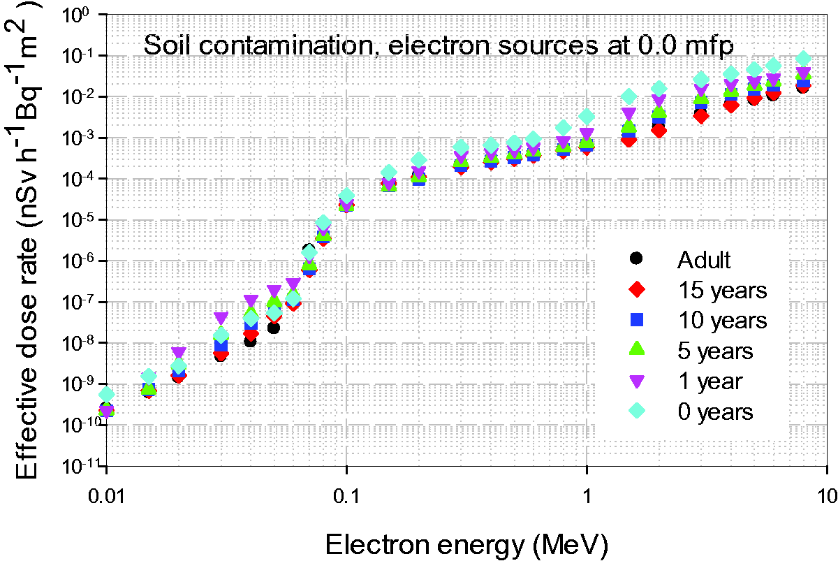
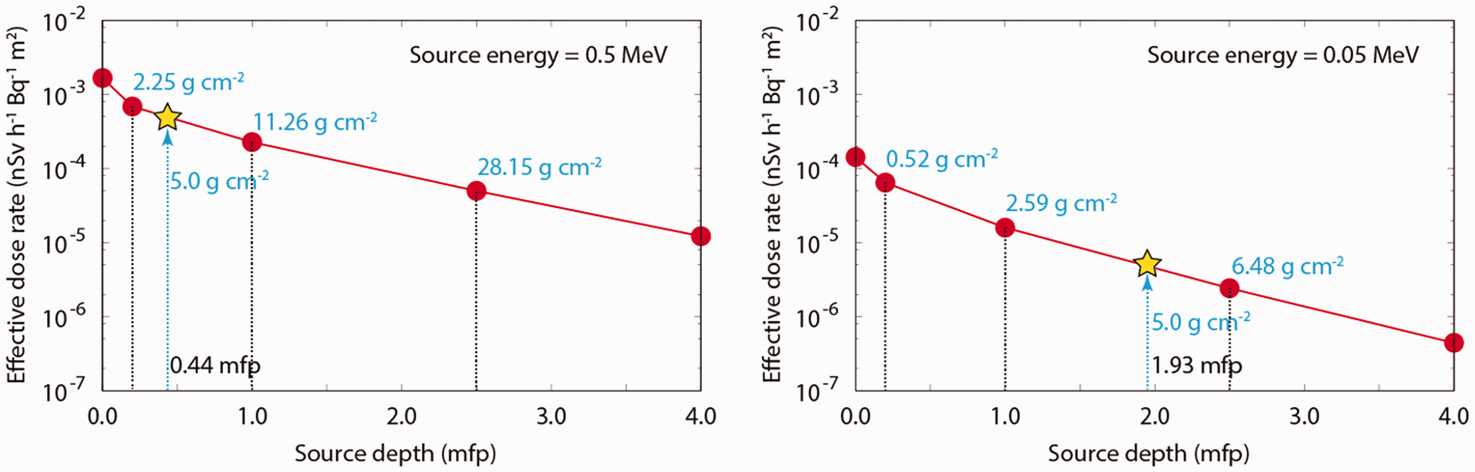
6.3. Dose-rate coefficients for air submersion
(103) The air submersion exposure geometry involves an individual standing in a large volume of uniformly contaminated air. It is assumed that the individual is standing on an uncontaminated flat surface of infinite area. The source for the submersion dose calculations is a semi-infinite cloud containing a uniformly distributed monoenergetic photon and electron emitter surrounding a human phantom standing on the soil at the air–ground interface. The right-hand image in Fig. 5.5 illustrates the irradiation geometry for the organ equivalent dose calculations. (104) Organ equivalent dose-rate coefficients in each phantom are computed using the environmental photon and electron data as recorded to the external ASCII file. It should be noted that for electron sources, the electrons do not only start from the surface of the coupling cylinder but also from within the volume of the cylinder, which is filled with contaminated air. Transport calculations for cylinder-surface source and cylinder-volume sources were performed separately. (105) The effective dose-rate coefficients of monoenergetic sources distributed uniformly in the atmosphere are shown in Figs 6.9 and 6.10 as a function of photon and electron energies, respectively. In total, 25 source energies were selected from 0.01 to 8 MeV. The unit of the dose-rate coefficients is nSv h−1 Bq−1 m3. Fig. 6.9 also shows (106) Regarding age dependency of the coefficients, it was observed that, in general, the smaller the body mass of the phantom, the higher the organ and effective dose due to the smaller amount of body shielding of internal organs in the younger and smaller reference phantoms. The difference in effective dose between the adult and the newborn is less than 50% above a photon energy of 0.06 MeV, while it reaches up to 160% below 0.03 MeV. (107) It can be seen in Fig. 6.10 that for electron sources and energies below 0.05 MeV, the effective dose-rate coefficients of the adult are higher than those of the newborn. In this energy region, the breast dose is the main contributor to effective dose, and the breast dose of the adult is higher than that of the newborn due to the larger volume of the breast region of the adult compared with the respective volume of the newborn. (108) Fig. 6.11 shows the effective dose-rate of the 10-year-old for monoenergetic electrons, together with the skin equivalent dose-rate multiplied by tissue weighting factor, wT, of 0.01, the equivalent dose rate of the breast multiplied by wT = 0.12, and the equivalent dose rate to the gonads multiplied by wT = 0.08. It should be noted that the skin equivalent dose has been computed with the PM-type phantoms in order to evaluate the dose at the radiosensitive region of the epidermis, which is considered to be 50–100 µm below the skin surface (see Annex C). It can be seen that up to 0.06 MeV, the dose to the breast is the main contributor to effective dose for electron sources in air submersion geometry. From 0.07 MeV to 1 MeV, the skin dose becomes dominant because the electrons can reach the radiosensitive region below the skin surface. Above 1 MeV, the breast dose is dominant again; in this energy region, the electrons penetrate the skin layer with partial energy deposition. Effective dose-rate coefficients and ambient dose equivalent rate coefficients, Effective dose-rate coefficients for monoenergetic electron sources distributed uniformly in the atmosphere. Comparison of effective, skin, breast, and gonad equivalent dose-rate coefficients for monoenergetic electrons distributed uniformly in the atmosphere for the 10-year-old phantom. For comparison, the skin, breast, and gonad dose-rates have been multiplied by their respective tissue weighting factors.


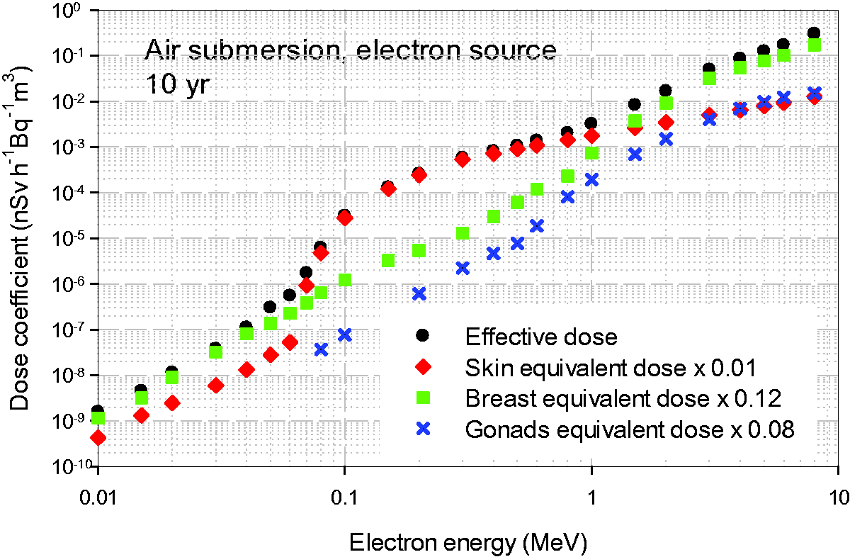
6.4. Dose-rate coefficients for water immersion
(109) Dose-rate coefficients for water immersion were calculated under the assumption that an individual is completely immersed in an infinite volume of uniformly contaminated water. For the water photon exposure, the whole spherical geometry is sampled, including those voxels in the phantom matrix outside the body that are identified as water. (110) Contributors to the organ equivalent doses from electron sources in the water immersion geometry are the primary electrons emitted from the water near the body surface and the bremsstrahlung photons generated by electron interactions in water. (111) Calculations were performed for 25 monoenergetic sources of photons and electrons ranging from 0.01 to 8 MeV and for all male and female adult and paediatric phantoms. Figs 6.12 and 6.13 present the evaluated effective dose-rate coefficients for photon and electron sources, respectively, distributed uniformly in water, as a function of particle energy. The data are given in nSv h−1 Bq−1 m3. (112) The age dependency of the effective dose coefficients is similar to the case of submersion in contaminated air, with the effective dose for newborns being up to 190% higher than for adults for photon energies of 0.015 MeV. (113) Fig. 6.14 shows the effective dose-rate of the 10-year-old for monoenergetic electrons, together with the equivalent doses of skin, breast, and gonads multiplied by the respective tissue weighting factors of 0.01, 0.12, and 0.08. The skin equivalent dose has been computed with the PM-type phantoms in order to evaluate the dose at the radiosensitive region of the epidermis which is considered to be 50–100 µm below the skin surface (see Annex C). It can be seen that above approximately 0.07 MeV, the dose to the skin is the main contributor to the effective dose for water immersion and electron exposures, whereas below this energy and above approximately 1 MeV, the contribution from the breast is the highest. Effective dose-rate coefficients for monoenergetic photon sources distributed uniformly in water (i.e. water immersion). Effective dose-rate coefficients for monoenergetic electron sources distributed uniformly in water (i.e. water immersion). Comparison of effective, skin, breast, and gonad equivalent dose-rate coefficients for monoenergetic electrons distributed uniformly in water (i.e. water immersion) for the 10-year-old phantom. For comparison, the skin, breast, and gonad dose-rates have been multiplied by their respective tissue weighting factors.


6.5. Verification of the calculations (spot-checks)
(114) The environmental fields specific for the exposure situations selected as being representative of the most common exposure scenarios were calculated for this publication by Daiki Satoh, Japan Atomic Energy Agency (JAEA) using the Monte Carlo code PHITS (see Section 4). The organ equivalent dose calculations for all geometries, particles, and phantoms were also performed by Satoh with PHITS (see previous sections). Separate calculations were performed for skin dosimetry by Yeon Soo Yeom (Hanyang University) using GEANT4 and the mesh format of the phantoms (see Annex C on skin dosimetry). The reference coefficients tabulated in this publication have been evaluated by the above data after smoothing and least squares polynomial fitting. (115) For quality assurance purposes, several organ equivalent dose data sets have been recalculated by different members of Task Group 90 using the same environmental fields and the same reference computational phantoms but different radiation transport codes. The Monte Carlo codes used were GEANT4 (Y.S. Yeom, Hanyang University), EGSnrc [H. Schlattl, Helmholtz Zentrum München (HMGU)], MCNPX [S.J. Yoo, Korean Institute of Nuclear Safety (KINS)], MCNP6 [J. Jansen, Public Health England (PHE)], MCNPX [C. Lee, National Cancer Institute (NCI)], and Visible Monte Carlo (VMC) [J. Hunt, Instituto de Radioproteção e Dosimetria (IRD)]. This section describes, in brief, the Monte Carlo calculations performed for the spot-checks.
6.5.1. GEANT4 (user Hanyang University)
(116) The GEANT4 code is a general-purpose Monte Carlo code, developed in the C++ programming language, exploiting software engineering and object-oriented technology (Agostinelli et al., 2003). Since the first public release in 1998, the GEANT4 code has been improved and maintained by the GEANT4 collaboration of various international research groups (http://geant4.cern.ch/). The GEANT4 code can simulate a large set of particles, covering a wide energy range from 100 eV to 10 TeV or, for some particles, 10 PeV (Allison et al., 2016). It is widely used in various applications including radiation dosimetry, medical application, space science, and accelerator physics. (117) GEANT4 Version 10.2 was used for the calculations of this publication for the spot-check to validate organ equivalent dose-rate coefficients for soil contamination, photon, and electron sources. The reference voxel phantoms were implemented in the GEANT4 code using the G4VNestedParameterisation class, which, among the GEANT4 classes, provides the best features for implementation of voxel geometry (Schümann et al., 2012). The physics library of the G4EmLivermorePhysics, including EPDL97 (Cullen et al., 1997), EEDL (Perkins et al., 1991), and EADL (Perkins et al., 1997), was used to simulate photons and electrons. A secondary production cut value for all the particles in all the media was set to a range of 1 µm for precise simulation. (118) Organ equivalent dose-rate coefficients for monoenergetic photons and electrons (0.03–3 MeV) of some soil contamination cases were calculated directly using the phase space source data generated in Step 1 of calculation method (see Section 5.1). During the calculations, from the source data, a particle was selected at random and its position and direction were rotated by an angle selected at random between 0° and 360° on the z axis (i.e. the centre axis of the coupling cylinder); this approach can avoid any undesirable direction bias at the given number of particles in the source data, considering that the irradiation geometry is cylindrically symmetrical. (119) For photons, relative statistical uncertainties of the calculated organ equivalent doses were generally below 1% for larger organs and 4% for smaller organs. For electrons above 0.2 MeV, the uncertainties were generally below 2% for larger organs and 10% for smaller organs, while for lower energy electrons, most of the calculated organ equivalent doses had large statistical uncertainties, with the exception of the skin doses whose uncertainties were all below 0.1%.
6.5.2. MCNP6 (user PHE)
(120) The Monte Carlo N-Particle code system MCNP (Los Alamos National Laboratory, Los Alamos, NM, USA) Version 6.1 (Pelowitz, 2013a,b) has been used in Fortran 90 code form. The source code has been patched according to Michael Lorne Fensin’s report on the MCNP Forum on 22 September 2014 to allow for convenient voxel sampling within a lattice. In addition, a Fortran 90 source routine has been inserted to allow reading of the source files describing the environmental field. This source routine reads the whole source file the first time and applies a source rotation over a sampled (random) angle during successive file reads. The Fortran 90 code has been compiled with the Intel Fortran compiler (Intel Corp., Santa Clara, CA, USA). For quality control, the executable was tested on the verification samples, and differences have been verified, documented, and forwarded to Los Alamos National Laboratory. (121) The cross-section libraries used were the MCPLIB04 for photons and EL03 for electrons, both being the MCNP6 default. For all organs, except active marrow and endosteum, and photon exposures, the organ equivalent doses are calculated without electron transport assuming electron equilibrium, except for air submersion where electron transport is performed for photon energies ≥1 MeV. For all organs, the tally 6 track length heating number estimator (i.e. a track length estimator with an internally calculated fluence-to-dose function) is used to derive the absorbed dose; an exception is the calculation of active bone marrow and endosteum absorbed dose-rate coefficients for photon exposures, where the dose enhancement factors are used to compensate for the lack of electron equilibrium and the tally 4 track length estimator is modified by a fluence-to-dose response function (see Annex B). The validation calculations performed were for air and water immersion, photon and electron sources, and all ICRP reference paediatric phantoms.
6.5.3. MCNPX (user KINS)
(122) The Monte Carlo particle transport code MCNPX 2.7.0 (Pelowitz, 2011) was used together with the cross-section libraries MCPLIB04 and EL03 for calculating the absorbed doses to the organs of the ICRP adult and paediatric reference phantoms due to unit source intensity of specified energies of photons for air submersion and water immersion exposure situations. For the specified photon energies, 25 energy bins are used for the range of 0.01–10 MeV. Absorbed doses for organs and tissues were calculated by applying the F6 tally in MCNPX code. The transport calculations were performed for the source volume within the converging of distances (Yoo et al., 2013a,b), which are determined by a simplified calculation model. (123) To resolve the poor statistics in small organs (e.g. the thymus and the lymph nodes), an approach, the ‘equivalent dose ratio method’ (Yoo et al., 2013a), was applied by assuming that the energy spectrum of photons entering the body would not change significantly with the geometrical ranges beyond a few mfp. The ratios of the absorbed doses for small organs to those received by the muscle (reference organ) were calculated at 50 m radius (reference distance) and for each energy bin. After confirming that the deviations of the ratios are within 10% while the radius of the air volume varies, these ratios were used to obtain doses to small organs. (124) The validation calculations performed were for air and water immersion, photon beams, and all ICRP reference paediatric phantoms.
6.5.4. MCNPX (user NCI)
(125) The Monte Carlo N-Particle eXtended (MCNPX) Version 2.7.0 (Pelowitz, 2011) was employed for the verification of calculations of organ equivalent dose coefficients at NCI. The verification was focused on soil contamination with a depth of 0.0 mfp (i.e. surface contamination) for 13 photon energy bins ranging from 0.01 to 5 MeV. The newborn and 15-year-old ICRP paediatric phantoms were included in the verification process. Organ equivalent dose-rate coefficients were calculated for over 30 organs and tissues, and delivered to JAEA for comparison with data from the PHITS code. (126) Source data for soil contamination computed at JAEA by PHITS, called ‘phase space data’, were delivered to NCI. As MCNPX writes and reads external source definition through the Surface Source Write/Read (SSW/SSR) routines, the source data from PHITS were not directly compatible with MCNPX. The source data from PHITS in ASCII format were converted into the binary format using an in-house script according to description of the SSW routine in MCNPX. (127) The cross-section libraries, MCPLIB04 and EL03, were adopted for photons and electrons, respectively, in the verification process. A total of 500 million particle histories were used to achieve acceptable statistical errors. A default energy cut-off (0.001 MeV) was used for both photon and electron transport. Absorbed dose to organs and tissues was calculated using F8 energy deposition tally. The high-performance computing server installed at NCI was utilised to facilitate the large number of Monte Carlo calculations.
6.5.5. EGSnrc (user HMGU)
(128) For calculations of photon organ equivalent dose coefficients, a code developed specifically for organ equivalent dose calculations (Schlattl et al., 2012) has been used, employing the electron-gamma-shower code system EGSnrc Version v4-2-3-1 (Kawrakow et al., 2009). EGSnrc is an extended and improved version of EGS4 (Nelson et al., 1985), maintained by the National Research Council of Canada (NRC). The transport of photons and electrons can be simulated for particle kinetic energies from a few keV up to several hundred GeV, although simulations performed in this study were only made for photons in the energy range of 0.01–8 MeV. (129) For photon transport, bound Compton scattering and secondary photo-electrons from K, L, and M shells are considered for all energies. In both cases, resulting fluorescence or Auger and Coster-Kronig electrons are followed. The input data for photon cross-sections agree with those of the XCOM database (Berger and Hubbell, 1987). (130) For the calculations performed for this publication, photon transport is terminated when the photon energy falls below 2 keV. Secondary electrons are followed until their kinetic energy drops below 20 keV. (131) The number of histories followed varied between 450 million at 0.01 MeV to 100 million at 8 MeV, resulting in coefficients of variance for most organs below 1%, and only in exceptions reached up to 4% (e.g. at low energies for gall bladder). (132) By assuming rotational symmetry, the phase-space source data of the coupling cylinder were converted into a discrete probability density function, φ(E, h, sin ϑ), with E being the particle energy, h being its source position on the cylinder, and ϑ representing its direction relative to the horizontal plane. At the lids of the cylinder, the probability density function was φ(E, sin ϑ). (133) The source sampling in the EGSnrc user code was performed by the cumulative density function obtained from the probability density function and enforcing rotational symmetry. (134) The validation calculations performed were for air submersion and ground contamination, photon beams, and the ICRP adult reference phantoms.
6.5.6. Visible Monte Carlo (user IRD)
(135) Visible Monte Carlo (VMC) (Hunt et al., 2004) has been developed at the Instituto de Radioproteção e Dosimetria from 1994 to the present date. VMC transports photons, electrons, alpha particles, and protons through voxel and general geometrical structures. Bremsstrahlung production and transport are not considered for low Z materials. The photon energy range considered for the spot-check calculations was 0.03–3 MeV. VMC benefits from an extensive graphical interface that shows all aspects of the simulated geometry and also the photon interactions with the environment and the phantom. (136) VMC Version March 2016 was used for the spot-check calculations to validate organ equivalent dose coefficients for water contamination and the adult phantoms. The cross-section library used was the NIST XCOM database (Berger and Hubbell, 1987), and the size of the water sphere considered for each photon energy was based on the maximum distance travelled by simulation of the transport of 108 photons in water. The photon transport is terminated when the photon suffers a photoelectric effect. The statistical uncertainties of the calculated organ equivalent doses were estimated to be below 1% for the larger organs and below 5% for smaller organs.
6.5.7. Comparison of dose-rate coefficients calculated with different codes and comparisons with other work
(137) Fig. 6.15 shows the effective dose for monoenergetic photons for the various age phantoms and ground plane surface source (left) and air submersion (right), as estimated by different calculators and codes. As can be seen, the agreement of computed dose-rate coefficient by the different Monte Carlo codes is within 10%, and in most cases, below 4%. Also shown are values of effective dose, as given in US Federal Guidance Report 15 (Bellamy et al., 2019). Note that the latter data have been obtained for environmental field data estimated by Bellamy et al. using stylised hermaphroditic models of the ICRP reference individuals (Cristy and Eckerman, 1987; Han et al., 2006). (138) Fig. 6.16 shows selected values of organ equivalent dose-rates for a ground planar source emitting monoenergetic electrons for the male adult phantom, as computed by PHITS and GEANT4 codes. It can be seen that, as mentioned in Section 6.1, the values which contribute to the effective dose less than 1% are set to zero. Fig. 6.17 shows organ equivalent dose-rates for the 15-year-old male phantom and photons, as computed by MCNPX and PHITS codes for ground planar source emitting monoenergetic photons. Similarly, Figs 6.18 and 6.19 show organ equivalent dose-rate coefficients for air submersion and water submersion, respectively, and photons. Effective dose-rate coefficients for photon ground plane surface source (left) and air submersion (right), as estimated by different calculators and codes. The PHITS data set shows the data after smoothing. For better visibility, the data were plotted multiplied by a factor of 10–105. mfp, mean free paths; FGR 15, Federal Guidance Report 15 (Bellamy, et al., 2019). Organ equivalent dose-rate coefficients for red bone marrow, testes, liver, and brain of the adult male phantom for a ground plane source emitting electrons, as calculated by PHITS and GEANT4 codes. mfp, mean free paths. Organ equivalent dose-rate coefficients for the 15-year-old male phantom, as calculated by PHITS and MCNP codes, for ground contamination on the surface (photons). The asterisks and open squares indicate calculations made at the Korean Institute of Nuclear Safety and Public Health England, respectively. Vertical lines indicate that data at lower energies have been set to zero. mfp, mean free paths. Organ equivalent dose-rate coefficients for the 15-year-old male phantom, as calculated by PHITS and MCNP codes, for submersion in contaminated air (photons). The asterisks and open squares indicate calculations made at the Korean Institute of Nuclear Safety and Public Health England, respectively. Vertical lines indicate that data at lower energies have been set to zero.
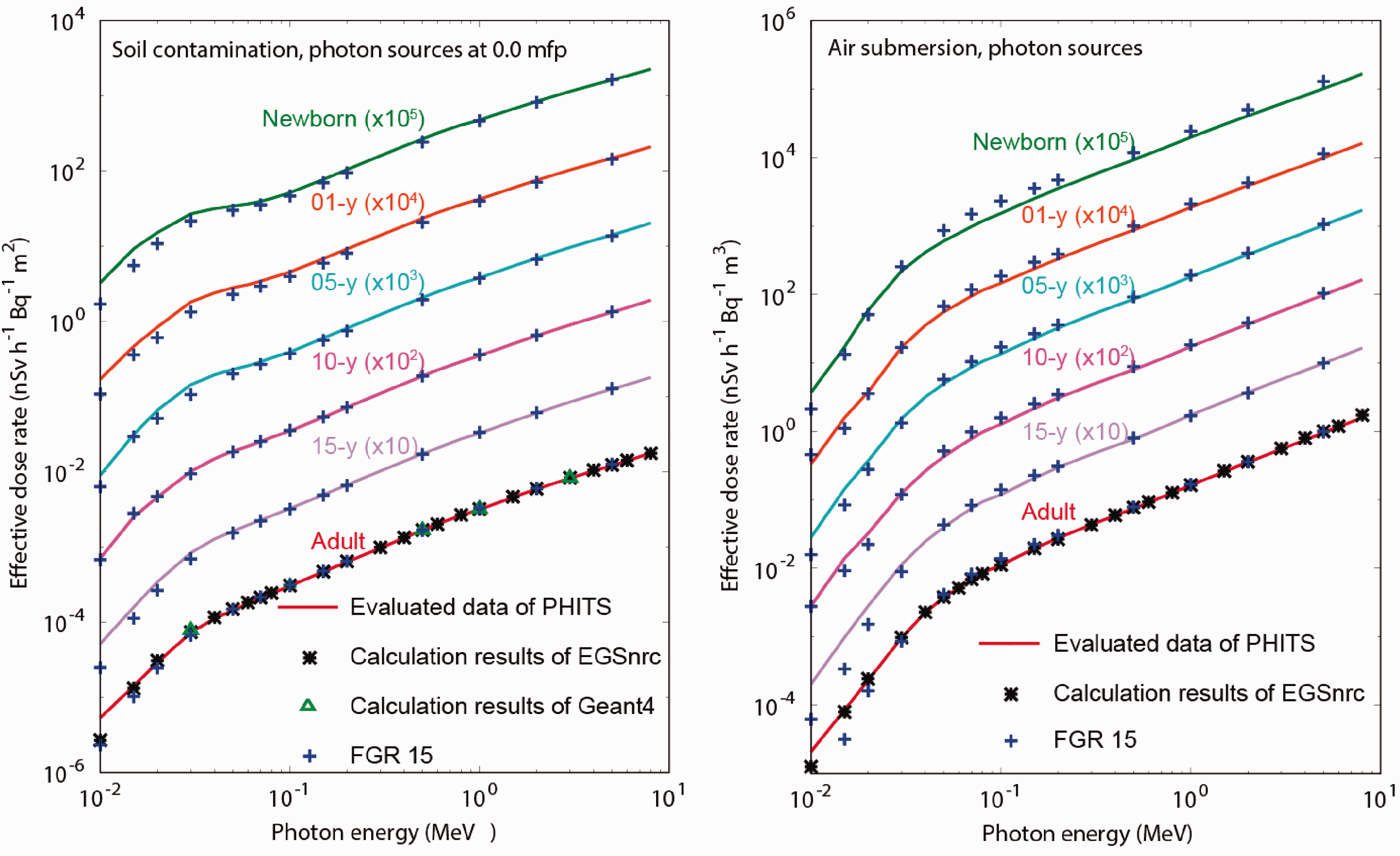
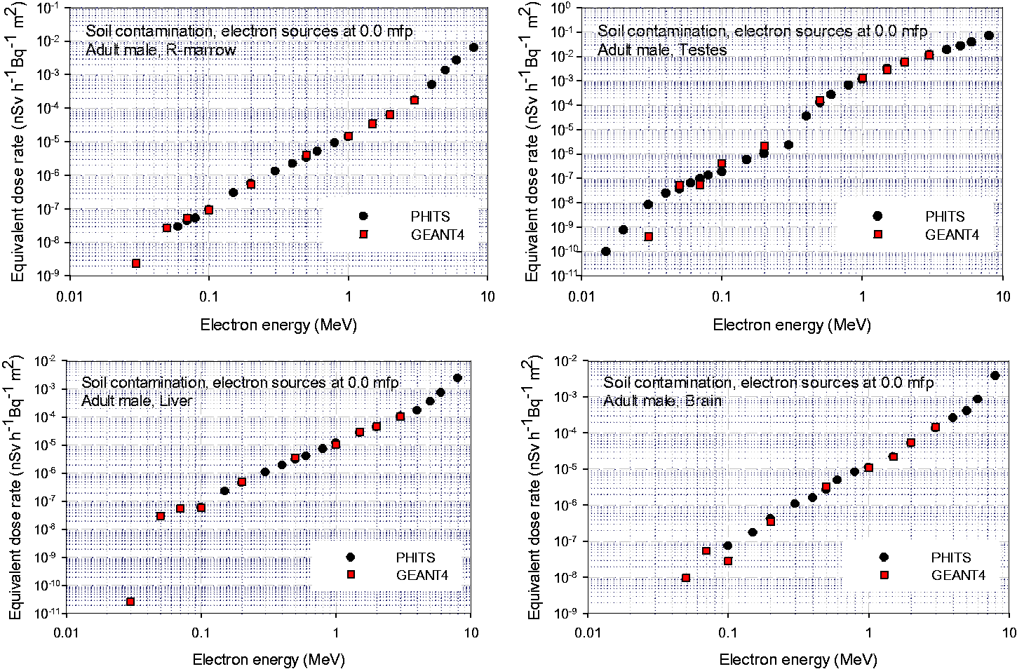
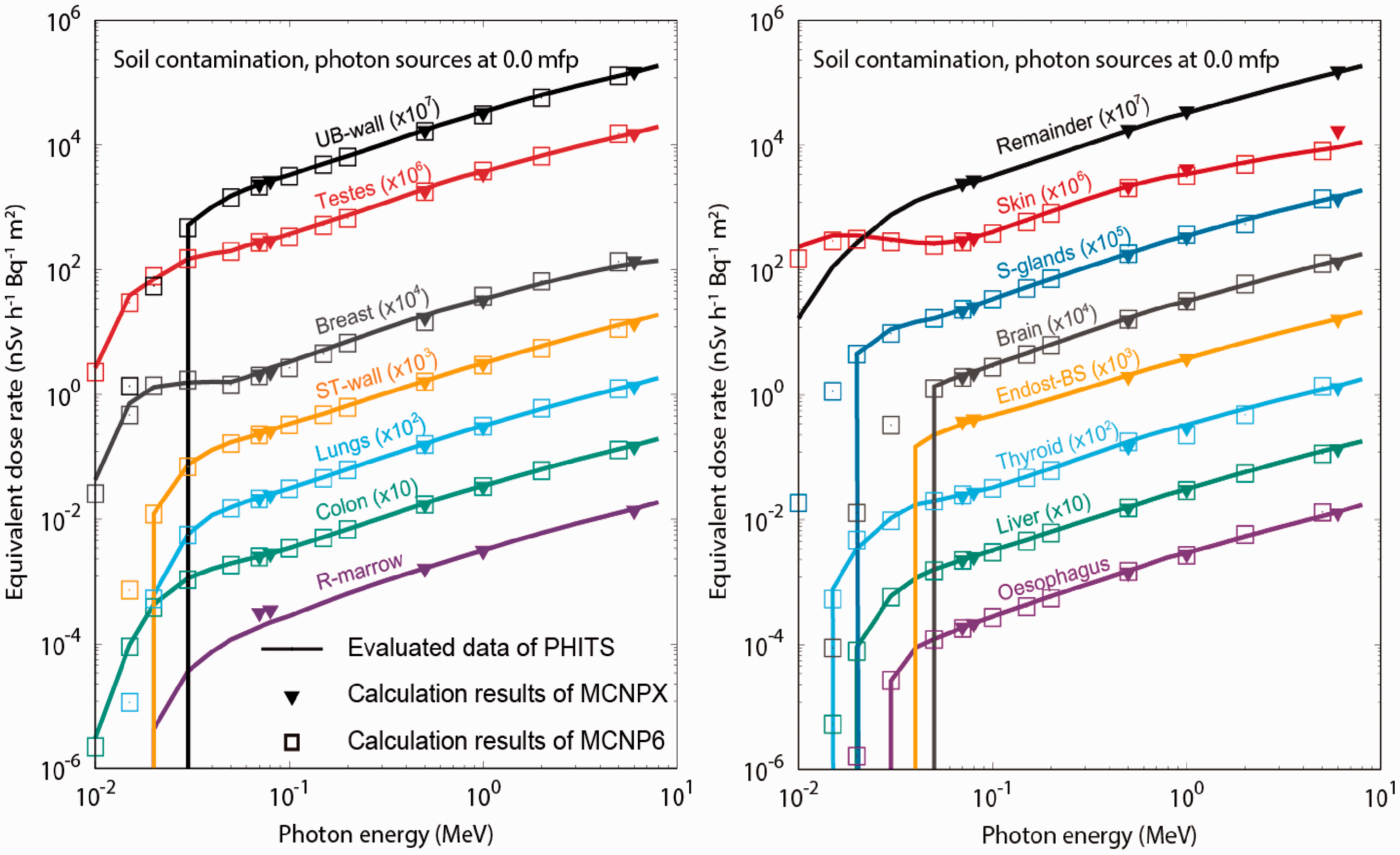

6.6. Dose-rate coefficients for monitoring – air kerma and ambient dose equivalent rates
(139) The ambient dose equivalent rates were compared with the effective dose rates for reference adult and reference newborn, 1-year-old, 5-year-old, 10-year-old, and 15-year-old phantoms (see Figs 6.2–6.6 and Fig. 6.9), as well as air kerma. It was shown that the ambient dose equivalent sufficiently overestimates effective doses, independent of age, for planar sources on and below the ground surface, as well as for submersion in a radioactive cloud and energies above 0.020 MeV. As mentioned previously, opposite trends are observed for ground contamination at energy of 0.01 MeV: at soil depths of 0.0, 0.2, and 1.0 mfp and for the newborn, 1-year-old, 5-year-old, and 10-year-old phantoms, the effective dose-rate coefficient is higher than the ambient dose equivalent rate, (140) In a previous study, Saito and Petoussi-Henss (2014) presented dose coefficients relating ambient dose equivalent rates to radionuclide density for sources exponentially distributed in the ground. The authors compared the ratio of ambient dose equivalent to air kerma obtained by simulation with the ratios measured at hundreds of locations in Japan which have been contaminated with radioactive 137Cs, 134Cs, 131I, 110mAg, and 129mTe after the Fukushima NPP accident in 2011. Good agreement was observed in all cases. (141) Figs 6.20 and 6.21 show the ambient dose equivalent rates and air kerma rates at 1 m above ground, respectively, for planar sources at different depths in soil. It can be seen that both quantities depend strongly on source soil depth, and as the depth increases, both the ambient dose equivalent rates and air kerma rates decrease because of the shielding effect of the soil. The ambient dose equivalent rates at 0.2 mfp depth are approximately 30–70% of that at 0.0 mfp. For 1.0 mfp, the reduction of the ambient dose equivalent rate coefficient is more pronounced, and the ambient dose equivalent is 80% less than the coefficient in case of surface contamination.
6.7. Comparison with new operational quantities for external radiation proposed by ICRU
(142) The operational quantities for external exposure in use at the time of compilation of this publication were defined in the 1980s and have been implemented into legal metrology worldwide. Nevertheless, the existing system has some limitations, particularly in application to fields containing very-high- and very-low-energy particles (Bartlett and Dietze, 2010; Endo, 2016). (143) ICRU Report Committee 26 (ICRU, in press) has examined the rationale for the operational quantities, and recommends redefinition of the operational quantities using coefficients that are based on protection quantities (Endo, 2016). Thus, consideration was given to define new quantities by the value of particle fluence (a radiometric quantity) at the point of interest, multiplied by values of the conversion coefficients to the protection quantities. This approach is justified because the reference values of the conversion coefficients for the protection quantities are available (ICRP, 2010). This change will avoid the use of different phantoms (anthropomorphic phantoms vs ICRU sphere or slab) and different forms of dose weighting for radiation quality (radiation weighting factor vs quality factor) between the protection quantities and the operational quantities. (144) In the proposed definitions, the ambient dose, (145) The ambient dose coefficients are given by (146) Fig. 6.22 shows the ambient dose (rate) as a function of photon energy for different soil depths. It can be seen that, generally, the values of ambient dose-rate are lower than those of ambient dose equivalent rate, and differences are more pronounced at energies below 0.015–0.07 MeV. However, it was shown that ambient dose-rate is also a good estimator of effective dose for this type of field. Organ equivalent dose-rate coefficients for the 15-year-old male phantom, as calculated by PHITS and MCNP codes, for submersion in contaminated water (photons). The asterisks and open squares indicate calculations made at the Korean Institute of Nuclear Safety and Public Health England, respectively. Vertical lines indicate that data at lower energies have been set to zero. Ambient dose equivalent rates for different depths in the soil, expressed as mean free paths (mfp). Air kerma rate at 1 m above ground for different depths in the soil, expressed as mean free paths (mfp). Ambient dose-rate, a quantity newly proposed by the International Commission on Radiation Units and Measurements (ICRU, in press), for different depths in the soil expressed as mean free paths (mfp).


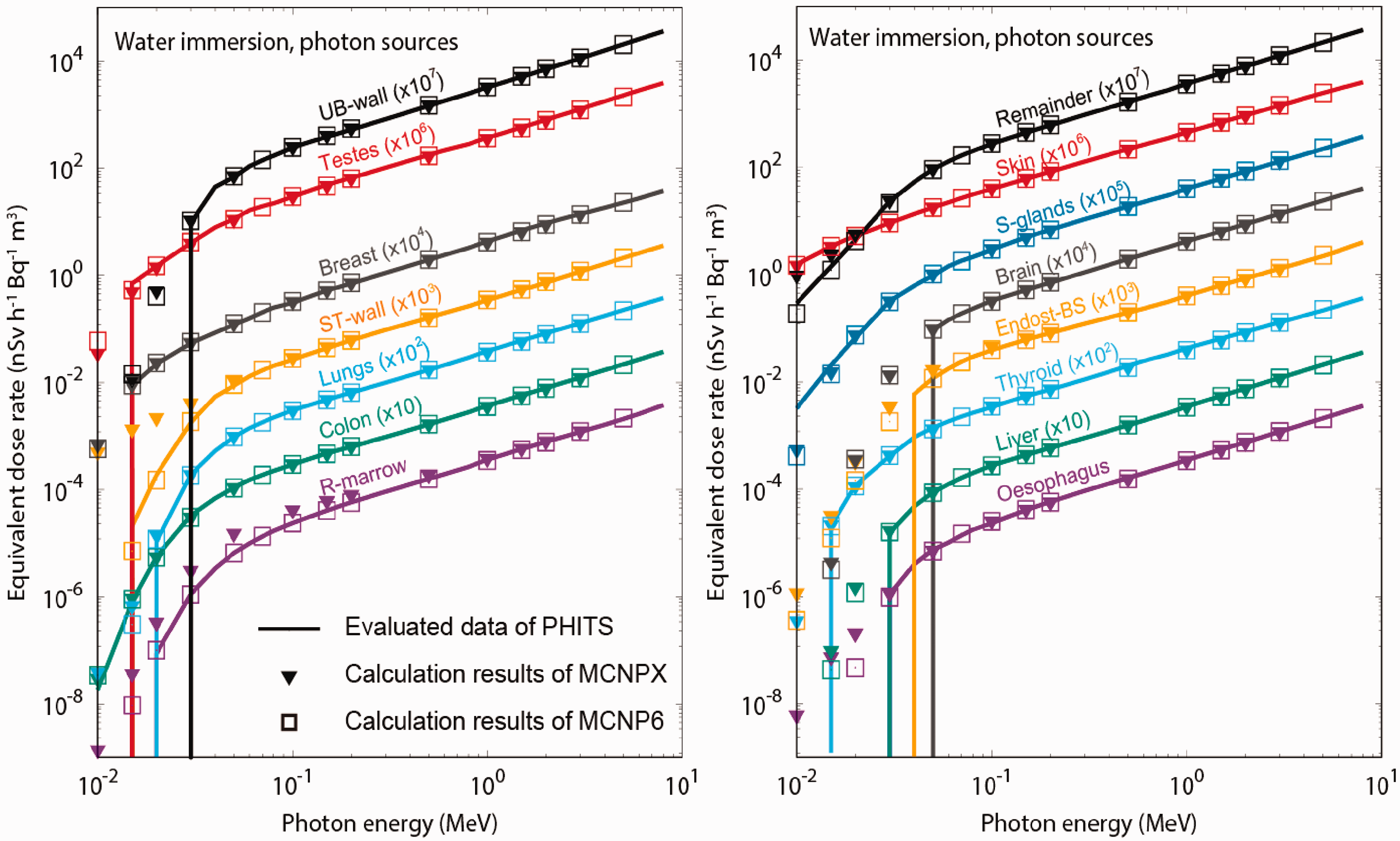
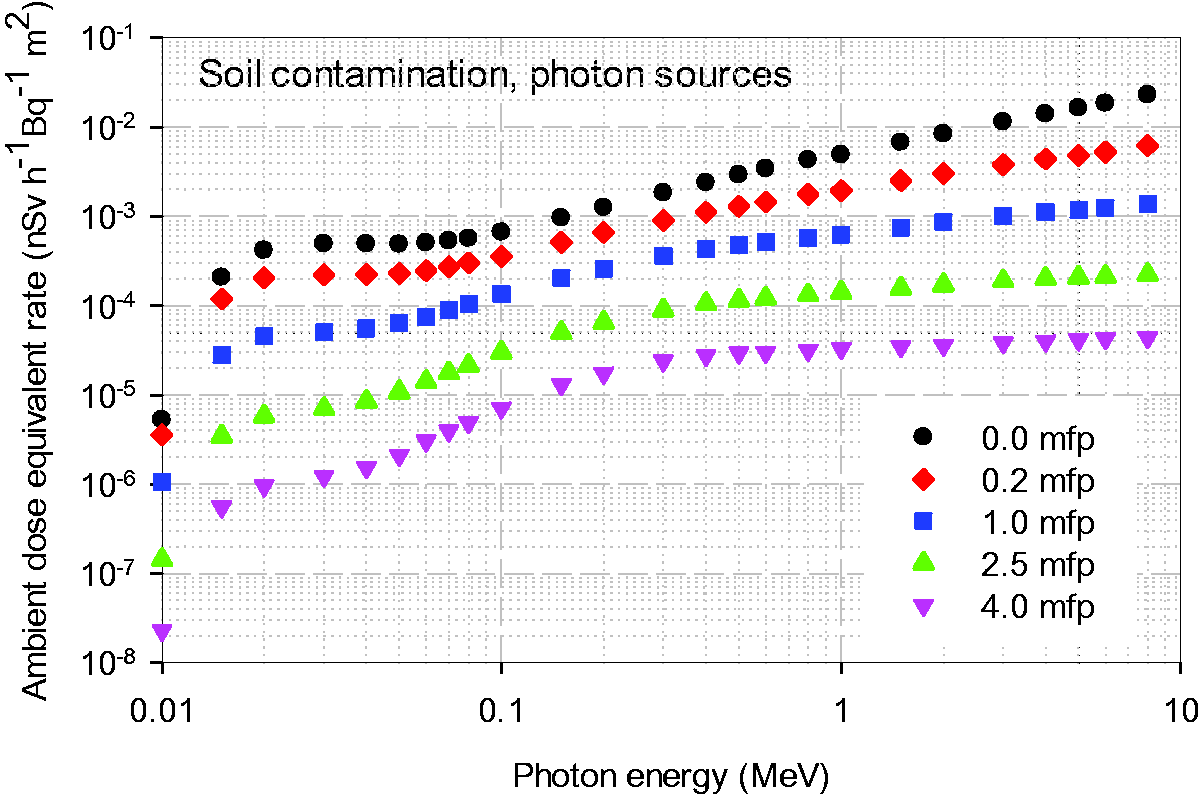

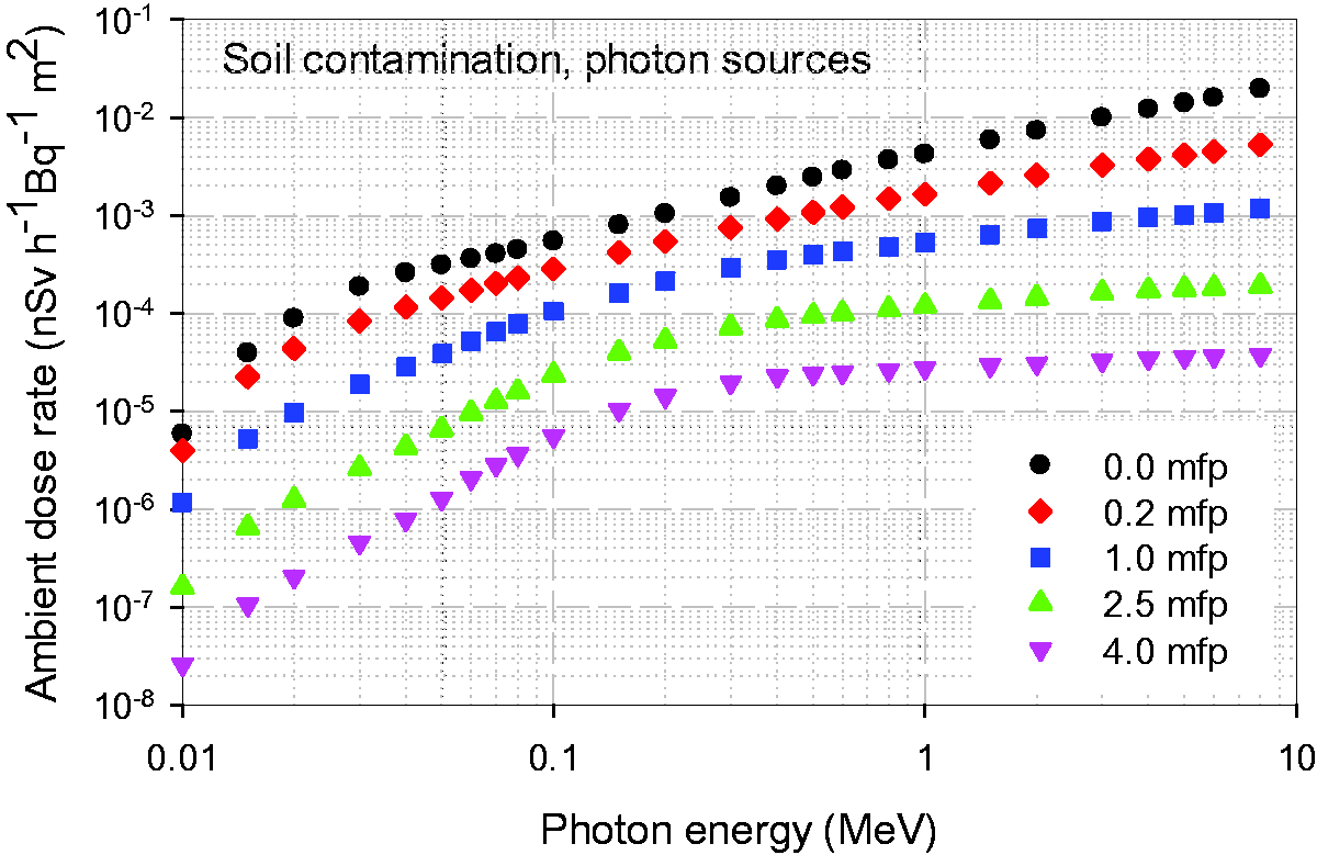
7. EQUIVALENT AND EFFECTIVE DOSE-RATE COEFFICIENTS FOR RADIONUCLIDES (STEP 3)
7.1. Coefficients for equivalent dose-rate to organs and tissues
(147) Radionuclide-specific equivalent dose-rate coefficients, (148) Interpolations of absorbed dose were carried out in a log-linear space using the piecewise cubic Hermite function (Fritsch and Carlson, 1980). As the coefficients for monoenergetic radiations obtained by Monte Carlo calculations only addressed photons and electrons of 0.01 MeV and higher energy, the values at energies less than 0.01 MeV are set to zero. (149) Radionuclide-specific organ equivalent dose-rate coefficients were evaluated for 1252 radionuclides of 97 elements compiled in Publication 107 (ICRP, 2008) distributed in soil, air, and water, and are given in tabular form in the electronic supplement accompanying this publication. It should be noted that dose-rate coefficients provided are calculated for the indicated radionuclide only and do not include radiations from daughter nuclides. A summary of the nuclear transformation of radionuclides can also be found in the electronic supplement (ICRP, 2008).

7.2. Coefficients for effective dose-rate
(150) As per the definition of effective dose in Publication 103 (ICRP, 2007), the organ equivalent doses of both male and female phantoms were used for its computation. Radionuclide-specific effective dose-rate coefficients were derived from the radionuclide-specific organ equivalent dose-rate coefficients discussed above. The effective dose-rate coefficient, (151) Table A.1 (Annex A) provides the effective dose-rates for the adult and paediatric ages considered, as well as the ambient dose equivalent and air kerma rate coefficients (see Section 7.3) for a planar source at a depth of 0.5 g cm−2 in soil (see Section 8.1.1.) and for some selected radionuclides. Similarly, Table A.2 gives the effective dose-rates for all ages and the ambient dose equivalent and air kerma rate coefficients for selected radionuclides distributed uniformly in the atmosphere (air submersion). Table A.3 shows the effective dose-rates for all ages and water immersion. Tables for all radionuclides can be found in the electronic supplement.

7.3. Coefficients for air kerma and ambient dose equivalent rate
(152) Radionuclide-specific coefficients of air kerma rate, (153) The values of (154) The radionuclide-specific air kerma and ambient dose equivalent rate coefficients for soil contamination and air submersion are listed in the folders ‘Soil contamination’ and ‘Air submersion’ of the electronic supplement, together with the data for radionuclide-specific effective dose-rates.


8. APPLICATION OF DOSE-RATE COEFFICIENTS
8.1. Application of dose-rate coefficients to various depth profiles of radionuclides in soil
8.1.1. Planar sources in specific depths
(155) As described in Section 5.1, the dose-rate coefficients for soil contamination were evaluated via Monte Carlo simulations for planar sources at five source depths expressed in mfp of the photons in soil (i.e. 0.0, 0.2, 1.0, 2.5, and 4.0 mfp). The source depth can be expressed as mass per unit area in g cm−2, which is independent of the soil density as absorption is only dependent on mass thickness. The mfp of photons depends on photon energy. For instance, a specific source depth of 3.0 g cm−2 corresponds to 72.3, 0.54, and 0.14 mfp of 0.01, 0.1, and 2.0 MeV photons in the soil, respectively. Dose-rate coefficients for monoenergetic photons emitted from a planar source at a specific depth (in g cm−2) can be reconstructed from the data at corresponding mfp using the piecewise cubic Hermite function (Fritsch and Carlson, 1980). Dose-rate coefficients were estimated for planar sources at the specific depths of 0.0, 0.5, 3.0, and 10.0 g cm−2 for monoenergetic photons and can be found in the electronic supplement. The nuclide-specific organ equivalent and effective dose-rate coefficients were evaluated by considering not only the dose contribution of photons, but also that of the electrons from electron emitters on the ground surface (if any) and bremsstrahlung photons generated by electron interactions in soil, as per Eckerman and Ryman (1993) and Bellamy et al. (2019), using the scaled bremsstrahlung cross-section data of Pratt et al. (1977). (156) The depth of 0.5 g cm−2 is considered to be representative of the surface roughness and initial migration following precipitation. It simulates the deposition of radionuclides in the ground in the first years after migration (ICRU, 1994). This source depth is consistent with earlier work by Saito et al. (1990) and Petoussi et al. (1991), and with recent work by Petoussi-Henss et al. (2012), Yoo et al. (2013a), and Bellamy et al. (2019). It has been shown that for a 137Cs/137mBa source distributed as a planar source at a depth of 0.5 g cm−2, the air kerma in air is reduced by a factor of 0.67 compared with a purely surface planar source on the ground (UNSCEAR, 2016). It should be noted that the planar source at depth of 0.5 g cm−2 of soil with density of 1.0 g m−3 is equivalent to a planar source at 3 mm of soil with density of 1.6 g m−3. The depth of 3 g cm−2 is the typical deposition depth for radiocaesium, and 10.0 g cm−2 is approximately the maximum depth where caesium has been observed. Table A.1 provides the effective dose coefficients for a depth of 0.5 g cm−2 for some selected radionuclides. Tables of coefficients – organ and effective dose-rates – for all specific depths considered and all radionuclides can be found in the electronic supplement.
8.1.2. Volumetric sources
(157) Measurements around the Fukushima area (Matsuda et al., 2015) revealed that the depth profile of radionuclides in soil changes over time due to terrestrial ecosystems. Calculating dose-rate coefficients for each depth profile is not practical; therefore, a method to obtain dose-rate coefficients for volumetric sources with arbitrary depth profiles is proposed. Note that the depth profile of volumetric sources indicates the vertical distribution of the activity concentration along the depth in the soil, whereas the horizontal distribution is assumed to be uniform. (158) Dose-rate coefficients for volumetric sources with any arbitrary depth profile can be obtained using the data for planar sources in depths given in g cm−2, and a weighted-integral method as described by Satoh et al. (2015, 2017). The weighted-integral method describes a depth profile in the soil with weights, w(ζ), regarding a radioactivity concentration distributed along a depth, ζ, described in g cm−2, and is applicable to any depth profile (e.g. exponential, Gaussian, or uniform). Note that w(ζ) expresses the depth profile of the radioactivity concentration as a relative value of the activity at a depth of ζ to the total activity in the volumetric source. (159) The coefficients for a volumetric source, (160) Matsuda et al. (2015) reported that the depth profiles of radioactive caesium in soil observed in the Fukushima area after the NPP accident in 2011 are fitted with an exponential function using the equation of weight (161) The weighted integration for volumetric sources distributed with exponential profiles was solved numerically using the trapezoidal rule with small strips with width of 1 µm. The values of (162) To examine the validity of the weighted-integral method, Satoh et al. (2015) incorporated an exponentially distributed volumetric source of 137Cs/137mBa with β = 1.0 into the PHITS code, and directly calculated the energy spectrum and the effective dose-rate. It was found that the reconstructed volumetric source was a good approximation of the source calculated directly via Monte Carlo methods. (163) Effective dose-rate coefficients were evaluated for each age for monoenergetic volumetric photon sources distributed with exponential profiles with relaxation masses per unit area, β = 0.5, 1.0, 2.5, 5.0, 10.0, 20.0, 50.0, and 100.0 g cm−2, respectively, and can be found in the electronic supplement together with the respective ambient dose equivalent, air kerma, and (sex- and age-dependent) organ dose-rate coefficients. The choice of the above values of β provide a useful grid for interpolation if evaluation for other profiles is desired. The nuclide-specific organ equivalent and effective dose-rate coefficients for these exponential sources were evaluated by considering the dose contribution of photons, as well as of electrons from electron emitters on the ground surface (if any) and bremsstrahlung photons generated by electron interactions in soil (Eckerman and Ryman, 1993; Bellamy et al., 2019). A full set of nuclide-specific organ equivalent dose-rate, effective dose-rate, ambient dose equivalent rate, and air kerma rate coefficients for these exponential volumetric sources can be found in the electronic supplement.


8.2. Radionuclide decay chain
(164) In this publication, external dose-rate coefficients are evaluated for 1252 radionuclides of 97 elements compiled in Publication 107 (ICRP, 2008). The electronic supplement enables interactive viewing of the dose coefficients and summary information on nuclear decay characteristics of the selected radionuclide. The nuclide-specific dose-rate coefficients are based on the radiations emitted by the indicated radionuclide and do not include radiations emitted by radioactive decay products. If the decay of the selected radionuclide yields radioactive decay products (progeny), the interactive viewer displays the decay chain. In addition, a table detailing the nuclear decay characteristics of the 1252 radionuclides is archived in the electronic supplement. Note this information is also available within Publication 107. (165) The dose contributions of a radionuclide and its progeny need to be evaluated considering the production and decay of radioactive progeny, and differences in environmental behaviour of the parent and daughter nuclides. Such consideration is required for evaluation of the effective dose-rate at a specified time and the effective dose integrated over a specified period (Eckerman and Ryman, 1993; Bellamy et al., 2019). (166) The serial transformation by radioactive decay of each member of a radioactive series is described by the Bateman equations (Bateman, 1910; ICRP, 1959; Skrable et al., 1974) and the following equations developed by Eckerman and Ryman (1993). Assume that at time zero, the concentration of the parent nuclide on the surface of the ground is (167) The activity at time t, (168) The effective dose associated with an exposure period of duration T following a contamination event at t = 0 that results in ground surface concentration of (169) If the parent radionuclide is long-lived relative to its progeny, then at times T such that


8.3. Relationship between radioactivity in soil, effective dose, ambient dose equivalent, and personal dose equivalent
(170) Operational quantities were originally developed for the protection of occupationally exposed workers. Use of the operational quantities has been extended to monitoring of radiation exposure of the public from natural and artificial environmental sources of radiation. One of the applications is radiation monitoring in environments contaminated by radionuclides released from nuclear facilities after an accident. (171) After the Fukushima NPP accident in 2011, a large-scale national environmental monitoring programme was carried out, and comprehensive data including radioactivity in soil and ambient dose equivalent rate, (172) The personal dosimeters indicate personal dose equivalent, (173) Satoh et al. (2017) investigated the relation of effective dose,
8.4. Application of dose-rate coefficients for remediation planning
(174) This publication provides radionuclide-specific dose-rate coefficients for members of the public resulting from environmental external exposures. These dose-rate coefficients could be utilised for planning of remediation from radioactive contamination of the environment. Remediation activities, including decontamination, reduce exposure of the public living in a contaminated area. Software has been developed to support decision-making and planning of remediation activities by optimisation of protective actions (Ulanovsky et al., 2011). The software addresses the annual effective dose of the population, and the dose-rate coefficients given in this publication are useful for this purpose. (175) Estimation of dose reduction by decontamination for a specific situation requires, among other factors, consideration of source size, inhomogeneity of source distribution, and decontamination factor. This estimation requires a different approach than that described in this publication; for that purpose, Satoh et al. (2014) have developed a methodology and respective software to estimate the effects of decontamination, and the dose reduction effect resulting from a decontamination scenario. To estimate the dose reduction for a specific contaminated site after decontamination measures, it is necessary to consider the inhomogeneity of the source distribution as well as the size of the source.
9. CONCLUSIONS
(176) This publication provides nuclide-specific reference effective and organ equivalent dose-rate coefficients to be used for the assessment of external dose from environmental exposure of the public for selected idealised environmental conditions. These are exposure to contamination on or below the ground surface and at different depths (soil contamination); submersion in a contaminated atmospheric cloud (air submersion); and immersion in contaminated water (water immersion). (177) For the first time, ICRP provides reference dose-rate coefficients for external exposures to radionuclides in the environment. These were computed using the ICRP voxel-based adult male and female reference computational phantoms (ICRP, 2009a), as well as for the 10 ICRP reference paediatric phantoms representing newborn, 1-year-old, 5-year-old, 10-year-old, and 15-year-old Reference Male and Reference Female (ICRP, 2020). Radiations considered include primary photons and electrons from environmentally dispersed radionuclides, scattered photons and electrons emitted within the environment, and bremsstrahlung photons produced via electron deceleration. The emitted particles include those of beta decay (electrons and positrons) and ejected orbital electrons due to internal conversion and Auger processes in the electron shell of the newly formed atom. Note that neutron emission associated with radionuclides undergoing spontaneous fission has not been addressed. (178) Organ equivalent dose-rates increase with decreasing age because of the reduced shielding effect of the smaller body and the closer vicinity of the source for ground contamination. It was found that for contamination in the ground, the effective dose-rate differs between the newborn and adult from 60% to 140% at a photon energy of 0.05 MeV, while the difference is less than 70% above a photon energy of 0.10 MeV. Effective dose-rates for the reference 15-year-old are close to those computed for the reference adult. For submersion in contaminated air, the difference in effective dose between the adult and the newborn is less than 50% above a photon energy of 0.06 MeV, while it reaches up to 160% below 0.03 MeV. For water immersion, the age dependency of the effective dose coefficients is similar to the case of submersion in contaminated air, with the effective dose for newborns being up to 190% higher than for adults for photon energies of 0.015 MeV. For realistic exposure situations of radionuclide environmental contamination, the difference is found to be more moderate. For example, for radioactive caesium (134Cs, 136Cs, 137Cs/137mBa) deposited on and in the ground, the difference in effective dose between an adult and an infant is in the range of 30–60%, depending on the radioactivity deposition depth within the soil. (179) The ambient dose equivalent rates and air kerma rates have been computed at 1 m above ground and are given for both soil contamination and air submersion for the environmental geometries considered. These data enable interpretation of monitoring data relating ambient dose equivalent rates or air kerma rates to effective dose-rates. Ambient dose equivalent and air kerma rates were found to provide conservative estimates of effective dose-rates for both the adult and the newborn (and thus for all ages) for most radionuclides. Exceptions are observed for radionuclides emitting low-energy photons (below approximately 0.015 MeV) and pure beta emitters. (180) The expected applications of the dose-rate coefficients are: (1) pre-accidental evaluations in order to predict the possible impacts to the public by postulated radiological accidents; (2) post-accidental evaluations to estimate doses in order to develop a radiological protection strategy for the exposed population; (3) evaluations following discharge of radionuclides from nuclear and radioisotope facilities or from naturally occurring radioactive materials from non-nuclear industries during routine operations; and (4) evaluations of naturally occurring radionuclides in the environment. The pre-/post-accident analyses are performed typically by software packages (e.g. codes for severe accidents). The software predicts the dispersion, migration, and distribution of radionuclides in the environment. The dose-rate coefficients of the present publication could thus be implemented within these codes. (181) It should be noted that dose-rate coefficients are calculated for idealised and hypothetical source geometries, such as semi-infinite and uniform distributions, for reference phantoms wearing no clothing and for idealised, upright postures, even for the exposed newborn. As a result, they do not fully reflect actual exposures for any particular situation or exposed individual. (182) This publication provides age-dependent organ and effective dose-rate coefficients; however, it should be noted that the current ICRP system of radiological protection uses a simplified set of tissue weighting factors in the calculation of effective dose, based on sex- and age-averaged relative detriment values, and specifies only two nominal detriment values: 5.7 × 10⁻² Sv−1 for the whole population and 4.2 × 10⁻² Sv−1 for adults (ICRP, 2007). Thus, recognised differences in detriment and relative detriment (the contribution of the various organs and tissues to total detriment) as a function of age at exposure are not taken into account other than in the differences between the two nominal detriment values (ICRP, 1991, 2007). This approach is adopted principally because application of the effective dose coefficients is in the protection of either the public or workers, and dose limits, constraints, and reference levels are set to apply to each of these groups. Thus, differences in effective dose-rate coefficients as a function of age shown in this publication relate only to differences in physical size and organ masses, and do not address differences in detriment per Sv. Similarly, differences in organ absorbed dose-rate coefficients do not inform on differences in stochastic risk per Gy as a function of age at exposure. (183) For ground contamination, this publication provides dose-rate coefficients for plane sources on the surface and at various depths in the soil. Although the respective data for concrete and asphalt, which are surfaces typically found in urban areas, are not directly provided, the data in this publication could be used for estimation of dose coefficients relevant to urban areas. This would require appropriate adjustment of the mass per unit area of the contamination depth profiles by considering the density of the respective medium, as per the discussion in Sections 8.1.1 and 8.1.2 where dose-rate coefficients for planar sources were convoluted to approximate any specific or desired radionuclide concentration soil depth profile. Above 0.2 MeV, the mass attenuation coefficients are insensitive to the elemental composition of the ground material, and an exponential distribution with relaxation mass per unit area of 0.1 g cm−2 is an appropriate depth profile for low-permeability urban surfaces such as roofs, asphalt, and concrete (ICRU, 1994). For 137Cs deposition, for example, the coefficients calculated for soil can be used for other ground materials, and a relaxation mass per unit area of 0.1 g cm−2 will be appropriate for asphalt and concrete. A caveat applies for low-energy photons (<0.2 MeV), where the mass attenuation coefficients are more sensitive to the elemental composition of the ground material (Saito and Jacob, 1995; Cresswell and Sanderson, 2012). (184) Effects of posture have not been studied for this publication. Previous work of Saito et al. (1998) examined the effective dose for phantoms in two prone positions (i.e. face-up and face-down on the ground) for soil contamination with plane photon sources at the depth of 0.5 g cm−2. The results indicate that the effective dose coefficients are lower (up to 25%) when the phantom is lying down compared with standing up (i.e. the standing posture leads to a conservative approach). This finding might seem unexpected, and can be explained by considering that photons from horizontal directions are dominant in this source configuration as shown in Fig. 5.3, and the contribution to effective dose is small for photons incident to the head, feet, and lateral directions. For air submersion, the findings show that effective dose for a person in a prone back-up position is higher than for a standing posture. In contrast, effective dose for a prone face-down posture is smaller. This is because more organs which contribute to effective dose are located in the front of the body. At 0.05 MeV, the differences in effective dose between the standing and prone positions are up to 30%, while for energies higher than 0.1 MeV, the differences are 5–15%. (185) External doses can be significantly lower indoors than outdoors due to the shielding effects of the building. This can be taken into account through the use of a so-called ‘shielding factor’ of 0.005–0.4 (IAEA, 2000b) which reflects structural shielding by building type (according to the country and building material) and an assumed occupancy factor of 0.6 (i.e. approximately two-thirds of the time per day spent indoors) which represents the fraction of time spent inside a house (IAEA, 2000b). This obviously can vary considerably according to the geographical distribution, profession, and population habits. This publication presents dose-rate coefficients for situations outside houses, and does not attempt to address issues of shielding or population behaviour. The selection and application of suitable shielding and occupancy factors is left to the user (i.e. the legislative authority or the developer of emergency programmes).
References
ANNEX A. TABLES OF NUCLIDE-SPECIFIC DOSE-RATE COEFFICIENTS
(A1) Annex A gives the reference coefficient rates for the effective dose for all ages considered, ambient dose equivalent, and air kerma for selected radionuclides for soil contamination at a depth of 0.5 g cm−2, air submersion, and water immersion. Tables for all radionuclides as well as for further planar sources and exponential volumetric sources can be found in the electronic supplement which may be downloaded from the ICRP and SAGE websites. Effective dose-rate, ambient dose equivalent rate, and air kerma rate coefficients for nuclides distributed at a depth of 0.5 g cm−2 in the soil. Ambient dose equivalent rate and air kerma rate coefficients were estimated at 1 m above ground. Effective dose, ambient dose equivalent, and air kerma rate coefficients for air submersion (volume source in air). Ambient dose equivalent rate and air kerma rate coefficients were estimated at 1 m above ground. Effective dose-rate coefficients for radionuclides distributed uniformly in water (water immersion).


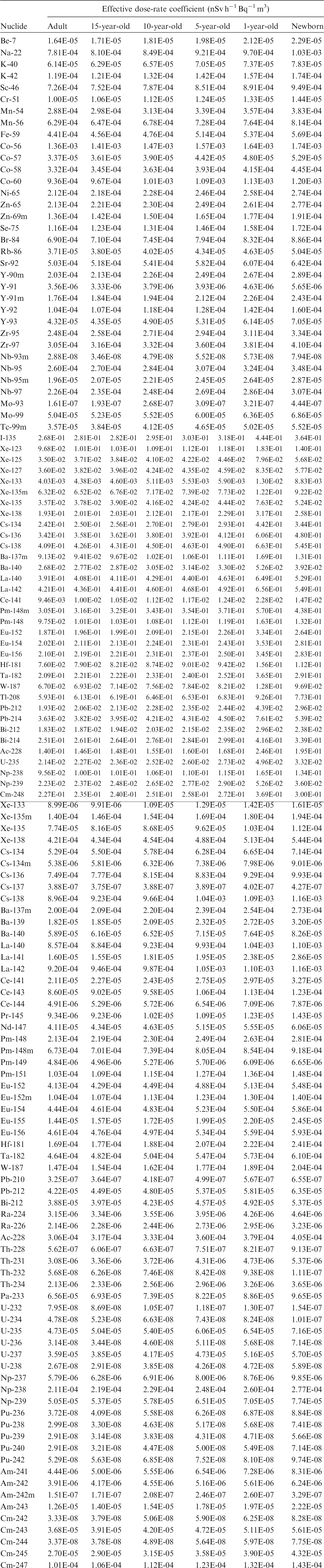
ANNEX B. SKELETAL DOSIMETRY
(B1) In this publication, the radiation absorbed doses to two different target tissues were assessed in computation of the effective dose: the haematopoietically active bone marrow (AM) and the skeletal endosteum (TM50). The former target region is taken as the non-adipose regions of the bone marrow cavities within both spongiosa and medullary marrow cavities of the phantom skeleton, while the latter target region is taken to be total marrow localised within 50 µm of the bone trabeculae surfaces and along the interior surfaces of the long bone medullary cavities. As described in Publications 110 and 116 (ICRP, 2009, 2010), the bone trabeculae and marrow cavities are tissue structures on the order of tens to hundreds of micrometres in thickness and extent, and thus cannot be fully modelled with the voxel resolution of either the reference adult or paediatric phantoms. Consequently, radiation absorbed doses, and therefore equivalent doses, to these two target tissues were determined employing the concept of the fluence-to-dose response function for photons as described and presented in Annex D of Publication 116 (ICRP, 2010). (B2) It should be noted that for this publication, energy deposition to the skeletal target tissues is almost exclusively by photons, either emitted directly from the environmental radionuclide sources (air, water, or soil) or indirectly by bremsstrahlung x-ray production by environmentally emitted beta particles and conversion/Auger electrons. In the rare instance that electron collisional kinetic energy is deposited within the marrow cavities of the phantom skeleton, radiation dose to spongiosa (or medullary marrow) is taken as a surrogate of the absorbed dose to either AM or TM50. (B3) The fluence-to-dose response function, (B4) As noted in Annex D of Publication 116, Eq. (D.2) was evaluated as described by Johnson et al. (2011). Electron absorbed fraction data were obtained through paired-image radiation transport calculations using micro-CT images of 32 bone sites extracted from the skeleton of a 40-year-old male cadaver (Hough et al., 2011). Values of electron absorbed fractions in the bones of the reference paediatric phantoms were taken from the UF doctoral dissertations of Pafundi (2009) and Wayson (2012). Micro-CT images of cadaveric newborn bones were used for radiation transport in the bones of the reference newborn phantom (Pafundi et al., 2009, 2010). Similarly, micro-CT images of cadaveric 18-year-old male bones were used in the construction of skeletal absorbed fractions for electrons in the reference 15-year-old phantom (Pafundi, 2009). Cadaveric bone samples were not available for the interior ages of the ICRP reference phantom series. Consequently, the linear path length distributions from the University of Leeds 1.7-year-old and 9-year-old cadavers were used, respectively, to assess electron absorbed fractions in the bones of the reference 1-year-old and 10-year-old phantoms (Beddoe, 1976). Values of skeletal electron absorbed fractions were then assessed via interpolation of the Leeds data to report values for the reference 5-year-old phantom (Pafundi, 2009). Charged particle equilibrium is typically established across bone sites at photon energies exceeding 0.200 MeV, and thus in this publication, values of the dose–response function above that energy are taken as their corresponding spongiosa kerma coefficients. (B5) In this publication, the absorbed dose in tissue, rT, in bone site x,


B.1. References
ANNEX C. SKIN DOSIMETRY
(C1) For environmental external exposures and dose to the skin, stochastic effects are relevant. In radiation protection, the mean value of the absorbed dose averaged over the specified organ, tissue, or cells at risk is correlated with the detriment due to stochastic effects. The skin cells at radiogenic risk have been identified and the equivalent dose to these cells has been assigned a tissue weighting factor wT = 0.01 (ICRP, 2007). The skin dose contributing to effective dose is the equivalent dose to the skin cells at risk averaged over the body. (C2) The skin cells at highest radiogenic risk are the basal cells, which are located between the epidermis and dermis of the skin. ICRP (2002) provides reference data for nominal epidermal depths as a function of age as follows: 45 µm for the newborn, 1-year-old, and 5-year-old phantom; 50 µm for the 10-year-old phantom; 60 µm for the 15-year-old phantom; and 70 µm for the adult phantom. However, it has recently been concluded that these data will not be used directly to specify target depths for skin cancer in the development of ICRP dosimetric phantoms. Recognising the substantial variation in epidermal thicknesses with body site and that hair follicles may also contain a substantial proportion of the epidermal stem cells, a simplified scheme will be adopted in which the 50–100-µm depth for the adult phantom (nominal 70 µm) will also be used for the 15-year-old phantom, and a slightly wider band of 40–100 µm will be used for the 10-year-old phantom and younger. (C3) For simplicity, for the calculations of the present publication, the target sensitive layer within the skin was defined at depths of 50 µm and 100 µm for all phantoms. The extent to which the mean value of the skin dose is representative of the absorbed dose to the critical region of the skin, located at 50–100 µm depth, depends, for external irradiation, on the homogeneity of the exposure and on the range of the incident radiation. For photon energies relevant for radionuclides in the environment, the assumption of the mean organ dose being representative of the dose to 70 µm can be considered valid due to their rather homogeneous dose distribution within the skin. For weakly penetrating radiations (e.g. electrons) which could exhibit a significant dose gradient within the skin, this approach could be invalid, underestimating or overestimating the doses to the basal cell layer at risk. (C4) For electron skin dosimetry of the adult phantoms, the voxel representation of 2.137 and 1.775 mm (for male and female phantoms, respectively) could underestimate or overestimate the doses, depending on the electron energy, as shown in Section C.1. In order to overcome this limitation, polygon mesh (PM) models were used: for the adult male and female phantoms, the skin models of the PM-type ICRP adult reference phantoms were employed for the calculations. These phantoms are the exact counterparts of the ICRP phantoms and have the advantage that they can model small tissues below the voxel phantom resolution. More information on these phantoms can be found in a forthcoming ICRP publication (ICRP, 2020b) and in Kim et al. (2011, 2016, 2017), Yeom et al. (2013, 2016a,b), and Nguyen et al. (2015). (C5) The mesh-type skin models of the adult phantoms were constructed by directly converting the skin models of the voxel-type ICRP adult reference phantoms (ICRP, 2009) to high-quality PM format. The PM skin models include a 50-µm-thick radiosensitive layer located at a depth 50–100 µm below the skin surface. Fig. C.1 shows a three-dimensional representation of the adult male and female PM skin models. The masses of the adult PM skin models are in accordance with the reference values (male 3300 g, female 2300 g) (ICRP, 2002). The average thicknesses of the skin models are 1.69 mm and 1.33 mm for the adult male and female, respectively, which are in good agreement with the reference values (male 1.6 mm, female 1.3 mm). The inner space of the skin PM models is filled with the average soft tissue for adults, as specified by ICRU (1992), but has slightly modified densities (male 1.024 g cm3, female 1.010 g cm3) in order to maintain the reference body weights of 73 kg (male) and 60 kg (female). (C6) For the paediatric phantoms, the mesh-type skin models were constructed from the outer surfaces of the non-uniform rational B-spline (NURBS) version of ICRP paediatric phantoms ICRP (2020a). These were the original phantoms from which the ICRP paediatric phantoms were derived (Lee et al., 2010). The NURBS-format outer surfaces were converted to the PM format using the tessellation procedure (Piegl and Richard, 1995). The PM outer surfaces were adjusted to match the total volumes of the ICRP paediatric phantoms. The outer surfaces were copied and their sizes were reduced to define the inner surface of the skin, matching the skin thicknesses to those (i.e. voxel sizes) of the ICRP paediatric phantoms. The inner space of the PM skin models was filled with average soft tissue (ICRU, 1989) but with slightly modified densities to maintain the reference body weights. The outer surfaces were copied to create two additional surfaces and reduce their sizes to define the target sensitive layer within the skin at depths of 50 µm and 100 µm. (C7) The skin dose-rate coefficients, shown in the electronic supplement of this publication and used for calculation of the effective dose-rates, for both electron and photon beams and all geometries were derived using the above PM phantom models and the Monte Carlo code GEANT4 (Agostinelli et al., 2003). For implementation, the skin phantoms in the PM format were converted to the tetrahedral-mesh (TM) format using the TetGen code (Si, 2015), and the converted TM phantoms were implemented in GEANT4 using the G4Tet class. Note that this tetrahedralisation maintains the original shape of the PM phantoms but improves computation speed significantly (Yeom et al., 2014). The electromagnetic physics library of G4EmLivermorePhysics was used to transport photons and electrons. Considering the 50-µm-thick target layer, a secondary production cut value of 1 µm was set for all particles. (C8) It should be noted that tissue reactions (sometimes referred to as ‘deterministic effects’) are correlated to the local skin dose (i.e. dose averaged over 1 cm2). The skin dose coefficients given in this publication are not correlated with tissue reactions as they have been evaluated for the entire skin in the whole body.
C.1. Electrons
(C9) Figs C.2 and C.3 show the skin equivalent dose-rate coefficients for the adult female phantom and contamination at the soil surface and submersion in contaminated air, respectively, as a function of electron energy and as calculated using the original voxel phantom coupled to the PHITS transport code (Sato et al., 2013), and the PM phantom and the GEANT4 code (Agostinelli et al., 2003). As mentioned above, the resolution of the voxel phantom does not allow targeting of the cells at risk (50–100 µm layer), but instead the dose is computed for the whole skin voxels. Using the mesh phantom, estimation of the dose-rate coefficients to the sensitive layer as well as to the entire skin is possible, and these are shown in Figs C.2 and C.3. It can be seen that the voxel approach overestimates the equivalent dose to the skin basal cell layer at electron energies below approximately 0.10 MeV and underestimates that same dose at energies between 0.10 and 1.5 MeV. (C10) For electron simulations in the case of water immersion, excessive computation times are needed because electrons emitted from the spherical water source of a diameter of 2 m hardly reach the phantom. To improve the efficiency of the calculation, a sampling source volume was limited from the skin surface to a certain distance in the water, depending on electron energies. For electrons with energies greater than 0.06 MeV, a distance, longer than the electron continuous slowing down approximation (CSDA) range in the water medium, was used to limit the sampling source volume because these primary electrons contribute to most of the energy deposited to the sensitive layer of the skin. On the other hand, for the lower energy electrons (≤0.06 MeV), a distance, longer than the mfp of the photon at the initial electron energy, was considered because these electrons, with a CSDA range less than 50 µm, cannot penetrate the 50-µm-thick dead layer to reach the sensitive layer of the skin, so only the secondary photons (e.g. bremsstrahlung photons) contribute to the dose. (C11) Figs C.4–C.6 show the skin dose-rate coefficients evaluated for the sensitive layer of the skin using the mesh phantoms for all ages and geometries considered. For the adult and 15-year-old phantoms, the male and female coefficients were averaged, whereas for the other paediatric ages, a single skin model was used. Representation of the adult male and female polygon mesh skin models. Red indicates the target sensitive layer of the skin, beige indicates the exterior skin surface, and black indicates the innermost skin surface. The dead skin layer between the exterior surface and the target layer is represented in green, as viewed from the left. Skin dose coefficients for the adult female phantom and contamination at the soil surface, calculated using the original adult voxel phantom and the PHITS code, and the polygon adult mesh phantom and the GEANT4 code. Skin dose coefficients for the adult female phantom and air submersion, calculated using the original adult voxel phantom and the PHITS code, and the polygon adult mesh phantom and the GEANT4 code.
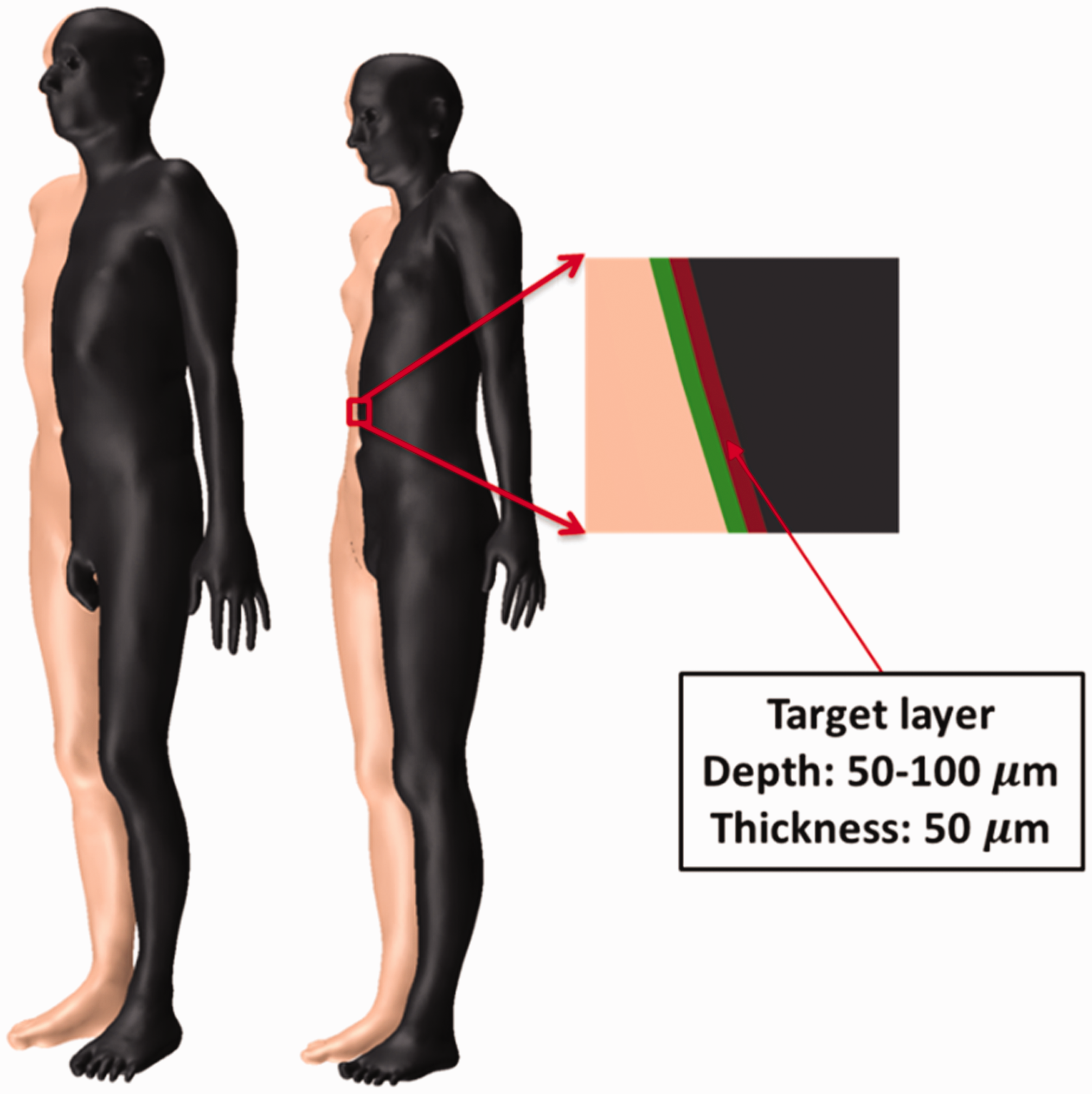
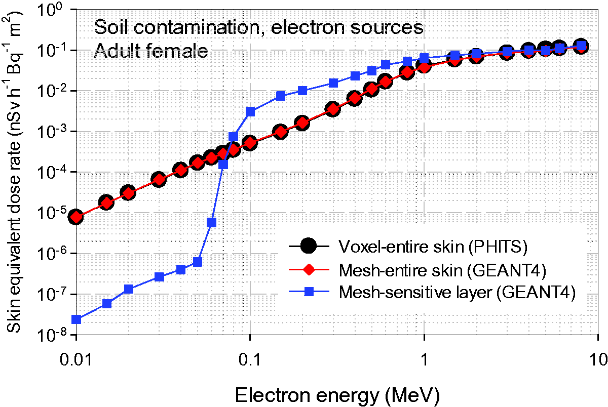
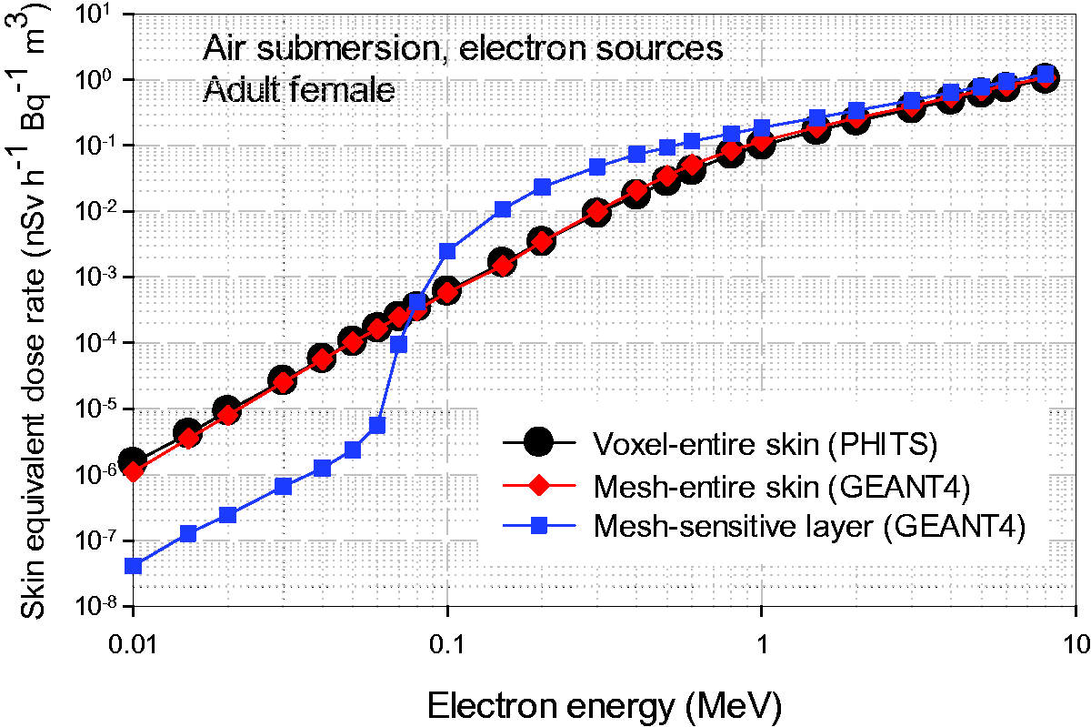
C.2. Photons
(C12) Figs C.7–C.9 show the skin dose-rate coefficients for the adult male phantom for monoenergetic photons and for the three environmental sources considered in this publication. For each plot, results are shown as calculated for the voxel-defined entire skin (i.e. averaged over all skin voxels), the entire skin as defined by the PM, and the sensitive layer of the skin, also defined by the PM and targeted between 50 and 100 µm below the skin surface. Similarly to the electron exposure simulations, the calculations for the voxel phantoms were performed with PHITS (Sato et al., 2013), whereas for the mesh phantoms, GEANT4 was used (Agostinelli et al., 2003). It can be seen that although the differences in evaluated coefficients are less pronounced than for electrons, the values of the coefficients for the sensitive layer of the skin are higher than those for the entire skin at energies below approximately 0.1 MeV. This pattern is seen because low-energy photons tend to establish their maximum dose near the skin surface, as the dose decreases rapidly with depth by exponential attenuation. However, the values of the coefficients for the sensitive layer of the skin are lower in the energy range of 0.2–0.6 MeV (depending on the environmental source). Photons penetrate the sensitive layer of the skin and partially deposit their energy, whereas they fully impart their energy to the voxel skin of the voxel phantoms, establishing the maximum dose at a depth deeper than seen within the sensitive layer of the skin. (C13) Moreover, it should be noted that while the skin dose-rate coefficients obtained with the adult voxel phantoms with PHITS and EGSnrc are in good agreement (see Section 6.5), GEANT4 gives slightly higher values in the low-energy region. This might result from differences in the cut-off algorithm during particle transport within each code. (C14) Figs C.10–C.12 show the skin dose-rate coefficients evaluated for the sensitive layer of the skin using the mesh phantoms, for all ages and environmental sources considered, for monoenergetic photons. As for the coefficients for electrons, for the adult and 15-year-old phantoms, the male and female coefficients were averaged, whereas a single skin model was used for the other paediatric ages. All skin dose-rate coefficients, including soil contamination at depths of 0.2–4 mfp, can be found in the electronic supplement. Skin dose-rate coefficients for monoenergetic electron sources distributed at the surface as a ground plane source. Skin dose-rate coefficients for monoenergetic electron sources distributed uniformly in the atmosphere. Skin dose-rate coefficients for monoenergetic electron sources distributed uniformly in water (i.e. water immersion). Skin dose-rate coefficients for monoenergetic photon sources distributed at the surface as a ground plane source. mfp, mean free paths. Skin dose-rate coefficients for monoenergetic photon sources distributed uniformly in the atmosphere. Skin dose-rate coefficients for monoenergetic photon sources and water immersion. Skin dose-rate coefficients for monoenergetic photon sources distributed at the surface as a ground plane source. mfp, mean free paths. Skin dose-rate coefficients for monoenergetic photon sources distributed uniformly in the atmosphere. Skin dose-rate coefficients for monoenergetic photon sources distributed uniformly in water (i.e. water immersion).

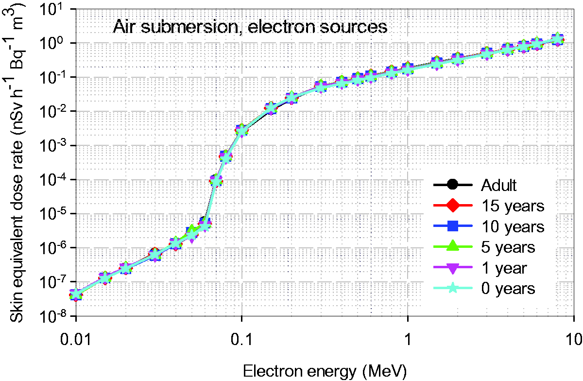
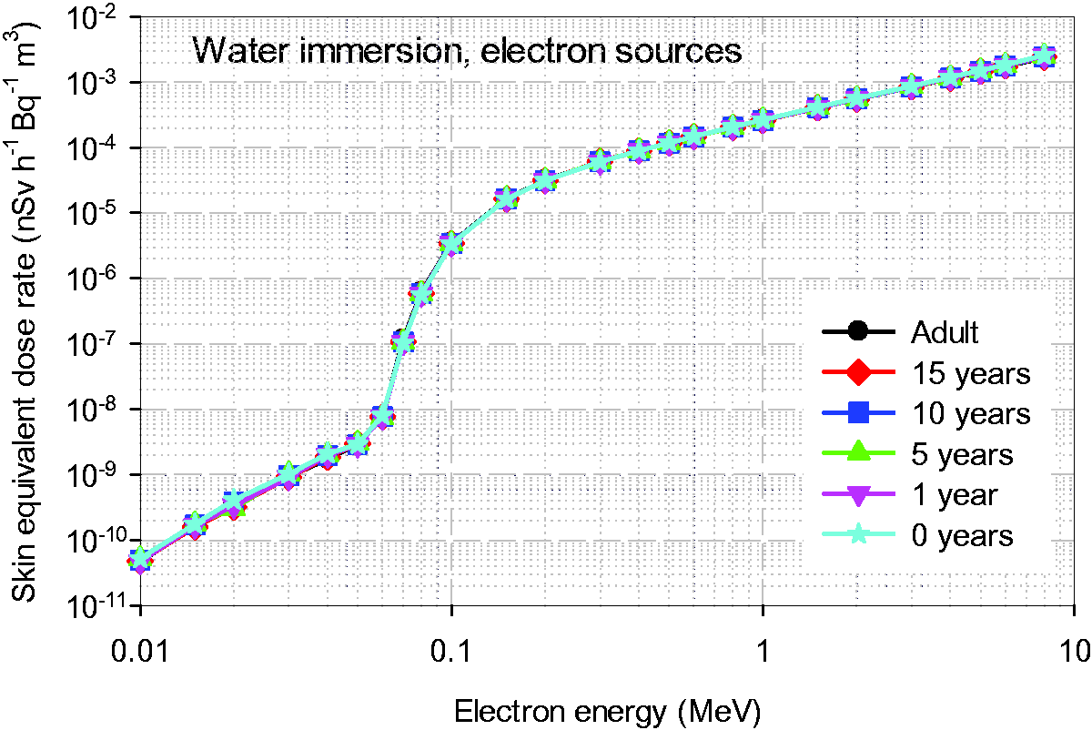


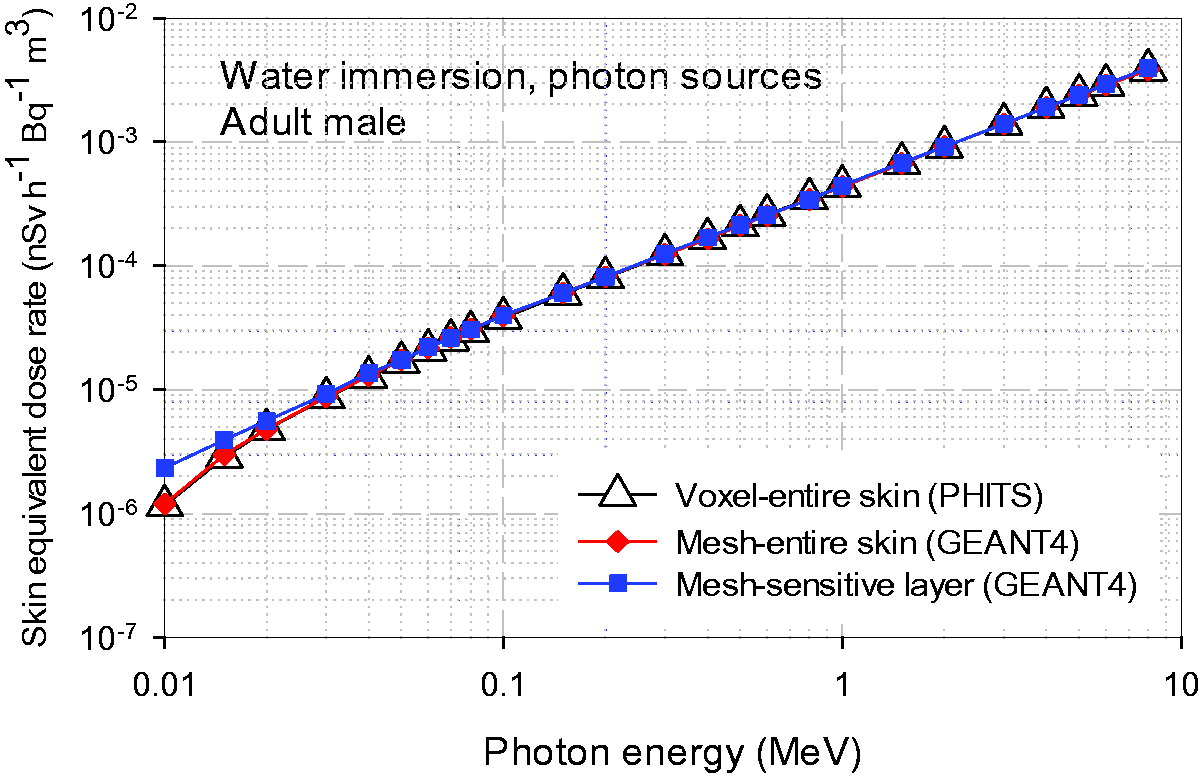
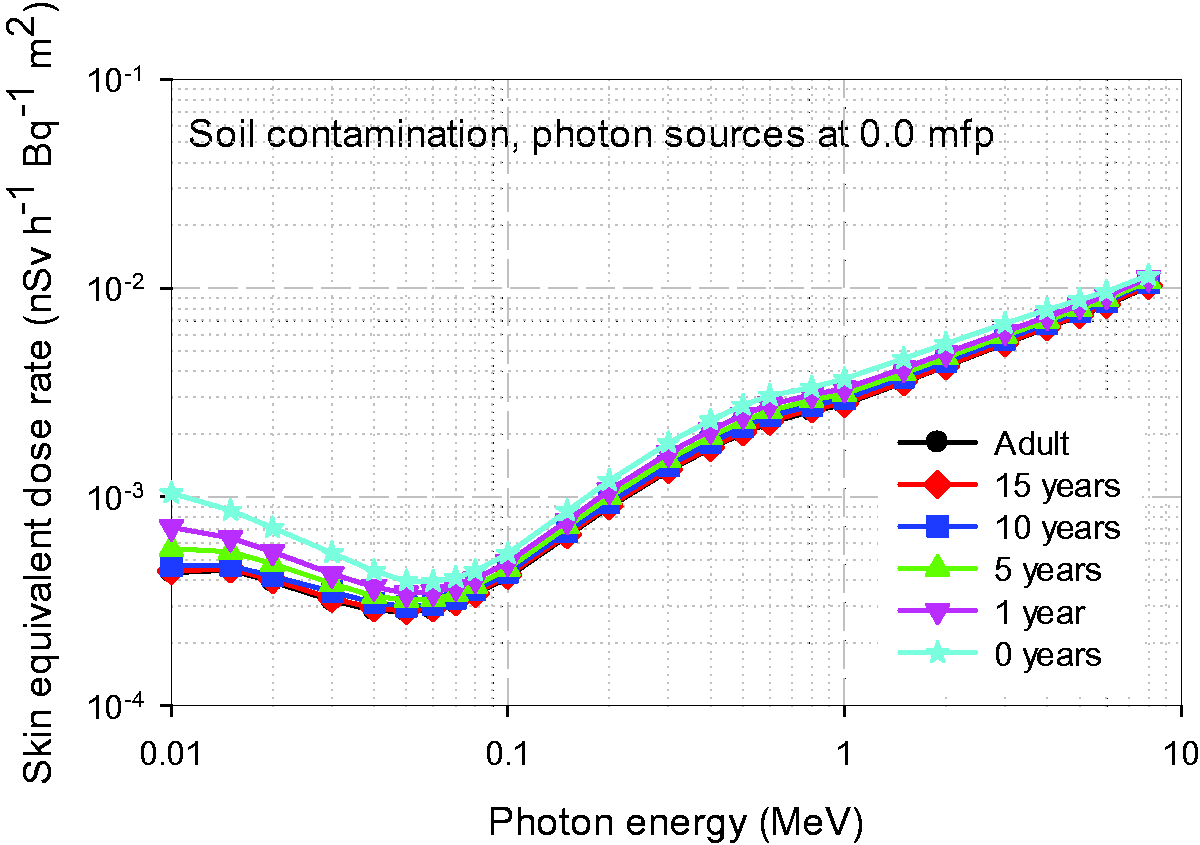


C.3. References
ANNEX D. EXAMPLES OF CALCULATIONS
(D1) This annex provides examples of estimation of effective dose-rates using the dose coefficients presented in this publication. Please note that neither shielding from housing or clothing, nor behaviour of the individual or considerations for cumulative dose over time are taken into account.
D.1. External exposure to 133Xe distributed uniformly in the atmosphere
(D2) Assume that the activity concentration, (D3) In the same manner, the effective dose-rate,


D.2. External exposure to 137Cs/137mBa distributed uniformly in the soil at a depth of 0.3 cm
(D4) 137Cs is discharged into the environment following an accident at a nuclear plant and subsequently deposited on the ground. Assume that 137Cs is distributed uniformly at a depth of 0.3 cm in the soil, the density, (D5) The physical depth, (D6) 137Cs has a physical half-life of approximately 30.2 years. It undergoes beta decay, and forms the radioactive progeny 137mBa with a branching fraction of 0.944. 137mBa has a half-life of 2.552 min, emits photons and electrons, and does not form any radioactive progeny (ICRP, 2008). As the half-life of 137Cs is sufficiently longer than that of 137mBa, 137mBa is in radioactive equilibrium with its parent 137Cs. Hence, the activity concentration of 137mBa, (D7) The effective dose-rate,


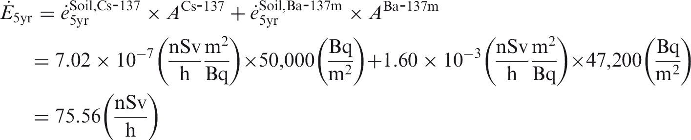
D.3. Reference
ANNEX E. CONTENT OF THE ELECTRONIC SUPPLEMENT
(E1) The electronic supplement of this publication, available for download from the ICRP and SAGE websites, presents age-dependent reference dose-rate coefficients of effective dose and organ equivalent doses for the three environmental exposures simulated: soil contamination, submersion in contaminated air, and immersion in contaminated water. The coefficients have been evaluated for the ICRP reference adult and paediatric phantoms using the methods described in Sections 4–8. (E2) Data are given separately for each age group and for the male and female phantoms. The effective and organ equivalent dose-rate coefficients are normalised to environmental radioactivity concentration and are given in nSv h−1 Bq−1 m2 (for soil contamination) or nSv h−1 Bq−1 m3 (for submersion in contaminated air and water immersion). (E3) Coefficients are given for monoenergetic values of photons and electrons, as well as for 1252 radionuclides of 97 elements compiled in Publication 107 (ICRP, 2008). The nuclide-specific dose-rate coefficients are based on the radiations emitted by the indicated radionuclide, and do not include consideration of the radiations emitted by radioactive decay products. (E4) Moreover, dose-rate coefficients are given for ambient dose equivalent and air kerma for monoenergetic photons, soil contamination, and submersion in contaminated air. For these geometries, nuclide-specific data are also given. (E5) The supplement is organised into three main folders (one for each exposure geometry): ‘Soil contamination’, ‘Air submersion’, and ‘Water immersion’. The ‘Soil contamination’ folder contains three subfolders:
a subfolder for planar sources at mfp, with five subfolders for each mfp considered: 0.0 mfp (i.e. surface contamination) and contamination in the soil at planar depths of 0.2, 1, 2.5, and 4 mfp; a subfolder for planar sources at specific depths, with four subfolders for each specific depth at 0.0, 0.5, 3.0, and 10.0 g cm−2; and a subfolder for exponential sources, with eight subfolders for sources exponentially distributed with eight different relaxation masses per unit area, (E6) The nuclide-specific dose-rate coefficients are given in ASCII format. The data for monoenergetic photons and electrons are given in ASCII format and Microsoft Excel format. (E7) Reference values of the organ equivalent dose-rate coefficients are given for the following organs: red bone marrow(R-marrow), colon, lung, stomach (ST-wall), breast, ovaries, testes, bladder (UB-wall), oesophagus, liver, thyroid, skeletal endosteum (Endost-BS), brain, salivary glands (S-glands), skin, remainder tissues, adrenals, extrathoracic (ET) region, gall bladder (GB-wall,) heart (Ht-wall), kidneys, lymphatic nodes, muscle, oral mucosa (O-mucosa), pancreas, prostate, small intestine (SI-wall), spleen, thymus, and uterus/cervix. (E8) An additional table summarises the nuclear decay characteristics of all 1252 nuclides of 97 elements as obtained from Publication 107 (ICRP, 2008). For each radionuclide, the radioactive decay products, if formed, are identified. (E9) Moreover, a data viewer is provided which allows interactive viewing and downloading of the nuclide-specific organ dose, effective dose, air kerma, and ambient dose equivalent rate coefficients. If the decay of selected radionuclide yields radioactive decay products (progeny), the interactive viewer displays the decay chain.
E.1. Reference
ICRP, 2008. Nuclear decay data for dosimetric calculations. ICRP Publication 107. Ann. ICRP 38(3).
GLOSSARY
Absorbed dose, D The absorbed dose is given by:
Active marrow is haematopoietically active and gets its red colour from the large number of erythrocytes (red blood cells) being produced. Active bone marrow serves as a target tissue for radiogenic risk of leukaemia. The number of nuclear transformations of a radioactive material during an infinitesimal time interval, divided by its duration (s). The SI unit of activity is the becquerel (Bq; 1 Bq = 1 s−1). Concentration of radioactivity per unit mass or volume. The SI unit of activity concentration is Bq kg−1 or Bq m−3. The activity of a specified radionuclide per unit mass, volume, or area of a specified substance. External exposure from radionuclides in the radioactive cloud or plume. The dose equivalent at a point in a radiation field that would be produced by the corresponding expanded and aligned field in the ICRU sphere at depth of 10 mm on the radius opposing the direction of the aligned field. The unit of ambient dose equivalent is J kg−1 and its special name is sievert (Sv). The coefficient to convert the activity concentration to the ambient dose equivalent rate. The unit of the ambient dose equivalent rate coefficient for the environmental exposures discussed in this publication is nSv h−1 Bq−1 m3 or nSv h−1 Bq−1 m2. The special name for the SI unit of activity. 1 Bq = 1 s−1, 1 MBq = 106 Bq. Bone marrow is a soft, highly cellular tissue that occupies the cylindrical cavities of long bones and the cavities defined by the bone trabeculae of the axial and appendicular skeleton. Total bone marrow consists of a sponge-like, reticular, connective tissue framework called ‘stroma’, myeloid (blood-cell-forming) tissue, fat cells (adipocytes), small accumulations of lymphatic tissue, and numerous blood vessels and sinusoids. There are two types of bone marrow: red (active) and yellow (inactive). See ‘Active (bone) marrow’ and ‘Inactive (bone) marrow’. See ‘Endosteum’. Charged-particle equilibrium in a volume of interest means that the energies, numbers, and directions of the charged particles are constant throughout this volume. This is equivalent to saying that the distribution of charged-particle energy radiance does not vary within the volume. In particular, it follows that the sums of the energies (excluding rest energies) of the charged particles entering and leaving the volume are equal. The cross-section of a target entity for a particular interaction produced by incident charged or uncharged particles of a given type and energy is given by:
Activity of a specified radionuclide per unit ground area integrated into depth direction, resulted from fallout. The unit is Bq m−2. See ‘Tissue reaction’. A coefficient relating a dose quantity to a physical quantity for both internal and external radiation exposure. For external environmental exposures, the quantities ‘activity concentration’, ‘ambient dose equivalent’, and ‘air kerma’ are chosen. In previous ICRP publications, ‘dose coefficients’ were often referred to as ‘dose conversion coefficients’. ICRP Committee 2 is currently making no distinction between these two terms. A coefficient relating a dose quantity to a physical quantity for both internal and external radiation exposure per unit time. The dose equivalent at a point is given by:
Dose equivalent per unit time. A particular function used in this publication to represent the absorbed dose in a target region per particle fluence in that region, derived using models of the microscopic structure of the target region geometry, and the transport of the secondary ionising radiations in those regions. The tissue-weighted sum of equivalent doses in all specified organs and tissues of the body, given by the expression:
The coefficient to convert the activity concentration to the effective dose-rate. The unit of the effective dose-rate coefficient for the environmental exposures discussed in this publication is nSv h−1 Bq−1 m3 or nSv h−1 Bq−1 m2. A 50-µm-thick layer covering the surfaces of the bone trabeculae in regions of trabecular spongiosa and those of the cortical surfaces of the medullary cavities within the shafts of all long bones. It is assumed to be the target region for radiogenic bone cancer. This target region replaces that previously introduced in Publications 26 and 30 – the bone surfaces – which had been defined as a single-cell layer, 10 µm in thickness, covering the surfaces of both the bone trabeculae and the Haversian canals of cortical bone. The equivalent dose in an organ or tissue T is given by:
The coefficient to convert the activity concentration to the equivalent dose rate in tissue T. The unit of the equivalent dose-rate coefficient for the environmental exposures discussed in this publication is nSv h−1 Bq−1 m3 or nSv h−1 Bq−1 m2. The quotient of dN by da, where dN is the number of particles incident on a sphere of cross-sectional area da, thus:
ICRU 4-element tissue-like material with density of 1 g cm−3 and a mass composition: 76.2% oxygen, 11.1% carbon, 10.1% hydrogen, and 2.6% nitrogen. The ICRU sphere has this assumed composition. In contrast to active marrow, inactive marrow is haematopoietically inactive (i.e. does not support haematopoiesis directly). It gets its yellow colour from fat cells (adipocytes) that occupy most of the space of the yellow bone marrow framework. Quantity for uncharged ionising particles, defined by the quotient of dE
t
r by dm, where dEtr is the mean sum of the initial kinetic energies of all the charged particles liberated in a mass (dm) of a material by the uncharged particles incident on dm, thus:
Kerma is sometimes used as an approximation for absorbed dose. The numerical value of kerma approaches that of absorbed dose to the degree that charged-particle equilibrium exists, that radiative losses are negligible, and that the kinetic energy of the uncharged particles is large compared with the binding energy of the liberated charged particles. The coefficient to convert the activity concentration to the kerma rate. The unit of the kerma rate coefficient for the environmental exposures discussed in this publication is nGy h−1 Bq−1 m3 or nGy h−1 Bq−1 m2. The fraction of bone marrow volume in a given bone that is haematopoietically active. Age- and bone-site-dependent reference values for marrow cellularity are given in Table 41 of Publication 70 (ICRP, 1995). As a first approximation, marrow cellularity may be thought of as 1 minus the fat fraction of bone marrow. The mean absorbed dose in a specified organ or tissue T is given by:
The average distance travelled by a particle without suffering a collision. Quantities used in practical applications for monitoring and investigating situations involving external exposure and intakes of radionuclides. They are defined for measurements and assessment of doses in the body. Short phrase for ‘mean absorbed dose in an organ or tissue’. Short phrase for ‘equivalent dose in an organ or tissue’. The period of time for one-half of the atoms of a radionuclide to disintegrate. Dose quantities related to the human body that ICRP has developed for radiological protection to allow quantification of the detriment to people from exposure to ionising radiation from both whole- and partial-body external irradiation and from intakes of radionuclides. The quality factor at a point in tissue, is given by:
A dimensionless factor by which the organ or tissue absorbed dose is multiplied to reflect the higher biological effectiveness of high-linear energy transfer (LET) radiations compared with low-LET radiations. It is used to derive the equivalent dose from the absorbed dose averaged over an organ or tissue. See ‘Active (bone) marrow’. An idealised male or female with characteristics defined by ICRP for the purpose of radiological protection, and with the anatomical and physiological characteristics defined in Publication 89 (2002) for both the male and female for newborn, 1-year-old, 5-year-old, 10-year-old, 15-year-old, and adult. Consequently, there are 12 reference individuals within the ICRP system of radiation protection. An idealised person for whom the equivalent doses to organs and tissues are calculated by averaging the corresponding doses of the Reference Male and Female. The equivalent doses of the Reference Person are used for the calculation of effective dose. There are six Reference Persons within the ICRP system of radiological protection, with one at each of the reference ages defined in Publication 89. The computational phantom of the human body (male or female voxel phantom based on medical imaging data) defined in Publication 110 (ICRP, 2009) for adults and in Publication 143 for paediatric ages, with anatomical and physiological characteristics defined in Publication 89 (ICRP, 2002). Value of a quantity recommended by ICRP for use in dosimetric applications or biokinetic models. Reference values are fixed and specified with no uncertainty, independent of the fact that the basis of these values includes many uncertainties. Activity concentrations in soil in many instances are described by a depth-dependent exponential function of the form A = exp(−z/β), where A is the activity concentration, z is the soil depth (expressed in g cm−2), and β is a parameter called the ‘relaxation mass per unit area’. The magnitude of β is an indication of the radionuclide penetration in the soil, with large values of β indicating sources distributed to a greater depth in the soil. The unit of relaxation mass per unit area is g cm−2. See ‘Dose–response function’. A source describing deposited radionuclides on the surface and in the soil. Term referring to the combined tissues of the bone trabeculae and marrow tissues (both active and inactive) located beneath cortical bone cortices across regions of the axial and appendicular skeleton. Spongiosa is one of three bone regions defined in the Publication 110 (ICRP, 2009) reference phantoms, the other two being cortical bone and medullary marrow of the long bone shafts. As the relative proportions of trabecular bone, active marrow, and inactive marrow vary with skeletal site and phantom age, the homogeneous elemental composition and mass density of spongiosa are not constant but vary with skeletal site and phantom age. Injury in populations of cells, characterised by a threshold dose and an increase in the severity of the reaction as the dose is increased further. Tissue reactions are also termed ‘deterministic effects’. In some cases, these effects are modifiable by post-irradiation procedures including biological response modifiers. A factor by which the equivalent dose in an organ or tissue T is weighted to represent the relative contribution of that organ or tissue to the total health detriment resulting from uniform irradiation of the body (ICRP, 1991). It is defined such that:
Computational anthropomorphic phantom based on medical tomographic images in which the anatomy is described by small three-dimensional volume elements (voxels). Collections of these voxels are used to specify the organs and tissues of the human body. See ‘Inactive (bone) marrow’. External exposure from radionuclides in the water.


ACKNOWLEDGEMENTS
ICRP Task Group 90 was established in April 2013 to develop age-dependent dose-rate coefficients for external exposure to radionuclides in the environment in soil, air, and water. Also, ambient dose equivalent and air kerma rates were estimated. Radiations considered included direct photons from radionuclide decays, scattered photons in the environment, beta particles and electrons, and bremsstrahlung x rays from beta particles and from conversion and Auger electrons. This is the first time that ICRP has developed reference dose-rate coefficients for these types of exposures, typical of areas contaminated from past practices and major accidents.
ICRP thanks all those involved in the development of this publication for their hard work and dedication over many years. The numerous and constructive suggestions raised during the public consultation of this publication are gratefully acknowledged.

H.G. Menzel and D. Santos

C.H. Clement (Scientific Secretary and Annals of the ICRP Editor-in-Chief)
H. Fujita (Assistant Scientific Secretary and Annals of the ICRP Associate Editor)


K. Eckerman
Chair: C. Cousins, UK
Vice-Chair: J. Lochard, France

†Although formally not a member since 1988, the Scientific Secretary is an integral part of the Main Commission.
Finally, thank you very much to all organisations and individuals who took the time to provide comments on the draft of this publication during the consultation process.
Supplementary Material
Please find the following supplemental material available below.
For Open Access articles published under a Creative Commons License, all supplemental material carries the same license as the article it is associated with.
For non-Open Access articles published, all supplemental material carries a non-exclusive license, and permission requests for re-use of supplemental material or any part of supplemental material shall be sent directly to the copyright owner as specified in the copyright notice associated with the article.