



Abstract

Introduction
While much has been written about the dramatic history of emergent tracheal incision, less is known about the evolution of the tracheotomy tubes that made the operation safe. In 1543, Vesalius described inserting a reed or cane tube into a pig’s trachea to maintain ventilation. Sanctorius (1561-1636), a Venetian physician, is credited with using a trocar and straight cannula to perforate the trachea (and the tracheo-esophageal wall), saving the life of an asphyxiating man. Around the same time, Giulio Casserio (Julius Casserius), an Italian anatomist suggested the use of a curved tube with an outer flange to intubate the trachea. Antoine Louis, the famous Parisian surgeon, illustrated the tracheotomy technique in his Memoire sur la bronchotomie (1759; Figure 1). 1 His tracheotomy tube included a 90° curve, distal fenestrations, and ringlets with tapes to secure the tube.
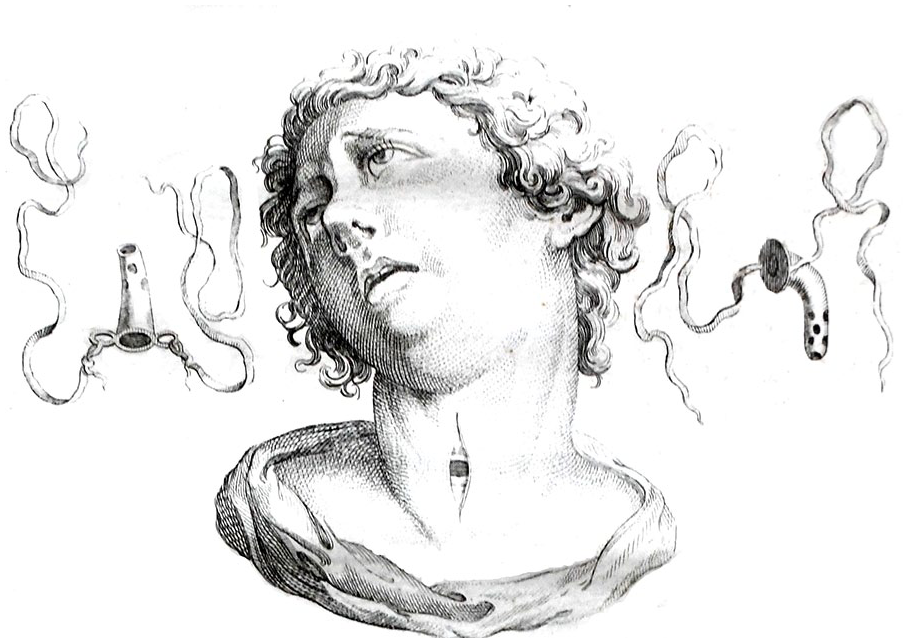
Antoine Louis’ tracheotomy tube included a 90° curve, distal fenestrations, and ringlets with tapes to secure the tube.
George Martin, a Scottish surgeon, is often credited with devising the tube-within-a-tube design. In a 1730 letter to his colleague William Græme, he describes using a lead pipe, originally designed for draining tissue fluid (dropsy) during an urgent tracheotomy. He had trouble clearing secretions from it and mentioned that a minister of his acquaintance had suggested designing an inner cannula.
The particles and steams arising from the lungs make a constant weeping of a thin slavery liquor from the mouth of the pipe part whereof thickening and stuffing its cavity. . . It must be acknowledged that there would be less hazard of a stoppage if our cannula had been shorter and wider especially at the mouth. I cannot but think it an ingenious proposal of one of our ministers here to make the pipe double or with one within another; that the innermost might safely and easily be taken out and cleaned when necessary without any molestation to the patient.
2
The greatest credit for the development of the tracheotomy cannula must go to Armand Trousseau (1801-1867), who improved existing technology during a continuing diphtheria epidemic in Paris. In 1839, he and Hippolyte Belloc described the insertion of a flexible curved gum cannula into a tracheal incision using a Trousseau dilator (Figure 2) to hold the anterior tracheal wall open.
3
Finding this cannula inadequate, Trousseau engaged in an intense exchange with his teacher Pierre Bretonneau. In the end, Trousseau took up Martin’s idea of an inner canula and built a silver double tube with the inner canula extending just beyond the outer one. A screw on the tube’s neck flange locked the inner canula in place.
Trousseau to Bretonneau-Charenton, July 3, 1826. I could introduce an equal cannula through a much smaller opening in the trachea; I could also introduce the inner cannula more easily and remove it more easily. . . . It is exactly your cannula cut in two. . . This mobile cannula would always remain in place: it would serve me to conduct the medications that I would like to introduce into the trachea, so that the internal cannula would never be blocked; it would serve me to let the diphtheritic concretions pass that the patient would want to expectorate. . . My dear master, are the cannular modifications that I propose to you; please tell me your opinion, and if you think them suitable. (From the French by Google Translate)
4

(Left) A Trousseau dilator for introducing the tube through the anterior tracheal wall. (Right) Trousseau built a silver double tube with the inner canula extending just beyond the outer one. A screw locked the inner canula in place.
Jacob Da Silva Solis-Cohen, in his 1872 American textbook Disease of the Throat, advocates for Trousseau’s technique and tracheotomy tube but also relates a battlefield story of hammering Minié balls (lead bullets) into a sheet and building a serviceable tube on the spot. 5
While most of the design concepts preceded him, the task of standardizing both tracheotomy and the tracheotomy tube fell to the American broncho-esophagologist Chevalier Jackson. His systematic approach and strong opinions became dogma in the 20th century. Jackson built many of his prototypes in his farmhouse workshop.
6
As to materials, silver was the answer (Figure 3):
Tracheotomic cannulae should be made of sterling silver. German silver plated with pure silver is good enough for temporary use, but the plating soon wears off under the galvanic action set up between the two metals. Aluminum becomes roughened by boiling and contact with secretions, and causes the formation of granulations which in time lead to stenosis. Hard rubber tubes cannot be boiled, the walls are so thick as to leave too little lumen, and the rubber is irritating to the tissues.
7

A Jackson sterling silver tracheotomy tube, circa 1920.
Jackson described fenestrations placed in older tubes as “pernicious things” producing bleeding and granulation. He introduced a standardized system of diameters and lengths to accommodate children, adults, and those with tracheal stenosis. His tubes included an olive-tipped “pilot” to facilitate insertion (Figure 4) and were engineered for respiratory efficiency, with thin walls and a large internal diameter to maximize airflow. They were also made long enough to prevent accidental decannulation from soft tissue swelling. Jackson thus incorporated the best innovations of his predecessors to create a design that lasted a century.

Properly shaped tracheotomic cannulae (according to C. Jackson). (Left) A cane-shaped cannula used for intrathoracic compression or other stenosis. (Right) Full-curved cannula for regular use. Pilots are made to fit in the outer cannula. Sterling silver is the best material for cannulae. 6
Footnotes

Level of Evidence
5 – expert opinion.
Ethical Considerations
This article does not contain any studies with human or animal participants.
Consent to Participate
There are no human participants in this article and informed consent is not required.
Funding
The authors received no financial support for the research, authorship, and/or publication of this article.
Declaration of Conflicting Interests
The authors declared no potential conflicts of interest with respect to the research, authorship, and/or publication of this article.
Data Availability Statement
This article contains no new data. All data included are available through conventional library sources. The authors would be pleased to assist interested researchers via the corresponding author’s email.