



Abstract

Significance Statement
This case underscores the dangers of nasogastric (NG) tube kinking in pediatric patients, where complications can lead to severe trauma, airway compromise, and significant distress. It highlights the necessity of extra precautions, including careful tube selection, accurate placement, and cautious management during retrieval. Early recognition of resistance and minimally invasive techniques are critical for preventing complications, emphasizing the need for heightened vigilance and specialized approaches in pediatric cases to ensure patient safety.
Clinic
A 2-year-6-month-old boy with a history of nephrotic syndrome was inserted with a NG tube three months ago to ensure accurate administration of prednisolone. The tube was routinely replaced every two weeks. During a recent replacement attempt, the tube could not be removed and appeared lodged at the 20 cm mark. The child cried during removal attempts and exhibited minimal bleeding from his left nostril, prompting referral for further treatment.
Further assessment revealed that an adult FG8-sized NG tube had been inserted 2 weeks prior and anchored at 70 cm, an excessive length for a toddler. The child was calm, breathing normally without respiratory distress, and showed no active bleeding in the nasal or oral cavity. Radiographic imaging confirmed the location of the tip of the NG tube at the vertebral level of T1-T2 with suspicious kinking of the NG tube in the nasopharyngeal space (Figure 1). A flexible endoscopy confirmed a kinked NG tube section in the nasopharynx, with unsuccessful attempts to untwist and retrieve it. Further endoscopic examination identified the tube tip at the cricopharyngeal level without any surrounding granulation or masses.
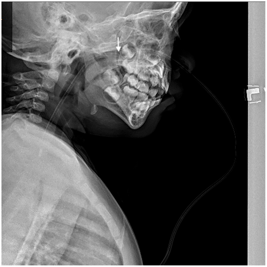
Lateral radiograph of the neck and upper thorax shows the tip of the nasogastric tube located at the T1-T2 vertebral level. An arrow highlights a suspected kink or knot within the nasopharyngeal space.
An examination under anesthesia revealed a significant kink over the NG tube at the nasopharyngeal level. To address this, the tube was carefully cut at the nasal level and the kinked distal portion was removed through the mouth (Figure 2). The child recovered smoothly after the procedure.

Distal end of the nasogastric tube exhibits a kink. A cut was made just proximal to the kinked area to facilitate safe retrieval of the tube.
Nasogastric tube insertion is a routine but blind procedure widely used for enteral feeding, medication administration, and gastric decompression. While straightforward, it carries potential complications, including nasopharyngeal discomfort, nasal septum erosion, sinusitis, pressure injuries, and, in rare cases, epistaxis, pneumothorax, and even death. 1 In this case, spontaneous kinking of the NG tube obstructed its removal, presenting a rare complication. Resistance during NG tube extraction may indicate knotting or kinking. Forceful removal of a kinked or knotted NG tube can cause trauma, especially within the narrow nasal passage, causing bleeding and possible airway compromise. This case involved minor bleeding due to the delicate nature of the posterior nasal cavity; in more severe cases, profuse bleeding can pose significant risks. Ensuring caution during NG tube retrieval is essential; resistance should prompt further investigation rather than forced extraction. A reported case describes a self-uvulectomy resulting from an attempt to remove a NG tube. 2
Radiography is often used as a noninvasive diagnostic tool to locate a lodged NG tube, especially in cases of knotting or coiling. However, imaging may offer limited information if it is obscured by surrounding structures. Flexible endoscopy offers clearer visualization, especially in the nasopharyngeal and cricopharyngeal regions, allowing direct observation of any kinking or knotting.
Various approaches have been described for removing kinked or knotted NG tubes. Endoscopic guidance is often effective; if retrieval fails, cutting the tube proximal to the kink and removing each end through the mouth and nose is an alternative approach.3-5 However, in toddlers, this can be challenging due to limited cooperation and resistance to oral procedures. In these cases, removal under anesthesia is preferred, minimizing distress and ensuring controlled management of any associated bleeding, while reducing the risks of airway compromise.
Preventive measures are essential to avoid such complications with the NG tube. Factors that contribute to kinking or knotting include the use of smaller diameter tubes, excessive length insertion, prolonged placement, or patient-specific anatomical factors such as small stomach capacity or previous trauma.2,6 In this case, while the diameter of the tube was appropriate, it was an adult tube with a more rigid structure, which may have contributed to kinking. Introducing excess tube length into a smaller capacity stomach increases the likelihood of coiling and subsequent kinking. Selecting a softer pediatric-specific tube of appropriate size and ensuring an accurate insertion length can help reduce this risk. The correct tube length is estimated by measuring from the nostril, tracing along the side of the face to the ear, and down to the xiphoid process. This typically allows the tip of the distal tube to reach the stomach fundus without excessive length, preventing tube coiling. 1
This case highlights the importance of the proper NG tube placement technique and appropriate tube selection to prevent complications. Awareness of potential complications and adherence to best practices significantly reduce risk. In cases of resistance during removal, a thoughtful approach with appropriate diagnostic and retrieval methods can prevent further trauma, bleeding, and airway compromise.
Footnotes
Acknowledgements
Not applicable.
Authors’ Contributions
All the authors have participated sufficiently in the conception and design of the work, in the analysis of the data, and in writing the manuscript to take public responsibility for it.
Availability of Data and Material
Not applicable.
Consent for Publication
Written informed consent was obtained from the patient’s guardian for the publication of the article and accompanying images.
Declaration of Conflicting Interests
The author(s) declared no potential conflicts of interest with respect to the research, authorship, and/or publication of this article.
Funding
The author(s) received no financial support for the research, authorship, and/or publication of this article.
Ethics Approval and Consent to Participate
Consent was obtained from the subject’s guardian of the case for the publication of his/her clinical data and photography.
