
Abstract
Congenital midline sinus of the upper lip are rare congenital malformations. We recently identified a case featuring a congenital midline sinus of the upper lip. The punctate opening was positioned at the midline of the philtrum, immediately below the base of the columella. Surgical removal of the sinus tract was conducted through an intraoral approach. Up to now, fewer than 70 cases have been reported. Several postulates, including the fusion theory, merging theory, and invagination theory, have been proposed to explain the formation of the congenital midline sinus of the upper lip. Nevertheless, the etiology of this uncommon abnormality remains unclear. This report details a case of a congenital upper lip sinus presenting as a congenital midline sinus of the upper lip and reviews the current literature on this condition.
Introduction
A congenital lip sinus is a rare condition; this epithelial anomaly has been reported in both the upper and lower lips. The majority of instances pertain to the lower lip, with an estimated incidence of lower lip sinus at 0.001% in the general population. 1 The occurrence of an upper lip sinus is even more infrequent, up to now, fewer than 70 cases have been reported.2,3 The earliest reported case of midline sinus of the upper lip dates back to 1891, as documented by Lanelongue. 4 Lip sinus may occur independently or in conjunction with cleft lip and/or cleft palate, as seen in Van der Woude syndrome. 5 Lower lip sinus is frequently associated with cleft lip and/or palate in Van der Woude syndrome; Van der Woude syndrome may be accompanied by a range of other anomalies, such as hypodontia, syndactyly, and congenital heart defects. 6 Upper lip sinus tends to be nonsyndromic epigenetic malformations. 6 Associations with other midline deformities have also been described, including double frenulum, frenulum sinus, nasal dermoid cyst, and hypertelorism have also been described. 6 The formation of the congenital midline sinus of the upper lip has been theorized using different postulates, such as the fusion theory, merging theory, and invagination theory. However, the underlying cause of this unusual anomaly remains unclear. 2 This report details a case of a congenital upper lip sinus presenting as a congenital midline sinus of the upper lip and review the current literature on this condition.
Case Report
A 17-year-old Chinese girl presented with a congenital midline sinus of the upper lip, the upper lip experiences swelling 3 to 5 times annually, accompanied by either pain or inflammation, at intervals, there is a flow of white fluid from this sinus, apart from this, there were no other reported discomforts. The patient is in excellent health, exhibiting no congenital anomalies apart from the congenital midline sinus of the upper lip. In addition, there is no reported family history of medical conditions. The patient’s mother denied taking medications during pregnancy that could potentially lead to fetal abnormalities, as well as any history of smoking, alcohol consumption, and other adverse habits. Examination findings indicated a pinpoint opening situated along the midline of the upper lip, positioned below the base of the columella (Figure 1A). There were no additional abnormalities noted in the craniofacial region.
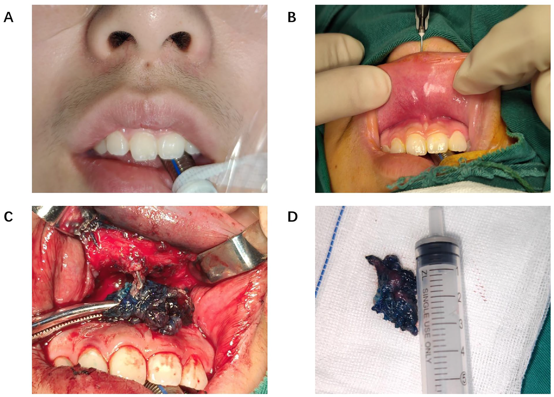
(A) A midline sinus on the patient’s upper lip. (B) The inserted probe verifies the existence of a sinus tract. (C) The excision of the sinus tract is performed through an incision in the upper buccal sulcus. (D) Excised sinus tract.
The patient and their family requested a procedure without skin incisions. Sinus tract removal through an intraoral approach was performed under general anesthesia. The probe was used for localization by inserting it into the opening on the surface of the midline of the upper lip. No mucosal opening was found, indicating no connection with the oral cavity (Figure 1B). Following this, methylene blue dye was injected to stain the sinus tract, and the dissection through the upper buccal sulcus approach began, proceeding from the blind end to the skin opening located 2 mm below (Figure 1C). The surgical procedure was uneventful, with the complete excision of the lesion (Figure 1D). The patient experienced a stable postoperative course, and there was no scarring on the skin.
Histopathological examination of the specimen measuring 2.2 cm × 1.5 cm × 0.6 cm revealed squamous epithelial lining within the tube wall. Three months postoperatively, the patient has not experienced a recurrence.
Discussion
The mechanism of congenital upper lip sinus formation remains not entirely clear, and several hypotheses currently exist: the “fusion theory” posits that these abnormalities arise from a failure in the anterior growth of the frontonasal process or an incomplete fusion between the maxillary and frontal processes.4,7,8 The “merging theory” posits that the congenital upper lip sinus is a result of aberrations in the normal mesenchymal merging process due to the deficiency of mesenchymal cells.9,10 The “invagination theory” suggests that the formation of upper lip sinuses results from the failure of ectodermal invagination of the nasal placodes during the frontonasal process.11-14 However, none of the aforementioned hypotheses can precisely elucidate the distorted process of congenital upper lip sinuses. In recent years, research on the interactions during facial embryogenesis has mainly focused on signaling pathways, mechanisms of gene interactions, and more. 15
Congenital sinus of the lip can be classified based on their location of occurrence into upper lip and lower lip. Most instances pertain to the lower lip, with an estimated incidence of lower lip sinus at 0.001% in the general population. 1 The occurrence of an upper lip sinus is even more infrequent; up to now, fewer than 70 cases have been reported.2,3 They traverse the orbicularis oris muscle, terminating just beneath the mucosal surface of the lip, devoid of any connection with the oral cavity. 16 We searched PubMed using the search term “congenital upper lip sinus,” reviewing case literature from the past 54 years (1970-2024). In this search, we identified 45 cases, including the case we are reporting (Table 1).
Congenital Midline Sinus of the Upper Lip Reported Between 1970 and 2024.
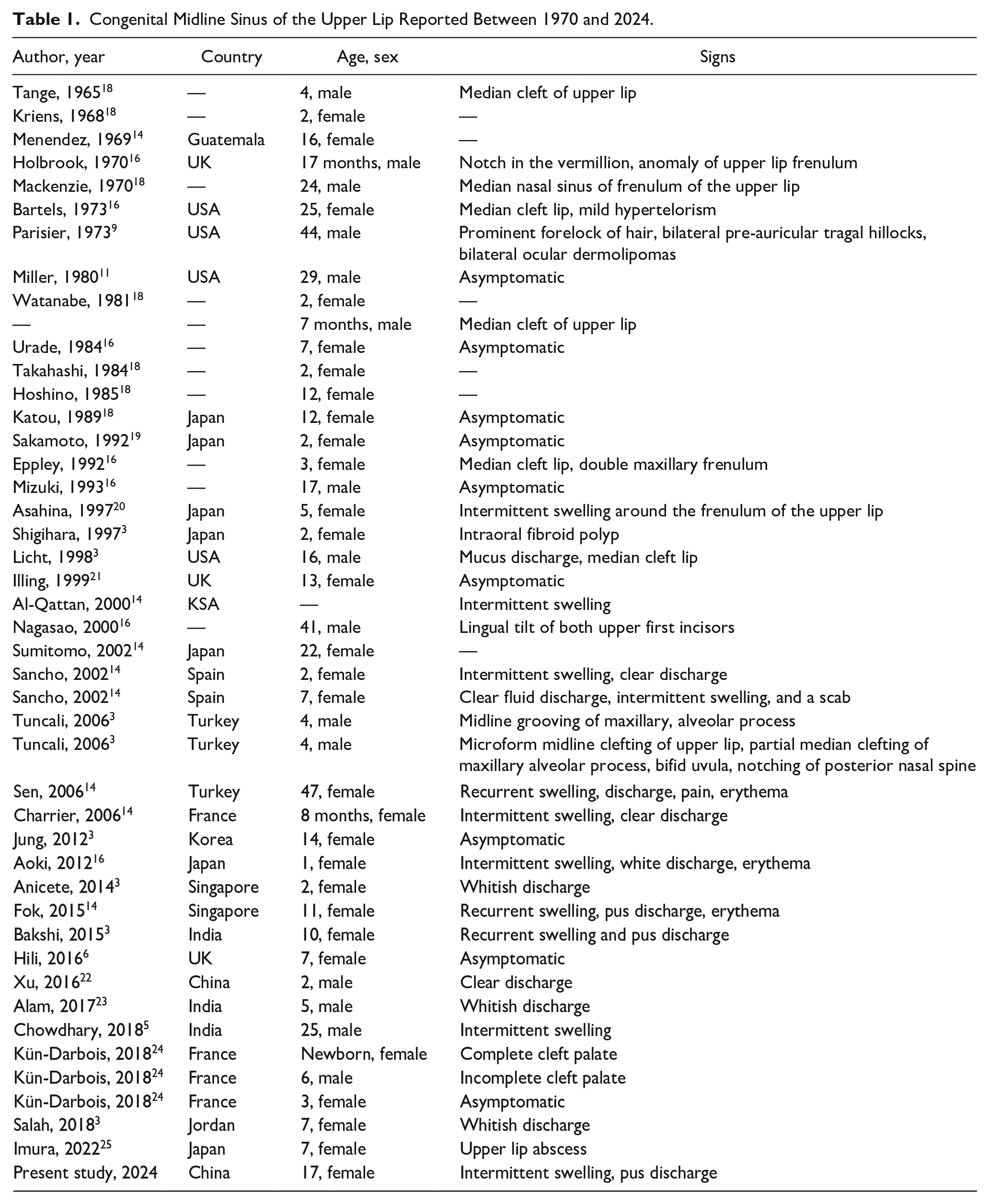
Aoki et al reviewed all reported cases of upper lip sinuses in English literature in 2011, classifying 31 cases into 3 categories: type 1 was a midline sinus without accompanying anomalies, type 2 was midline sinus with accompanying anomalies, type 3 was lateral sinus with or without accompanying anomalies. 16 They found that type I upper lip sinuses are more common in females (12/13 females). 16 This is also consistent with the literature we reviewed (7/9 females). They reported that the distribution of type II sinuses is more uniform (5 males, 4 females), 16 which is also consistent with the literature we reviewed (13 males, 16 females). Previous reviews have indicated that the majority of patients were of Asian descent, with a higher prevalence among females,5,14 consistent with our reviews. However, conclusive analysis cannot be performed due to the limited number of patients.
While the exact etiology remains unclear, surgical excision is the primary approach for treating upper lip sinus tract. Complete excision is necessary to minimize the potential for postoperative recurrence. It is noteworthy that congenital midline upper lip sinuses should be carefully distinguished from nasofrontal dermoid cysts with an opening in the philtrum. The latter has only been reported in 2 cases so far. 17 Donnell et al mentioned in their article that for all patients with a nasal midline or philtrum sinus who do not achieve positive results after initial excision, consideration should be given to intracranial extension, because the skin sinus tract may only constitute a portion of a deeper dermoid cyst. Therefore, early identification and accurate diagnosis require evaluation through computed tomography scan and magnetic resonance imaging. 17
Conclusion
For unusual congenital abnormalities of the midface, even those that may seem clinically insignificant, such as congenital midline upper lip sinuses, it is imperative to investigate their underlying causes. This contributes to the advancement of the field, enabling specialists to independently assess various theories related to embryonic development. Therefore, the documentation of congenital midline upper lip sinuses is a crucial observational outcome.
Footnotes
Author Contributions
PL and JW helped gather all of the necessary materials and were responsible for writing the manuscript. LL revised the manuscript. All authors contributed to manuscript revision, read, and approved the submitted version.
Data Availability
The data sets for this article are not publicly available due to concerns regarding participant/patient anonymity. Requests to access the data sets should be directed to the corresponding author.
Declaration of Conflicting Interests
The author(s) declared no potential conflicts of interest with respect to the research, authorship, and/or publication of this article.
Funding
The author(s) received no financial support for the research, authorship, and/or publication of this article.
Ethical Approval
The ethics committee of the Linyi People’s Hospital approved the research protocol, and written informed consent was obtained from participant.