
Abstract
Lipoma of the tongue is a rare benign tumor that accounts for approximately 1% to 5% of all oral cavity tumors while 0.3% of tumors are of the tongue. Notably, it is rarer in children. In this article, we report the case of a 4-year-old girl with lipomas of the tongue. The lipomas were found at age 1 year by her parents, located on the tip, ventro, and dorsum of the tongue, and presenting with a trend to increase gradually. At the time of presentation to the hospital at age 4 years, the articulatory function of the patient was partially affected, and surgical excision was performed. The surgery was uneventful, and no evidence of recurrence was noted at 3 month follow-up.
Introduction
Lipoma is the most common among the benign tumors in human body, and it can occur at any site with or without adipose tissue. Oral cavity lipoma is rare and present in multiple sites of the oral cavity, including mostly the salivary gland, buccal mucosa, lips, palate, tongue, floor of mouth, and vestibule. Lipoma of the tongue is relatively rarer, comprising approximately 1% to 5% of all oral cavity tumors1,2 while 0.3% of tumors are of the tongue.3,4 Notably, it is rarer in children. Lipoma is a neoplasm composed of adipose tissue usually with intact capsule. However, lipomas that occur in intermuscular or intramuscular fat area, known as infiltrating lipomas, have no capsule and poorly defined boundaries. 5 In the presence of other tissue components, there may be some isoforms such as angiolipoma, angiomyolipoma, and fibrolipoma. There are some other classifications: (1) familial lipoma, which is a multiple lipoma associated with genetic factors and potentially accompanying nervous system disease; (2) multiple painful lipoma; (3) diffuse lipoma of the neck in the elderly population, which presents with diffuse fat accumulation in the entire neck, occipital, and upper back areas, and significantly affects neck activity and even respiration 6 ; (4) retroperitoneal lipoma, which is large and usually presents with an abdominal mass with abdominal distension; (5) malignant liposarcoma, in the context of abrupt lipoma growth in a short term or >10 cm tumor diameter 7 ; (6) brown lipoma, which is also known as hibernoma.
Lipoma can occur in any sites that contain adipose tissue and rarely in sites, such as the tongue, without adipose tissue. Lipoma of the tongue is usually asymptomatic, painless, and solitary in clinical practice. It is commonly found as a lobulated round or oval mass in the submucosal and sublingual tissues, which has a clear boundary, soft texture, yellowish color, high activity, and a slow growth pattern. According to the thickness of mucosa overlying mass, the mass usually has a smooth surface without ulceration, unless being affected by trauma, infection, chronic irritation, hormonal imbalance, metabolic conditions, and so on. 2
The diagnosis of lipoma is based on mainly clinical and imaging findings, while histopathology is a must for a confirmed diagnosis.
Treatment should be surgical excision (without major surgical difficulties), and postoperative pathology review is required to confirm the nature of the lesion.
In this article, we report a rare case of lipoma of the tongue in a 4-year-old child.
Case Report
A 4-year-old girl was diagnosed with a mass on the tip, ventro, and dorsum of the tongue, which appeared as a longitudinal, long, yellowish, soft, and sessile streak. The mass was found by chance at age 1 year but was left untreated. Over the next 3 years, the mass gradually increased to a size of 1.0 cm × 1.0 cm × 2.0 cm with a clear boundary at this medical visit (Figure 1A).
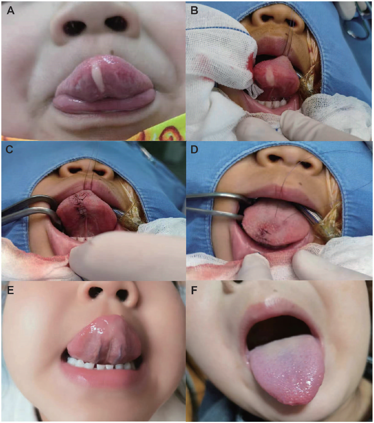
Preoperative, intraoperative, and postoperative manifestations of lingual lipoma. Preoperative images of the lipoma on the tongue of the patient (A). Intraoperative images of the patient (B, C, D). Recovery of the patient’s tongue 3 months after surgery (E, F).
At age 4 years, speech of the patient was poorly articulated. After initial physical examination, subtotal excision of the mass was decided. Under general anesthesia, a longitudinal incision was created on the mucosa overlying the mass. Blunt dissection was applied throughout this process. The mass popped out from underneath the mucosa, and it did not invade the muscle tissue of the tongue after the capsule was dissected. The mass was yellowish and well-capsuled, and en bloc excision was performed (Figure 1B). Following local hemostasis, the defect of the tongue was sutured using absorbable thread. The surgical site healed well post the surgery (Figure 1C and D). Cut surface of the mass demonstrated yellowish adipose tissue composed of a lot of fiber ropes and spaces laterally and longitudinally (Figure 1E and F). The lipoma tissue floated on the surface of formalin rather than getting deposited at the bottom of the container, as all types of adipose tissues.
Pathology review indicated mature adipocytes and erythrocytes, and the adipocytes were arranged in lobules, consistent with the characteristic of lipoma (Figure 2). The patient’s ability to eat was normal, and her speech has improved without loss of sensory or motor function of the tongue at present.
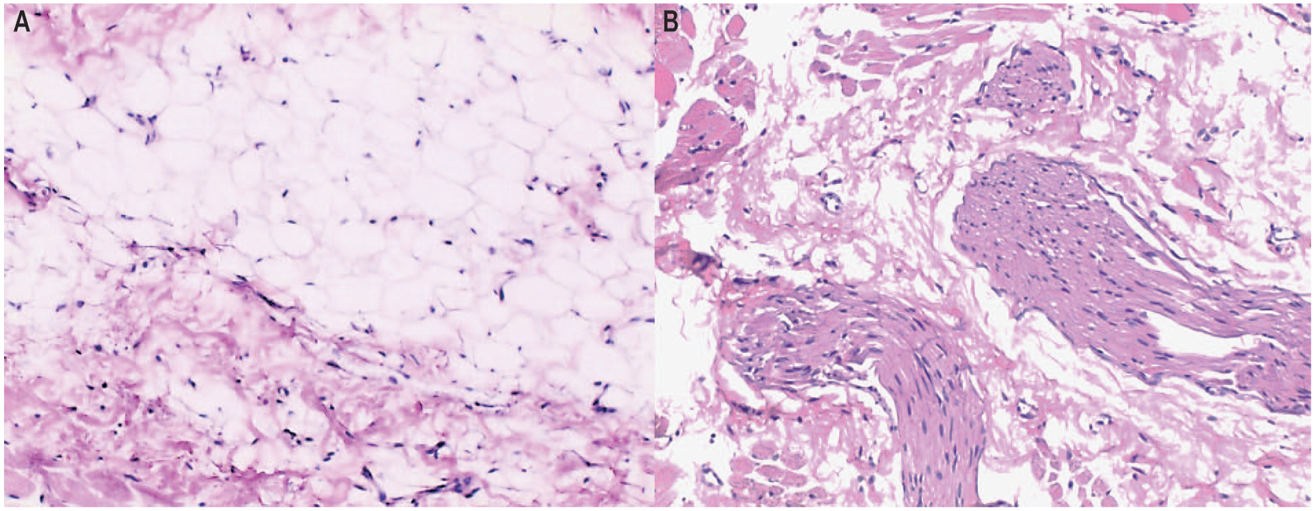
Histopathology of specimen showing mature fat cells. Pathological diagnosis: (tongue) Squamous epithelium with parakeratosis cells, local squamous epithelial hyperplasia, vacuolar degeneration of spinous cells, hyperplasia of adipose tissue under the lamina propria, and the presence of nerve fibers and blood vessels in the skeletal muscle layer. Benign lesion, considered lipomatous-like hyperplasia (A, B). Immunohistochemistry: S-100 (+) , CK (+) , SMA (-) , desmin (+).
Conclusion
Venkateswarlu et al 8 reported a rare case of intraoral lipoma. The patient is a 6-year-old boy who cannot close his mouth for a week after an injury. An oral examination showed that a pedunculated tumor with a pale yellow surface was visible in the mucosal area of the right cheek, with tooth marks around it. There was no tenderness on palpation, and its size was 3 cm × 1.5 cm. The histopathology diagnosis of surgical resection was considered as intraoral lipoma. 8
The case we reported is different. Lipoma of the tongue in children is extremely rare. In this case, the lipoma was found at age 1 year. Whether it is congenital cannot be confirmed as the specific time of onset is unknown. Most common, lipomas in children are familial and congenital diffuse lipomas. Congenital diffuse lipomas occur immediately after birth. They are poorly demarcated and increase with age, potentially accompanying bony hypertrophy, musculature hypertrophy, or venous angioma (hemangioma racemosum).
The differential diagnosis of lipoma of the tongue includes mucocele, lymphangioma, hemangioma, rhabdomyoma, neurofibroma, neuroma, pleomorphic adenoma, salivary gland tumor, adenocarcinoma, liposarcoma, and so on. 2 Of these, liposarcoma can easily be misdiagnosed and often with lipoma in clinical practice, while liposarcoma can originate from the malignant transformation of lipoma. Comparatively, liposarcoma has a harder texture and a stronger relationship with adjacent tissues. It has to be noted that lipoma of the tongue must be distinguished from liposarcoma. 9
Surgical excision is often needed for lipomas with symptoms or causing functional disorder, such as diffuse multiple congenital lipoma, generalized intestinal hemangioma, neurofibroma, and symmetric lipoma. Infiltrating lipomas are difficult to remove due to their indistinct boundaries. Similarly, it is also difficult to complete en bloc resection of multiple lipomas, and they tend to recur with a rate of up to 62.5%. 10 Simple lipomas can be easily resected and may not be followed by recurrence. 11 To prevent recurrence, surrounding normal tissues will be excised by some operators. 2
Footnotes
Declaration of Conflicting Interests
The author(s) declared no potential conflicts of interest with respect to the research, authorship, and/or publication of this article.
Funding
The author(s) disclosed receipt of the following financial support for the research, authorship, and/or publication of this article: This research was funded by Tianjin Key Medical Discipline (Specialty) Construction Project (TJYXZDXK-040A).
Informed Consent
Written informed consent to participate in this study was provided by the participants’ legal guardian.