
Abstract

Introduction
Temporal bone otopathology laboratories have long clarified the anatomy, histology, and pathology of temporal bone and inner ear disorders but have become a scarcity in the United States. 1 These collections contain arrays of information, however, and can support multidimensional analyses that provide extraordinary interconnection and nuance: Temporal bone otopathology laboratories and archives provide a three-dimensional overview of patient sociodynamic situations, medical conditions, otologic conditions, their audiometric and vestibular testing, and the anatomy and molecular biology of their physical ears. To illustrate these extraordinary features of one otopathological collection, we review the data and analysis of three of the famous Radium Dial Painters. 2
The Temporal Bones of Three Radium Dial Painters
The radium girls were a group of young female factory workers who painted watch dials with luminescent radium paint in the early 20th century. They sharpened the points of their brushes with the “lip, dip, paint” technique, and most of them later suffered from radiation poisoning and various forms of cancer. Their struggle and sacrifice played a quintessential role in establishing workers’ rights and occupational safety. 2
By 1965, 300 cases of radium dial poisoning were being followed by the University of Chicago Hospitals. Eight of these patients had developed carcinoma of the mastoid, and six had died of their tumors. 3 In the John Lindsay Otopathology Laboratory and Archives are the records of three people who had been radium dial painters 30-50 years earlier.
Case 1 was a radium dial painter from 1925 to 1945, using the “lip, dip, paint” technique for her first 2 years—1925-1926. Her right ear showed radiation osteitis and a destructive adenocarcinoma involving the entire mastoid, extending to the sigmoid sinus and dura, filling her entire tympanic cavity and surrounding the ossicles (Figure 1). Her left ear’s apical region contained adenocarcinoma, with some areas of squamous cell carcinoma. The endochondral bone showed only radiation necrosis without tumor invasion (Figure 2). She developed palsies of right cranial nerves 5-12 and died in 1961 of aspiration pneumonia at the age of 54, 36 years after her initial exposure.
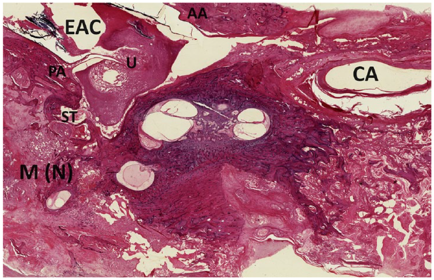
Case 1’s right endochondral labyrinthine structures are spared of tumor. The mastoid air cells and incus are involved with adenocarcinoma. AA, anterior annulus; CA, carotid artery; EAC, external auditory canal; M, mastoid air cell system; N, neoplasm; PA, posterior annulus; ST, stapedial tendon; U, umbo of the malleus.
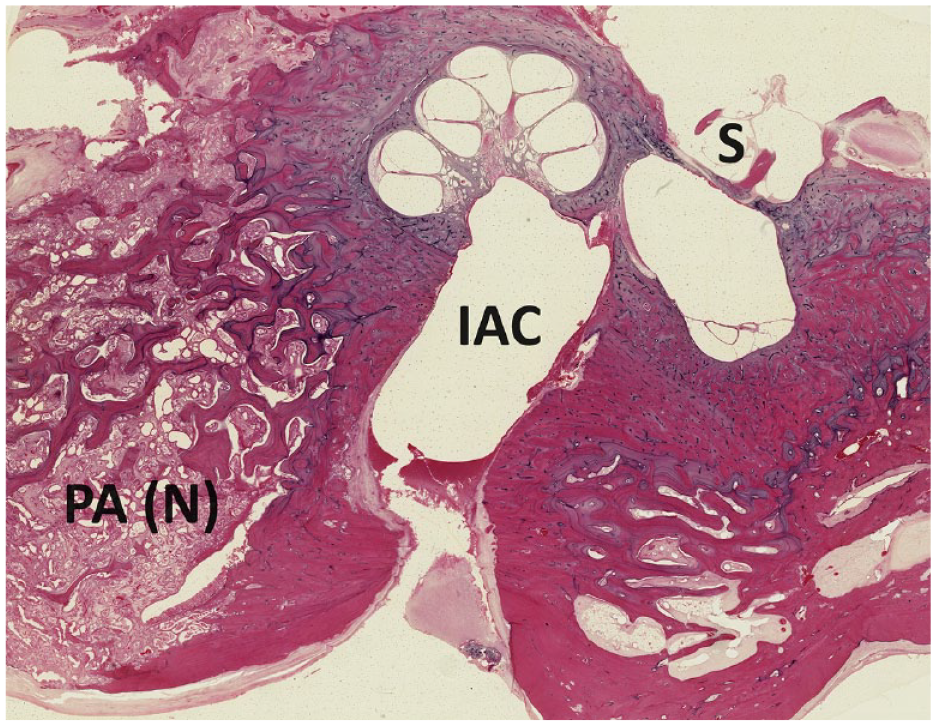
Case 1’s left endochondral labyrinthine structures are spared of tumor. The apical region shows adenocarcinoma intermixed with squamous cell carcinoma. IAC, internal auditory canal; PA, medial air cells, contiguous with the petrous apex; N, neoplasm; S, stapes.
Case 2 was a radium dial painter for 2 years from 1923 to 1924. Her right ear showed radiation necrosis of the mastoid, with tumor cells of adenosquamous mixed tumor type. Once again, the endochondral bone predominantly resisted tumor invasion, although one area of the right lateral semicircular canal showed a tumor breach. Her left mastoid showed some areas of radiation necrosis, but no tumors. On autopsy, she had osteosarcoma of the left femur and osteolytic lesions of her upper cervical vertebrae. She died in 1956 of a subdural hematoma caused by extension of her mastoid tumor through her dura, at the age of 53, 33 years after her initial exposure.
Case 3 was a radium dial painter for 13 years—1922-1935. Her right ear showed radiation necrosis in the mastoid, and nests of squamous cell carcinoma in the apex, internal canal, and neural canals. Her left ear showed radiation necrosis in the mastoid, and squamous cell carcinoma in the tympanic cavity, middle ear, and mastoid. The left endochondral bone was neither necrotic nor invaded. She died in 1972 with widely metastatic carcinoma, bronchopneumonia, and myocardial infarction, at the age of 74, 50 years after exposure.
Discussion
Observational and narrative medical reporting has become unfavored. Oliver Sacks published his book Awakenings 4 about the unexpected effect of L-dopa on encephalitis lethargica, to tremendous popular response. 5 But Sacks noted a “strange mutism” of scientific professionals toward Awakenings—one sole editorial in the British Clinical Journal reviewed it. 5 The depth and type of information in temporal bone archives is also observational, rather than “hypothesis driven.” The special features of temporal bone otopathology laboratories and archives provide intersectionality of science, commerce, industrialization, politics, and culture. The narrative of the radium dial painters and their suffering provides insight into past social shortcomings. We can notice the deep-rooted patriarchy and sexism that disregarded all issues of protection and occupational safety for these young female workers.
Many otopathological features are also of interest. The endochondral bone escaped radium-induced changes in these cases. The protected labyrinth would have reduced audio-vestibular symptoms in these painters. The predilection of the mastoid to house these carcinomas perhaps arises from an anatomical peculiarity—the mastoid air cells are among the few areas of the body where the mucosa is directly applied to bone. 3 The latency between exposure to radium and its progression to cancer can be explained by radium’s very long half-life and total body burden required for the development of neoplasia. 3 (The estimated half-life of Radium (Ra226) was reported as 1577 ± 9 years. 6 ) The histopathology of Case 1 shows the presence of two different tumor types within the same person.
Temporal bone archival laboratories provide cognizance not only regarding scientific principles but also the social, cultural, and political circumstances of the past. This makes them a comprehensive and quintessential tool for learning and advancement of otopathology.
Footnotes
Declaration of Conflicting Interests
The author(s) declared no potential conflicts of interest with respect to the research, authorship, and/or publication of this article.
Funding
The author(s) received no financial support for the research, authorship, and/or publication of this article.