
Abstract
Kimura disease is a benign subcutaneous chronic inflammatory disease of unknown etiology that is usually seen in young males. A 26-year-old Syrian adult, has suffered from focal segmental glomerulosclerosis for 10 years with no history of renal transplantation, complained of swellings in his preauricular area which was diagnosed as Kimura disease. There is no consensus regarding the optimal treatment for Kimura disease and the chosen treatment was surgery in the young patient with localized lesions. During 9 months of following up after surgical removing of the lesions, no recurrence was noticed.
Keywords
Introduction
Kimura disease is a benign subcutaneous chronic inflammatory disease of unknown etiology that is usually seen in young males. 1 It usually affects people of Asian race. 2 It is usually associated with autoimmune diseases, especially kidney diseases. 3 Classically, the disease manifests with slow-growing masses, especially in the head and neck areas 4 with or without satellite lymphadenopathy. 1
Case Report
We present a case of a 26-year-old Syrian adult, who came to the ENT clinic complaining of progressively enlarging swellings in his left preauricular and parotid areas that associated with intermittent headache from approximately 3 months. The lesions are very itchy but without any pain or tenderness.
Our patient did not complain of any nasal, oral, pharyngeal, or laryngeal complaints. He is known to have a significant history of focal segmental glomerulosclerosis (FSGS) (Figure 1) for 10 years, treated with oral prednisolone 10 mg daily. He denies any significant surgical or allergic history. The patient smokes only on occasions and is nonalcoholic.
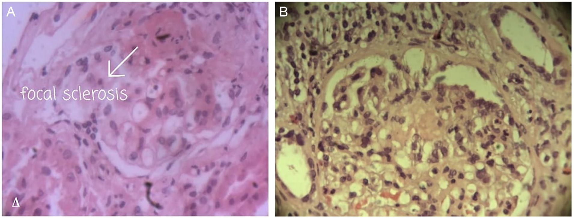
Microscopic examination for the renal biopsy of our patient reveals (A) areas of collagenous sclerosis within the glomerulus and (B) focal glomeruli are affected and just part of the affected glomerulus is involved (segmental) with the sclerosis.
The patient has tried many skin creams and ointments containing antifungals and steroids in their composition, and many herbal remedies but without any benefit. Several sessions of liquid nitrogen have been applied to the lesions with partial benefit and very little regression of the lesions. The lesions tend to regress when the patient took a dose larger than usual of oral prednisolone (i.e., more than 10 mg/day), but the lesions recurred if the patient returned to the usual dose of prednisolone for his underlying renal disease.
Examination of the right ear was within normal limit whereas, examination of the left ear revealed pink, relatively soft, granular lesions covering the tragus and anterior pinna, measuring about 5 × 3 cm for the entirety of the lesions (Figure 2). No important palpable lymph nodes were noted on physical examination.
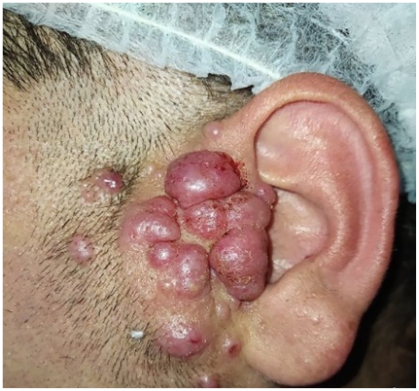
Pink, relatively soft, granular lesions covering the tragus and anterior pinna of the left ear.
Apparently, the appearance of masses in the patient was accompanied by an increase in the severity of his underlying renal disease, and therefore the patient, without consulting the doctor, increased his dose of corticosteroids to 15 mg daily. The patient’s self-medication led to a partial regression of his lesions for a short period only, then the lesions gradually increased in size until they reached the size at which the patient visited the ENT clinic.
Investigations revealed hemoglobin level of 10.5 g/dL, white blood cell count of 10.47 cells/L × 109 cells/L with 15% eosinophil, and normal platelet count. His blood urea and serum creatinine were within normal limits. The ESR was 22 mm/hour. Routine Chest adiograph (CXR) and Electrocardiograph (ECG) were within normal limits. After making the definite diagnosis by doing several incisional biopsies, we decided to do a surgical removal of the masses (Figure 3). The patient was followed up for about 9 months without any recurrence (Figure 4).
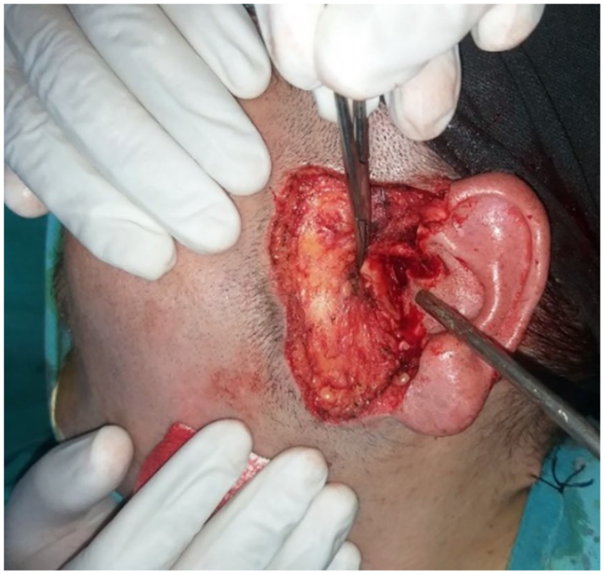
Intraoperative view of surgical removal of the masses.
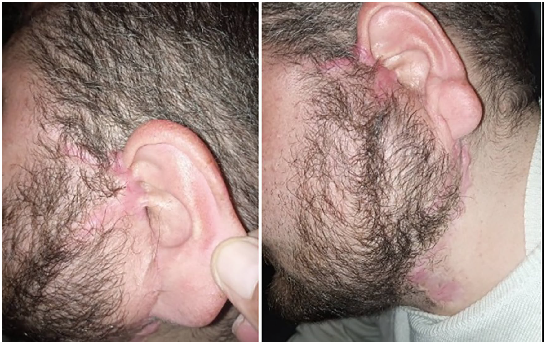
View of surgical site after 3 months of the resection.
The diagnosis of Kimura disease is definitively made based on the histopathological findings of an excisional biopsy of the suspected mass. Microscopically, vascular proliferation, dense inflammatory infiltrates containing especially eosinophil and granuloma formations containing histiocytes and lymphocytes can be seen (Figures 5 and 6).
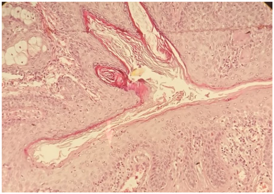
Histopathological section of skin showing pleomorphic inflammatory infiltration.
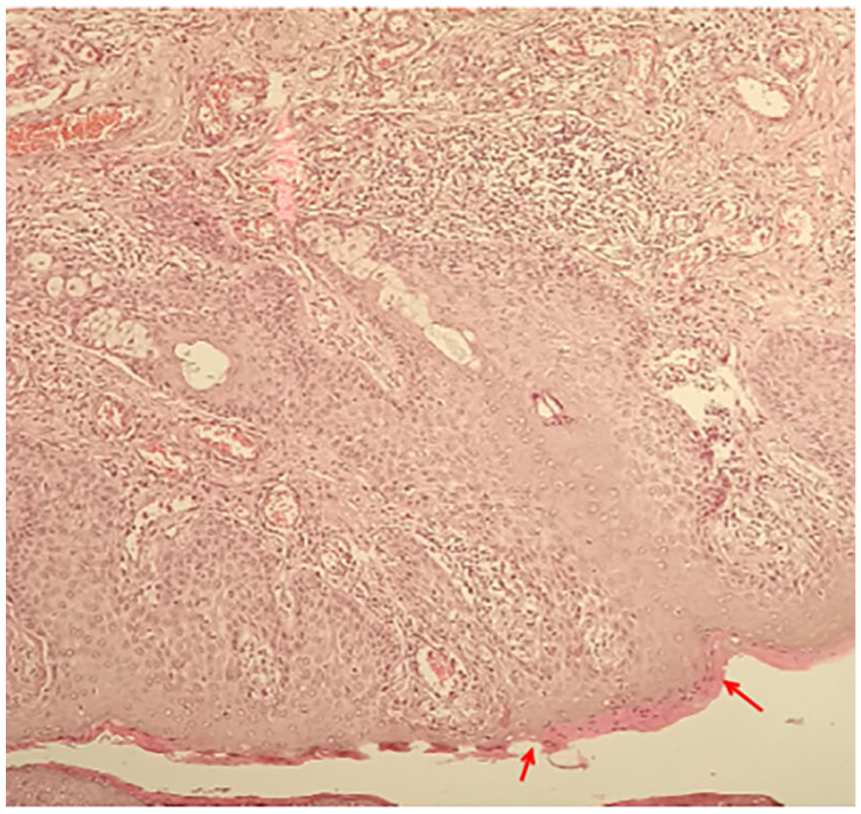
Histopathological section of skin showing parakeratosis (red arrows).
Longevity of the lesions and expensiveness and difficulty in obtaining immunosuppressive drugs such as cyclosporine and other therapies that may be effective in treating Kimura disease in our young patient with the localized lesions certainly made us prefer the application of surgical treatment.
Discussion
To our best knowledge, this is the first diagnosed case of Kimura disease in Syria. Our case is one of the very rare cases of Kimura disease with FSGS patient without a history of renal transplantation. Kimura disease is an inflammatory condition that sometimes manifests as painless subcutaneous asymmetric nodules in the head and neck regions. 5
In some cases, Kimura disease is misdiagnosed as a mass in the context of hematological or lymphatic malignancy due to the presence of a local mass in the cervical region, which may sometimes be accompanied by lymphadenopathy. 6
The presence of lymphadenopathy raises suspicion for an underlying malignancy, especially if the history and initial clinical examination were directed toward malignancy in the initial presentation. Moreover, Kimura disease can mimic other chronic and malignant diseases on radiological examination.
Kimura disease is diagnosed based on the histopathological findings. Microscopically, vascular proliferation, dense inflammatory infiltrates containing especially eosinophil and granuloma formations containing histiocytes and lymphocytes can be seen. 7
There is a well-studied role of T lymphocytes, especially helper cells, in the pathogenesis of Kimura disease, which explains some of the association between Kimura disease and renal diseases, especially nephrotic syndrome. 8
Even at this point, there is no consensus regarding the optimal treatment for Kimura disease. The advantage that is clearly observed in Kimura disease patients with renal disease is that both diseases usually respond to corticosteroids and immunosuppressants. The preferred initial treatment for localized Kimura disease is surgical excision. 9
One report has mentioned a remission of secondary membranous nephropathy in a patient with Kimura disease after surgical removal of an axillary mass. 10 Although Kimura disease has a chronic relapsing course, the general prognosis is good. The prognosis for patients with associated renal involvement varies depending on the form and severity of nephropathy.
We have searched the medical literature for cases about Kimura disease with FSGS similar to our case and we have summarized the results in Table 1.
Comparison of our case with the medical literature for patients with Kimura disease in combination with FSGS.
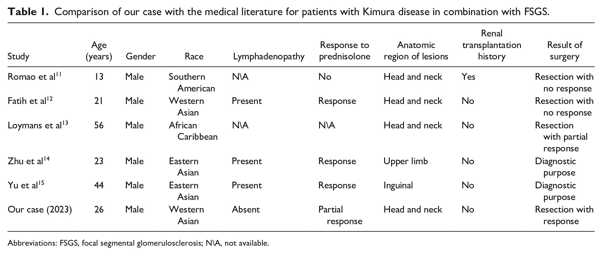
Abbreviations: FSGS, focal segmental glomerulosclerosis; N\A, not available.
Conclusion
We present the case of Kimura disease to highlight this underdiagnosed disease in our country. We also emphasis on the possibility of Kimura disease to affect patients without history of renal transplantation. We need more studies to declare the actual prevalence of this disease and to detect the optimal management.
Footnotes
Acknowledgements
The authors are grateful for their colleges in Al-Mouwasat Hospital in the ENT-HNS department.
Data Availability Statements
The data that support the findings of this study are available from the corresponding author upon reasonable request.
Declaration of Conflicting Interests
The author(s) declared no potential conflicts of interest with respect to the research, authorship, and/or publication of this article.
Funding
The author(s) received no financial support for the research, authorship, and/or publication of this article.
Ethical Approval
Written informed consent was acquired from the patient for publication of this article.