
Abstract

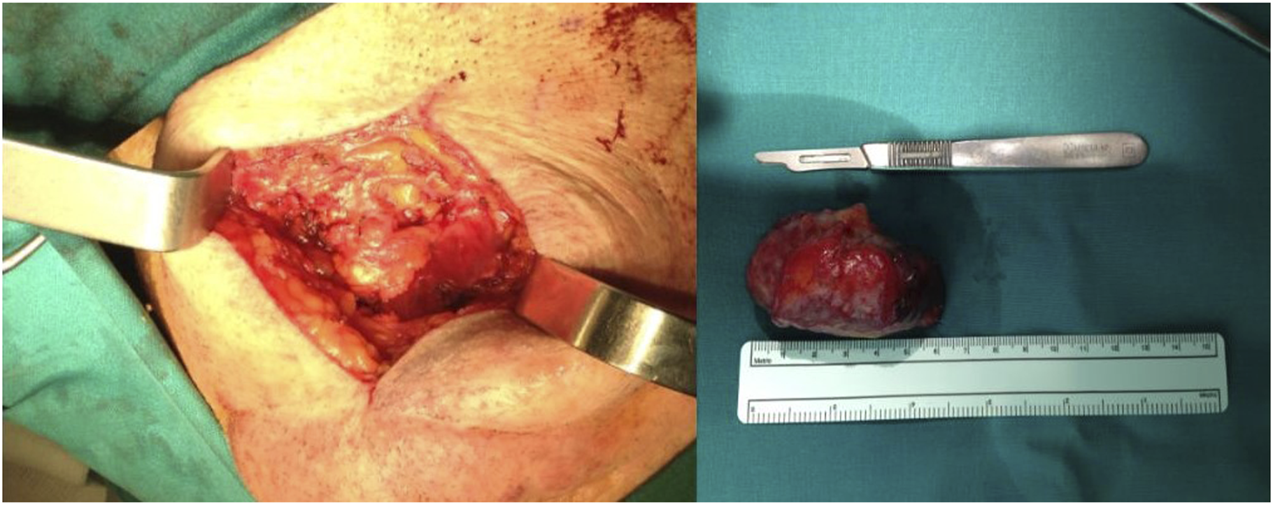
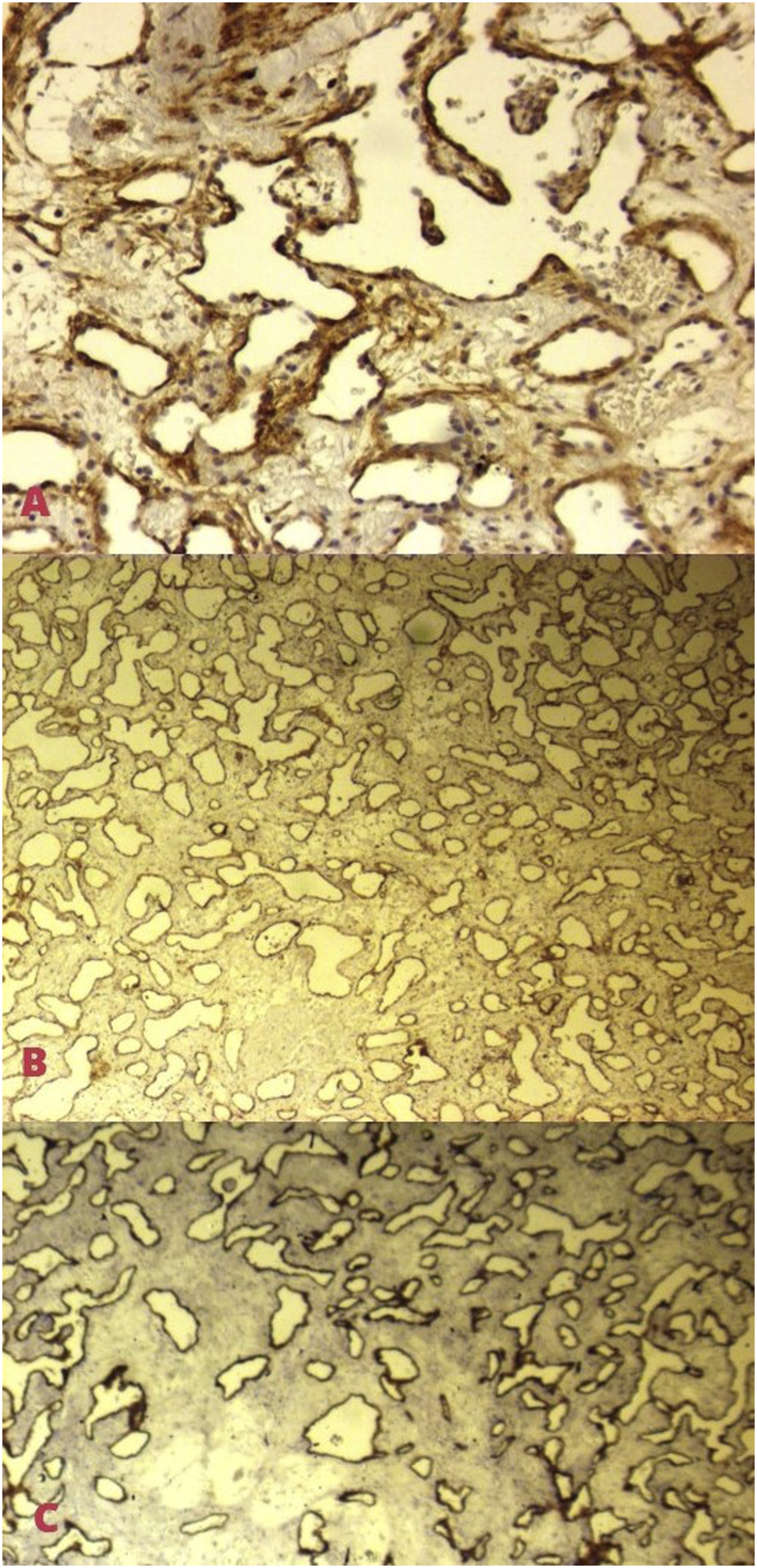
An 85-year-old patient presented to the outpatient Ear, Nose, and Throat (ENT) clinic with a submental swelling that had been present for at least one year. There was no history of trauma, surgery, radiation exposure, smoking or associated clinical symptoms such as fever, pain, dyspnoea, hoarseness or dysphagia. On clinical examination, a well-circumscribed, painless, and mobile mass was found in the left submental region without any changes in the overlying skin or motor nerve deficiency. The examination of the upper aerodigestive tract was unremarkable. Routine blood tests were normal. Computed tomography (CT) with intravenous contrast showed a smooth clearly circumscribed mass of soft tissue density, with a maximum diameter of 6.5 cm, located within the subcutaneous tissue of the submental area and exerting pressure effects in the right submandibular gland (Figure 1). Subsequently, a fine needle aspiration cytology (FNAC) was performed, but was not diagnostic due to mainly bloody specimen without containing adequate cellularity. The procedure also caused minor bleeding which stopped after applying firm pressure over the injured area. Under general anaesthesia, the patient underwent open surgery and the mass was completely excised (Figure 2). Histological examination showed macroscopically an ovoid circumscribed tissue of dimensions 5.7 × 3.2 × 2 cm, which was completely surrounded by a thin capsule. Microscopically, the texture of the vascular lesion was observed that was surrounded by several papillary vascular formations, lined by an endothelial cell layer without atypia. Collagenization was also observed in the upper layer (Figure 3). All findings indicated Masson’s tumor. The post-operative recovery was uneventful and no recurrence was observed at 6-months’ follow-up. Computed tomography (CT) with intravenous contrast (saggital and axial view) demonstrates a smooth and clearly circumscribed mass of soft tissue density within the subcutaneous tissue of the submental area, with a maximum diameter of 6.5 cm (white arrow). Image of the lesion intraoperatively and after complete excision. Microscopy (2.5x magnification) and immunochemistry with dyes Κi-67 (A), CD-31 (B), and CD-34 (C) show a texture of a vascular lesion, surrounded by several papillary vascular formations, and lined by an endothelial cell layer without atypia. Collagenization was also observed in the upper layer.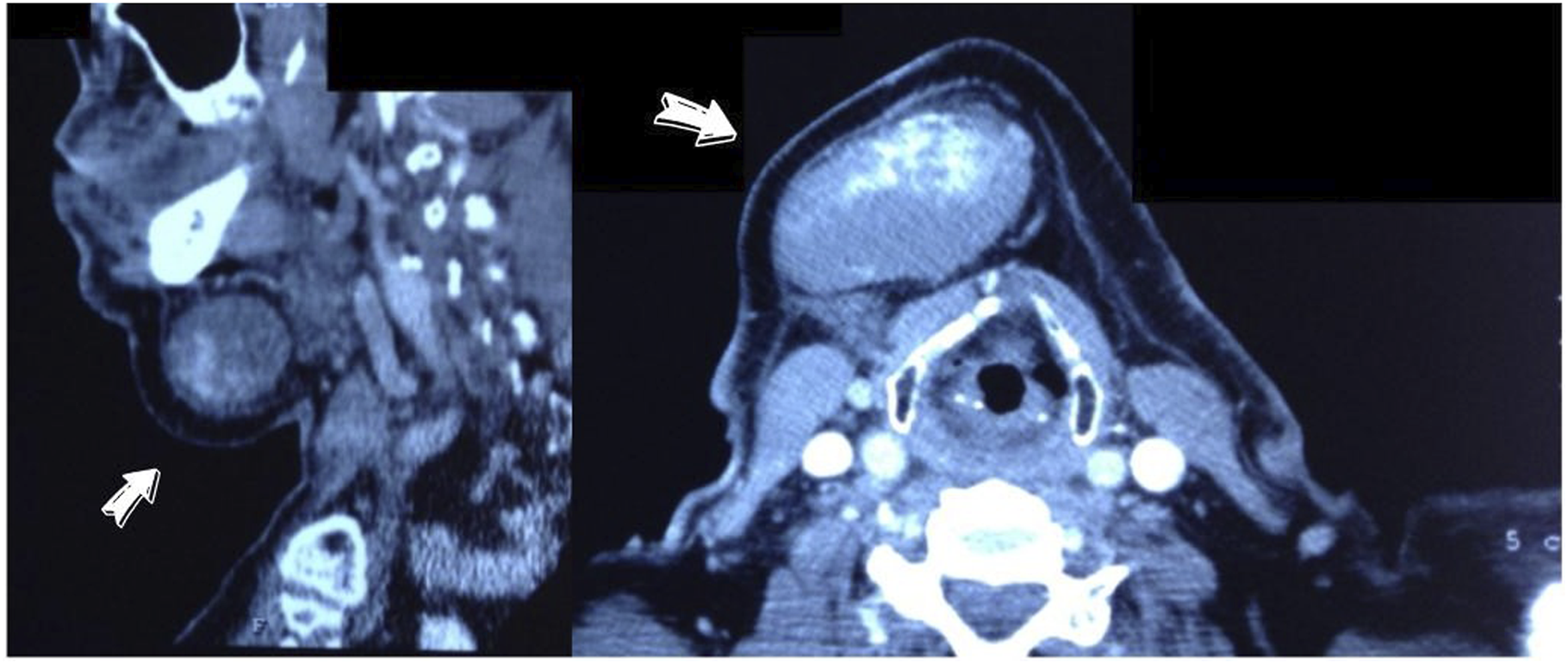
IPEH was first described by Masson in 1923 and was assumed to be a type of haemangioma. 1 It usually develops in medium veins, but may also be observed in all veins and sporadically in the arteries. 2 It commonly appears in fingers, trunk, head, and neck, with a predilection for skin and subcutaneous tissue of the scalp and face. 3 Despite several theories, the exact pathogenesis of IPEH remains unknown. 4 This abnormality is usually related to irradiation or chronic trauma which can transform blood flow leading to papillary endothelial proliferation, which is considered as a secondary reaction due to vascular stasis. The classification of IPEH is consisted of three types. The first type is the most common (56% of cases) and arises de novo in dilated vascular spaces, the second is a mixed type owing to focal changes in pre-existing vascular lesions, and finally, the third type is an unusual one resulting from an organized haematoma in extravascular location. 2 The clinical significance of Masson’s tumour is its similarity to different lesions, benign, such as pyogenic granulomas, haemangiomas, lymphangiomas, thrombosed veins, haematomas, and malignant, including Kaposi’s sarcomas and angiosarcomas. 5
The prognosis of Masson’s tumor after complete surgical excision is excellent with an exceptionally low recurrence rate. 2 To our knowledge, this is the first case of IPEH presented as a submental swelling. Doctors should be aware of this rare non-neoplastic entity that can mimic other abnormalities, since its misdiagnosis can lead to incorrect management. 4
Footnotes
Declaration of Conflicting Interests
The author(s) declared no potential conflicts of interest with respect to the research, authorship, and/or publication of this article.
Funding
The author(s) received no financial support for the research, authorship, and/or publication of this article.
Significance Statement
Masson’s tumor, also known as intravascular papillary endothelial hyperplasia (IPEH), is a rare benign vascular lesion that is usually found in the soft tissue and skin of different parts of the human body. We present an unusual case of IPEH presenting as a submental swelling. This non-neoplastic lesion can mimic other entities, and thus it is essential that physicians maintain a high index of suspicion to diagnose IPEH and prevent mismanagement.