
Abstract
Mucocele is rarely seen in the sphenoid sinus. It may cause compression of neighboring structures due to significant destruction of the bony structure and its expansile nature. Although headache is the main symptom, it may present with different symptoms. Imaging methods play an important role in the diagnosis and differential diagnosis.
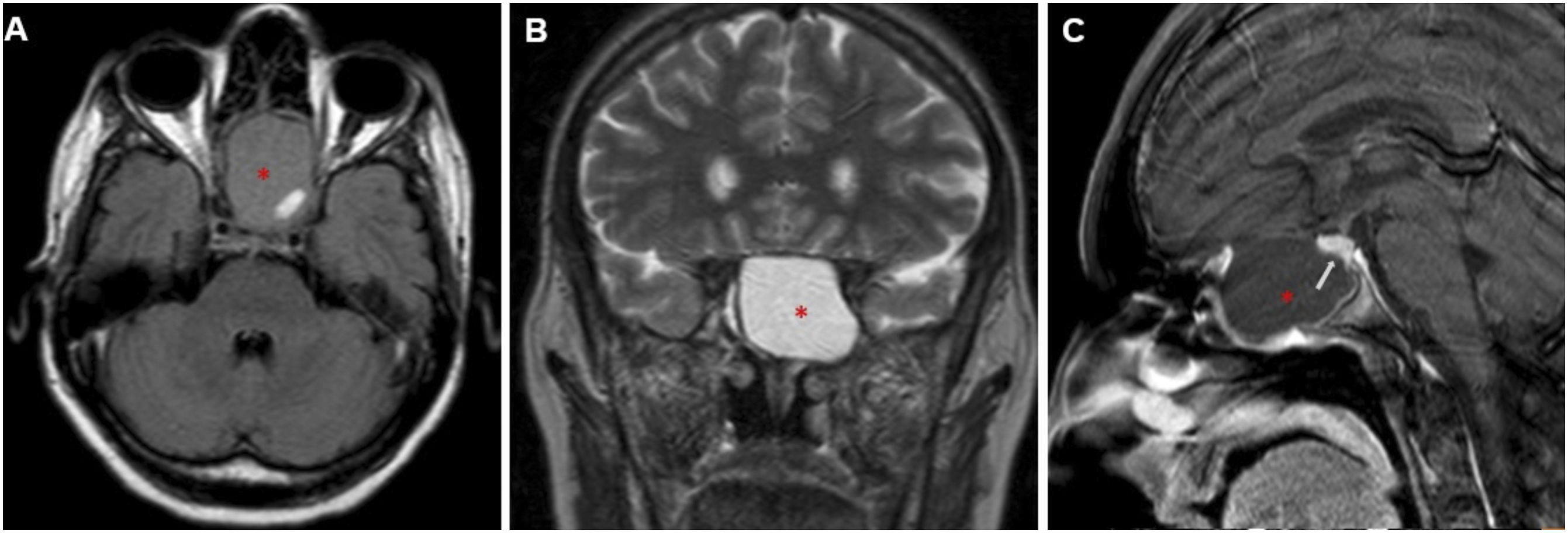
A 45-year-old woman was admitted to our center with complaints of severe headache, anorexia, constipation, and sensitivity to cold. She had no history of trauma or surgery but had a diagnosis of migraine made 7 years ago in an external center. Physical examination was unremarkable. In the blood tests, hemoglobin, follicle stimulating hormone, luteinizing hormone, testosterone, and adrenocorticotropic and thyroid stimulating hormone values were below the normal range. She underwent computed tomography (CT) of the brain. Images showed a hypo dense, expansile lesion measuring 4 × 3 × 3 cm in the left part of the sphenoid sinus. The mass was causing significant destruction of the dorsum sella posterior-superiorly (Figure 1). On contrast-enhanced brain magnetic resonance imaging (MRI), the lesion was heterogeneously hypo intense on T1-weighted images, hyperintense on T2-weighted images, and peripherally minimally contrast enhanced. The pituitary gland appeared pushed and compressed secondary to the expansion of the lesion (Figure 2). The patient was diagnosed with sphenoid sinus mucocele and hypopituitarism secondary to compression. Surgery was planned and performed. The patient who did not develop any postoperative complications and whose hormone levels gradually increased was discharged with recommendations. Axial (A) and sagittal (B) contrast-enhanced brain CT images showing a hypo dense lesion (asterisk) expanding the left sphenoid sinus and marked pressure erosion of the sellar floor (arrows). MR images show a heterogenous hypo intense lesion in T1-weighted images (A) and a hyperintense lesion in T2-weighted images (B) located in the sphenoid sinus (asterisk). Sagittal plane contrast-enhanced MR image (C) shows minimal peripheral contrast enhancement of the sinus mucosa and upward compression of the pituitary gland (arrow).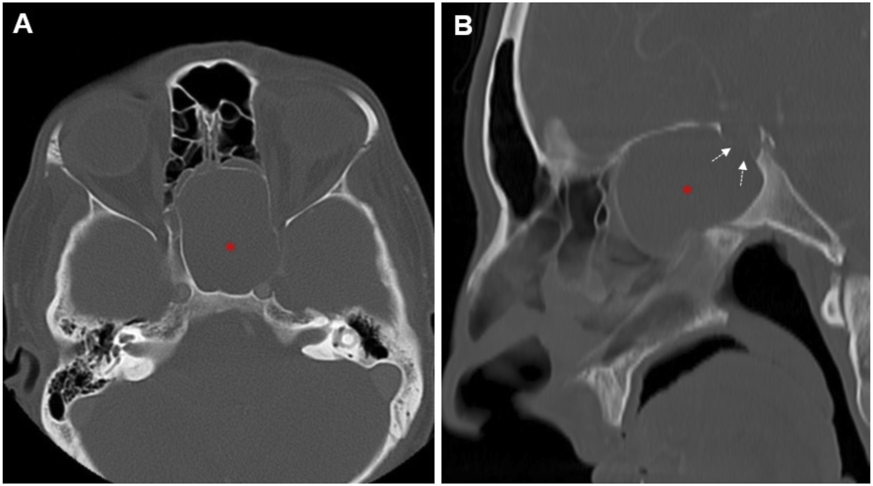
Mucoceles are mucus-filled cystic lesions surrounded by epithelium that are common in the paranasal sinuses. It is very rare in the sphenoid sinus and its incidence has been reported as 1–2%. It is more common in men and most common in the 3rd-4th decade. Although the pathophysiology is not clearly known, it is thought to occur as a result of obstruction of the sinus ostium or cystic dilatation of glandular structures. It is benign in nature, but its marked expansile and destructive behavior can cause different and severe symptoms. 1,2 The most common complaint is headache. In the literature, optic nerve and cranial nerve compression findings and hypopituitarism have been reported as cases. 3,4
Imaging modalities are crucial in the diagnosis and differential diagnosis of mucocele and in showing its relationship with surrounding tissues. Due to its fluid content, it is usually hypo dense on CT, hypo intense on T1-weighted images and hyperintense on T2-weighted images on MRI. However, these may vary depending on the proteinaceous fluid or superinfection. Contrast-enhanced images may show peripheral contrast enhancement. The destruction of the bony structure by the lesion can be evaluated on CT and its relationship with the orbit, cranial nerves, and pituitary can be evaluated on MRI. Endoscopic trans nasal marsupialization is the preferred treatment option. 1,3,5
Footnotes
Declaration of Conflicting Interests
The author(s) declared no potential conflicts of interest with respect to the research, authorship, and/or publication of this article.
Funding
The author(s) received no financial support for the research, authorship, and/or publication of this article.