
Abstract
Juvenile nasopharyngeal angiofibroma is a benign vascular tumor seen predominantly in adolescent males in the second decade of life. Extranasopharyngeal angiofibroma includes vascular fibrous masses that occur outside the nasopharynx. The diagnosis of an angiofibroma is based on the clinical presentation and imaging, with biopsies being avoided to avoid excessive bleeding. Computed tomography scan is considered sufficient for the diagnosis of extranasopharyngeal angiofibroma as it clearly delineates and identifies the tumor.
Introduction
Nasopharyngeal angiofibroma previously known as juvenile nasopharyngeal angiofibroma is a benign vascular tumor of the nasopharynx. It was previously considered to arise from the following sites: the roof of the nasopharynx, the anterior wall of sphenoid bone, and the superior part of the nasal cavity. 1,2 Angiofibromas originating from areas other than the nasopharynx are termed extranasopharyngeal angiofibromas. These are most commonly found in the maxilla, but may also be found in sites such as the ethmoid sinuses, the nasal cavity or the nasal septum. The 2 angiofibromas differ significantly in characteristics and are therefore considered separate entities.
The current literature does include cases involving the nasal septum; however, the clinical characteristics and management protocols differ in terms of setting and available resources. We therefore present a case of a nasal angiofibroma arising from the nasal septum managed in a developing world setting.
Case Presentation
A 23-year-old male was presented to the ENT clinic with a slow-growing, nontender nasal mass that had been present for a year. There had been episodes of epistaxis that were gradually becoming worse. The rest of the clinical enquiry was satisfactory. Nasal endoscopy revealed a 5 mm by 5 mm mucosal mass arising from the right nasal septum (Figure 1).
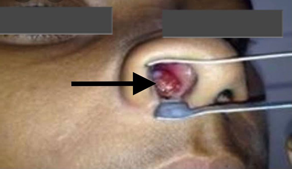
Nasal septal mass (arrow).
The nasal septum was intact and there was no extension to the nasopharynx. A contrast computed tomography (CT) scan confirmed the clinical diagnosis of a vascular soft tissue mass limited to the nasal cavity (Figure 2). Endoscopic excision of the entire mass was done under general anesthesia. Histology of the mass demonstrated an angiofibroma.
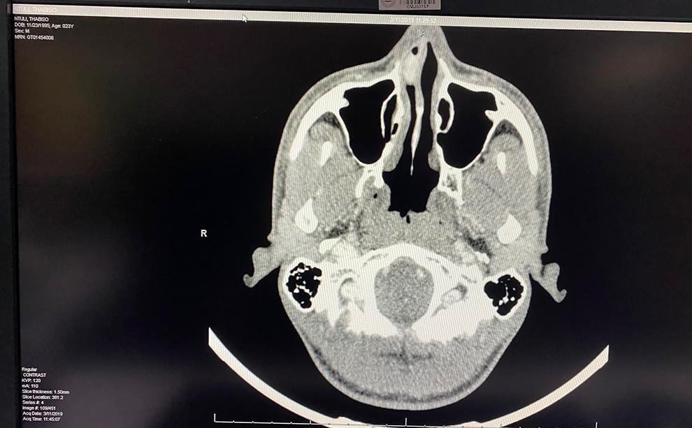
Axial computed tomography (CT) scan nasal septal mass.
Discussion
Juvenile nasopharyngeal angiofibroma is a benign vascular tumor seen predominantly in adolescent males in the second decade of life. 1 The site of origin of tumor was thought to be the roof of the nasopharynx or the anterior wall of sphenoid bone but now it is believed to arise from the posterior part of nasal cavity close to the superior margin of sphenopalatine foramen. 2 -6
The term extranasopharyngeal angiofibroma includes vascular fibrous masses that occur outside the nasopharynx. The clinical characteristics between the nasopharyngeal angiofibroma and extranasopharyngeal angiofibroma differ in terms of prevalence, age, affected site, pathogenesis, and recurrence. The extranasopharyngeal angiofibroma grows quicker, mainly occurs in older patients and is seen equally in males and females. 5,7
Patients typically present with recurrent epistaxis and progressive nasal obstruction as is the case with this patient when the tumor is originating from the nasal cavity. 5,7,8 The symptoms are less specific for those arising in other sites. 8 The diagnosis of an angiofibroma is based on the clinical presentation and imaging, with biopsies being avoided to avoid excessive bleeding.
Computed tomography scan is considered sufficient for the diagnosis of extranasopharyngeal angiofibroma as it clearly delineates and identifies the tumor. Contrast-enhanced CT and magnetic resonance imaging determine the tumor site and its extension, with special attention being focused on skull base involvement, intracranial spread, and relationship to important vascular structures. 9 Endoscopic surgical excision of the mass is the treatment of choice with there being a low risk for recurrence is rare. 5,7,10
Footnotes
Declaration of Conflicting Interests
The author(s) declared no potential conflicts of interest with respect to the research, authorship, and/or publication of this article.
Funding
The author(s) received no financial support for the research, authorship, and/or publication of this article.