
Abstract
Angiomatous polyps are an uncommon subtype of sinonasal polyps, characterized by extensive vascular proliferation and ectasia. The authors report the first case of angiomatous polyp originating from the inferior turbinate, which is a variant of the sinonasal polyp.
A 65-year-old man with a 6-month history of a left-sided, slow, progressive nasal obstruction was referred to our hospital. Nasal endoscopy revealed a polypoid nasal mass obstructing the left nasal floor (Figure 1). The mass appeared to have originated from the left inferior turbinate. The right nasal cavity and nasopharynx appeared normal. Allergic skin test was negative. There were no other relevant findings. Coronal computed tomography (CT) revealed a soft tissue density in the left nasal cavity (Figure 2). We suspected an inverted papilloma or a simple polypoid mass and decided to perform an excisional biopsy for confirmation.
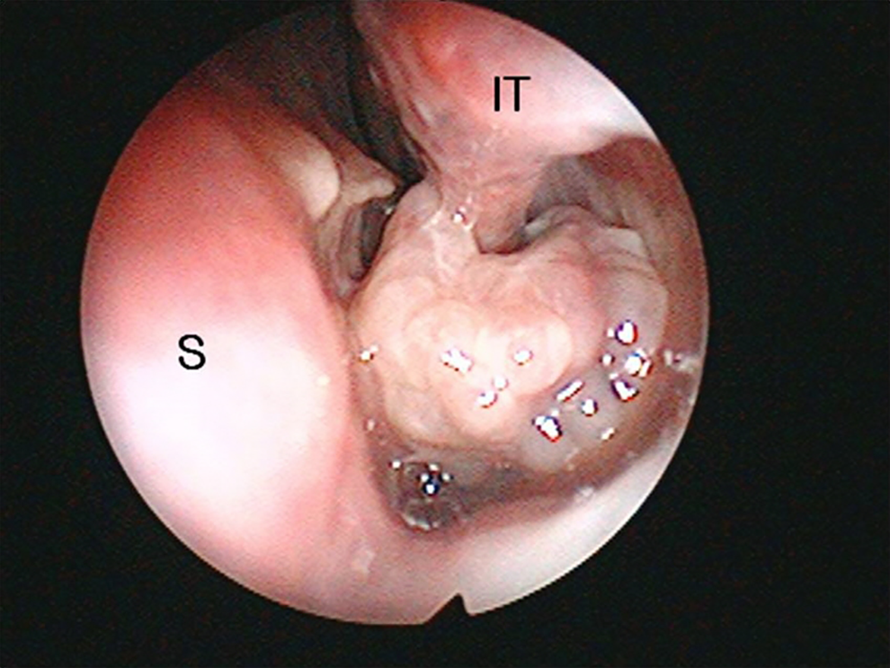
Endoscopy revealed a mass in the left nasal cavity. IT indicates inferior turbinate; S, septum.
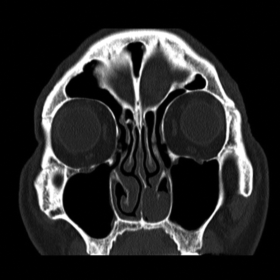
A coronal computed tomography (CT) scan revealing soft tissue density in the left nasal cavity.
The mass was removed endoscopically under local anesthesia. It originated from the middle and posterior portion of the inferior turbinate. During the removal of the mass, the patient exhibited a more severe bleeding than we had expected. To control the bleeding, we used a suction coagulator and packing. The patient had a mild intermittent bleeding for 3 days after the procedure.
The mass comprised an inflammatory polyp and numerous small and large vessels in a fibrous stroma (Figure 3). On the basis of the tumor’s histopathology, a diagnosis of angiomatous polyp (AP) was made. The postoperative course was uneventful, and the patient exhibited no further symptoms.
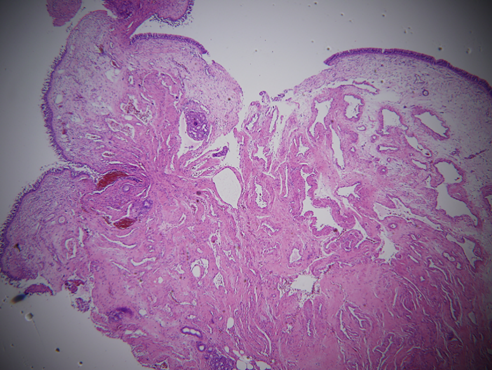
The tumor comprises an inflammatory polyp and numerous small and large vessels in a fibrous stroma (×40).
Angiomatous polyps are an uncommon subtype of sinonasal polyps, characterized by extensive vascular proliferation and ectasia. 1 The most common site of origin of sinochoanal polyps with angiomatous changes is the maxillary sinus. 2,3 The clinical, radiological, and pathological findings of an AP obstructing the choana may be confused with those of a vascular neoplasm, including juvenile nasopharyngeal angiofibroma. 4 Imaging studies such as CT and magnetic resonance imaging studies may be helpful in the differential diagnosis of juvenile nasopharyngeal angiofibromas and APs obstructing the nasopharynx. In the preoperative differential diagnosis of these cases, inverted papilloma is among the most frequently considered conditions. 5 In our case, inverted papilloma could not be excluded on the basis of the clinical or radiological study results alone. In this case, we strongly suspected an inverted papilloma or a simple polypoid mass after examining the patient during his first visit to our hospital. To our knowledge, APs originating from the inferior turbinate have not been reported previously. The authors report the first case of AP originating from the inferior turbinate, which is a variant of the sinonasal polyp.
Footnotes
Declaration of Conflicting Interests
The author(s) declared no potential conflicts of interest with respect to the research, authorship, and/or publication of this article.
Funding
The author(s) received no financial support for the research, authorship, and/or publication of this article.