



Abstract

A 4-year-old male child tracheostomized for Guillain-Barre syndrome 4 months ago reported to Ear Nose Throat outpatient department with complaints of pain, pus oozing from tracheostomy site, and occasional creeping out of some crawling insects. The tube of the patient had not been changed for the past 1 month and patient had developed these symptoms for the past 15 days. Immediately, in the emergency operation theatre, the tracheostomy tube was removed: a swarm of maggots partially blocking the tracheostoma was seen (Figure 1A). Fortuitously, using turpentine oil, the larvae were removed and airway secured. This was followed by repeated use of turpentine oil with some debridement and surgical exploration to mechanically remove these extensive larvae present across adjoining intermuscular planes, under general anesthesia (Figure 2). Patient underwent 3 such sittings under strict asepsis and antibiotic cover of amoxicillin + clavulanic acid and metrogyl. It would be pertinent to note that there was no use of any antiparasitic drug like ivermectin in the management of this case. Patient was subsequently discharged at the end of 3 weeks as his clinical condition improved dramatically: healthy granulations were seen in the tracheostoma (Figure 1B). The patient is in regular follow-up with us for the past 6 months with no untoward incident to report and has been successfully decannulated (Figure 3).

A, Clinical photograph showing maggots in tracheostomal site. B, Clinical photograph showing healthy granulations in the tracheostomal wound after removal of maggots.
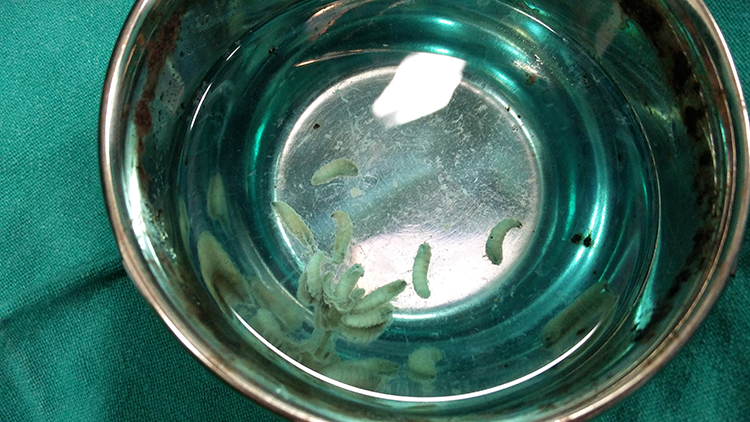
Removed maggots: Oestrus ovis.

Clinical photograph of the decannulated patient.
Entomologically, the microscopic examination of the specimen revealed 10-mm larvae with features consistent with Oestrus ovis (Figure 2). The specimen demonstrated anterior hooks, spiracles with a flat side medially, and respiratory holes arranged radially. 1
Myiasis is the invasion of dipterous insect larvae into tissues. Though described worldwide, it is mostly prevalent in developing countries of tropics and subtropical regions.2-4 Tracheostomal myiasis is a rare clinical entity and extremely rare in paediatric population.2-7 In a massive internet search using PubMed/Medline services, authors could find only 3 cases reported in children on the cited subject.6,8,9 Myiasis is classified as specific (obligate), semi-specific (facultative), and accidental (pseudomyiasis). 2 Open wounds and bodily orifices that emit odors of natural secretions are major factors in susceptibility to myiasis, as they provide a favorable environment for attraction and oviposition of flies. 4 Myiasis causing flies in India belong to the important genus Chrysomya and Lucilia (Phaenicia). 2 There are some interesting facets to this case which merit mention:
Firstly, myaisis is regarded as a disease associated with unhygienic conditions and poor socioeconomic environment. 6 Of note, these factors were not associated in this case as the said patient is a resident of an urban city, living in a decent house with good surrounding environment. It would also be prudent to note that most of the indexed cases of tracheal wound myiasis in literature have been reported from the developed world.3,6,9
Secondly, small dimension of the surgical tracheostomy (as seen in children), poor care of the tracheostomy, like not changing the tube for days, and absence of covering of stoma opening (the child did not tolerate this and often removed the covering playfully) probably were the important predisposing factors for myiasis infection in this case.2-4,10
Lastly, Oestrus ovis (sheep nasal bot fly) identified in the present case has seldom been reported in otorhinolaryngology and is usually associated with ophthalmomyiasis. 1 Human infestation is usually seen in shepherds or people who come in close contact of these shepherds. Contrary to the above facts, we recorded this infection in a child with no history of contact with shepherds/sheeps. To the best of our knowledge, this clinical record is the first reported case of Oestrus ovis in tracheostomal myiasis.
There are no fixed guidelines for management of tracheal myiasis. Removal of maggots with turpentine oil, chloroform, ether, or Vaseline solution is recommended under antibiotic cover. In addition, surgical debridement is also done if there is extensive spread of infection in adjoining tissue planes. Maintaining strict antisepsis in general care interventions is an integral part of treatment. Only, if this treatment fails, antiparasitic drugs like ivermectin and doramectin are given.2,6,10 Maggots warrant aggressive management for there is a chance of maggots creeping down or being aspirated in air passages like trachea and bronchi thus acting as an animate foreign body and causing aspiration pneumonitis.6,8 Moreover, if not treated timely, tracheostomal myiasis may cause severe complications, including secondary infection, respiratory failure, tracheal stenosis, trachea-esophageal perforations, and thromboembolization of adjoining vessels.5,10 In conclusion, tracheostomal myiasis can occur in children with long-term tracheostomy, albeit rarely.
Footnotes
Declaration of Conflicting Interests
The author(s) declared no potential conflicts of interest with respect to the research, authorship, and/or publication of this article.
Funding
The author(s) received no financial support for the research, authorship, and/or publication of this article.
