
Abstract
Introduction
A cervical rib is a supernumerary rib that occasionally projects from the lateral mass of the seventh cervical vertebra. 1 Rarely it may arise from the sixth or fifth vertebra. Clinically, it can cause obscure nervous or vascular symptoms and it is difficult to diagnose. 1 Studies have shown the prevalence of cervical ribs to lie between 0.03% on radiographs and 3% in postmortem investigations, depending on the sex and race of the population studied. 1 In children with cervical ribs are often asymptomatic and present as neck mass and mild pain. 1 Cervical rib is one of the most important bony factors which leads to thoracic outlet syndrome due to the displacement and compression of the neurovascular structures while crossing the thoracic outlet to the upper limb. 1
Case Report
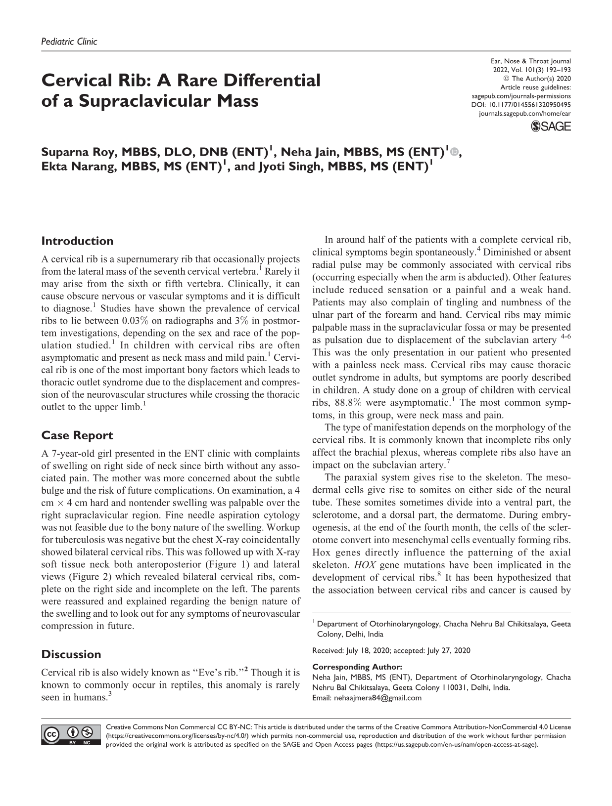
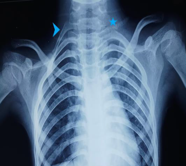
A 7-year-old girl presented in the ENT clinic with complaints of swelling on right side of neck since birth without any associated pain. The mother was more concerned about the subtle bulge and the risk of future complications. On examination, a 4 cm × 4 cm hard and nontender swelling was palpable over the right supraclavicular region. Fine needle aspiration cytology was not feasible due to the bony nature of the swelling. Workup for tuberculosis was negative but the chest X-ray coincidentally showed bilateral cervical ribs. This was followed up with X-ray soft tissue neck both anteroposterior (Figure 1) and lateral views (Figure 2) which revealed bilateral cervical ribs, complete on the right side and incomplete on the left. The parents were reassured and explained regarding the benign nature of the swelling and to look out for any symptoms of neurovascular compression in future.
Chest radiograph (A-P view) demonstrating bilateral cervical ribs, complete cervical rib on the right (arrow head) and an incomplete rib on the left side (star).
X-ray neck lateral view showing a fully formed cervical rib (white arrow) at the level of seventh cervical vertebra.
Discussion
Cervical rib is also widely known as “Eve’s rib.” 2 Though it is known to commonly occur in reptiles, this anomaly is rarely seen in humans. 3
In around half of the patients with a complete cervical rib, clinical symptoms begin spontaneously. 4 Diminished or absent radial pulse may be commonly associated with cervical ribs (occurring especially when the arm is abducted). Other features include reduced sensation or a painful and a weak hand. Patients may also complain of tingling and numbness of the ulnar part of the forearm and hand. Cervical ribs may mimic palpable mass in the supraclavicular fossa or may be presented as pulsation due to displacement of the subclavian artery 4-6 This was the only presentation in our patient who presented with a painless neck mass. Cervical ribs may cause thoracic outlet syndrome in adults, but symptoms are poorly described in children. A study done on a group of children with cervical ribs, 88.8% were asymptomatic. 1 The most common symptoms, in this group, were neck mass and pain.
The type of manifestation depends on the morphology of the cervical ribs. It is commonly known that incomplete ribs only affect the brachial plexus, whereas complete ribs also have an impact on the subclavian artery. 7
The paraxial system gives rise to the skeleton. The mesodermal cells give rise to somites on either side of the neural tube. These somites sometimes divide into a ventral part, the sclerotome, and a dorsal part, the dermatome. During embryogenesis, at the end of the fourth month, the cells of the sclerotome convert into mesenchymal cells eventually forming ribs. Hox genes directly influence the patterning of the axial skeleton. HOX gene mutations have been implicated in the development of cervical ribs. 8 It has been hypothesized that the association between cervical ribs and cancer is caused by a common underlying genetic defect that causes abnormal Hox gene expression. 8
A cervical rib may vary in structure ranging from a fully formed bony rib to a thin strand of tissue fibers, which may or may not show up on radiological scans. A cervical rib is present in 0.5% to 0.7% of the population and appears more commonly in females than males, in a ratio of 2:1. 1 The cervical rib is found more often on the left side, but right-sided cervical ribs are more symptomatic.
In view of the above reported case, an ENT surgeon should always keep a cervical rib as a differential diagnosis while dealing with supraclavicular masses. Though mostly asymptomatic, the presence of an unexplained neck swelling can be a cause of anxiety and curiosity to patients. Hence, a swift diagnosis and reassurance could be the best medicine for such patients. We used a simple radiological tool to give the patient’s family the correct diagnosis and a boost of confidence, given the benign nature of the cervical rib. Also, this will aid the future consulting doctors in making a diagnosis if any neurovascular signs and symptoms arise in the patient.
Footnotes
Declaration of Conflicting Interests
The author(s) declared no potential conflicts of interest with respect to the research, authorship, and/or publication of this article.
Funding
The author(s) received no financial support for the research, authorship, and/or publication of this article.