



Abstract

Case Presentation
A 57-year-old male with a history of previous right alveolus squamous cell carcinoma after reconstruction using a right supraclavicular island flap presented to the head and neck clinic for 3-month follow-up with complaint of new protuberant bony growth in his right inferior neck. The growth was causing him substantial discomfort, and he reported sharp pain specifically with head rotation to the left. Physical examination revealed a 3-cm vertically oriented bony protuberance originating from his right clavicle (Figure 1). The lesion was tender to palpation and visibly tented his overlying neck skin. He underwent computed tomography of his neck, which revealed a 2.7-cm bony spicule emanating off his right clavicle (Figure 2). Given the patient’s discomfort and additional cosmetic concerns, the patient was taken to the operating room for surgical excision. A small incision was made in the skin overlying the lesion. Brief blunt dissection was easily able to expose the bony spicule, which was further exposed down to its base along the clavicle (Figure 3). The calcified lesion was not adherent to the overlying cutaneous tissue of the neck; however, it was firmly affixed to the underlying clavicle. The lesion was grasped at its base and fully removed using a rongeur. The overlying skin was closed in the standard manner. Pathologic evaluation revealed benign-appearing bone with medullary fibrosis, osteosclerosis, and remodeling consistent with heterotopic ossification. The patient subsequently presented for follow-up 3 months later with complete resolution of his symptoms and no concerns on physical examination and has remained symptom-free for the past 18 months.
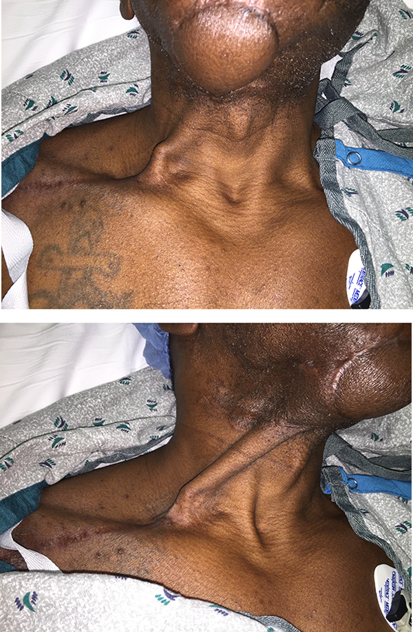
Preoperative appearance of clavicular lesion (A). Note the marked skin tenting which occurred with head rotation to the left (B).

Computed tomography of the neck without contrast revealing a 2.7-cm vertical bony growth originating from the right clavicle: (A) coronal and (B) sagittal views.

Intraoperative view of heterotopic calcification as it was exposed down to the right clavicle.
Discussion
Heterotopic calcification is the abnormal growth of bone in nonskeletal tissues and can occur as the result of trauma, infection, or iatrogenic surgical interventions as seen in this case.1,2 This patient’s history of recent supraclavicular island flap reconstruction was likely the inciting event given the location of the lesion and postoperative time course. Elevation of the clavicular periosteum with the supraclavicular flap has been advocated to protect the vascular pedicle as it crosses the clavicle. 3 This was the technique used with this patient, and the displaced periosteum was the likely catalyst for the development of this bony protuberance. While vascular pedicle calcification is a recognized phenomenon following osseous free flap reconstruction, the location of the heterotopic calcification seen in this case was not along the trajectory of the previously elevated supraclavicular pedicle.4,5 Heterotopic calcification seen in other parts of the body are often only followed clinically but can be surgically removed in symptomatic cases. Heterotopic calcification requiring additional surgery should be considered a potential sequela of supraclavicular flap harvest when the clavicular periosteum is incorporated.
Footnotes
Declaration of Conflicting Interests
The author(s) declared no potential conflicts of interest with respect to the research, authorship, and/or publication of this article.
Funding
The author(s) received no financial support for the research, authorship, and/or publication of this article.
