
Abstract
Keywords
Ectopic thyroid is an uncommon condition defined as the presence of thyroid tissue at a site other than the pretracheal area. Ectopic thyroid in the anterior midline of the neck has been reported in the lingual, sublingual, perihyoid, and thyroglossal region, and occasionally in the larynx, trachea, esophagus. 1 Ectopic thyroid is rare in the lateral neck such as submandibular or carotid sheath region.1-9 Furthermore, there has been ectopic thyroid tissue rarely involved in cervical lymph node. 10 To our knowledge, there are few reports of lateral ectopic thyroid of cystic formation. Therefore, we report a rare case of an ectopic thyroid cyst of the lateral neck.
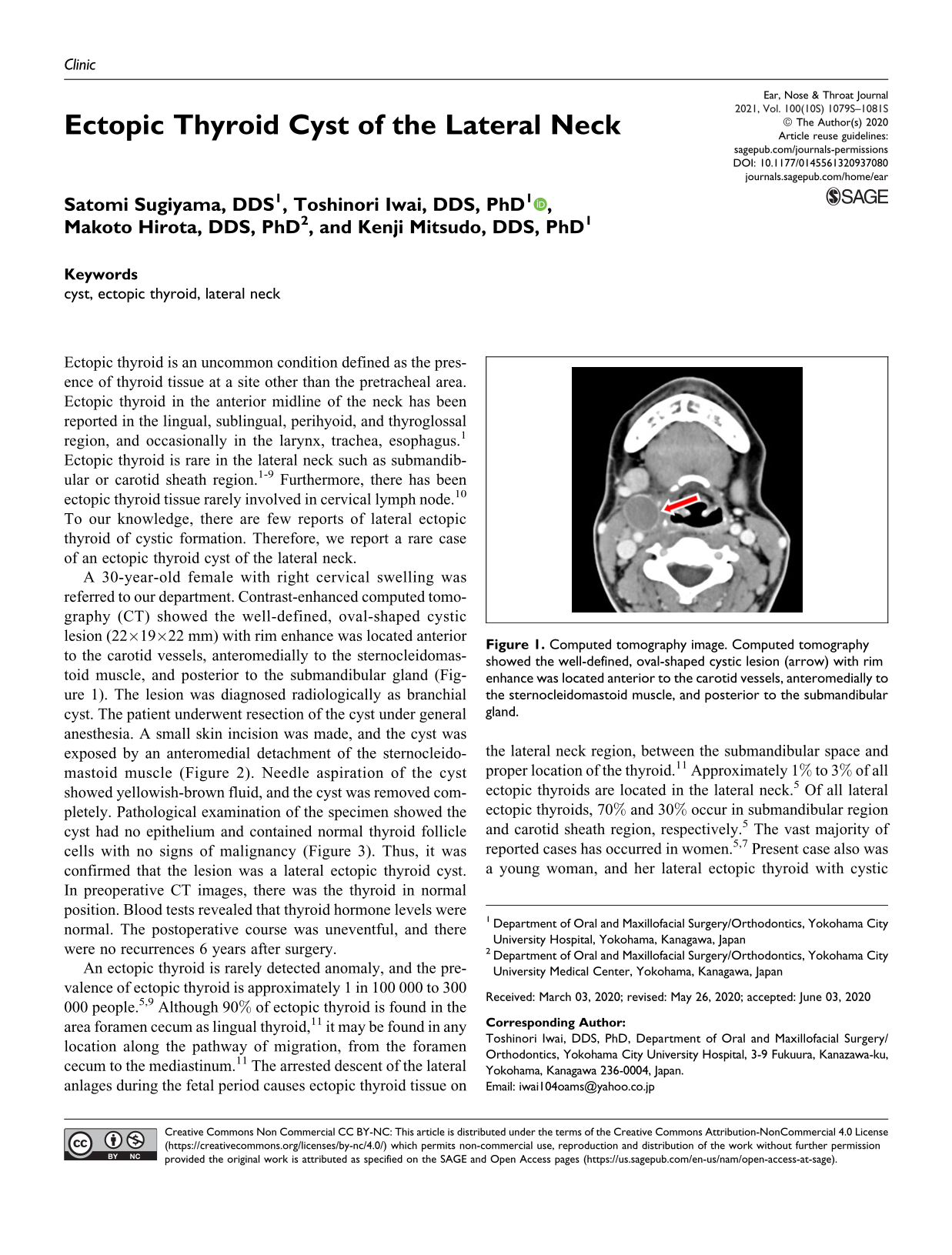
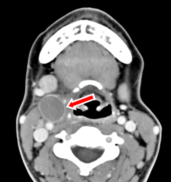
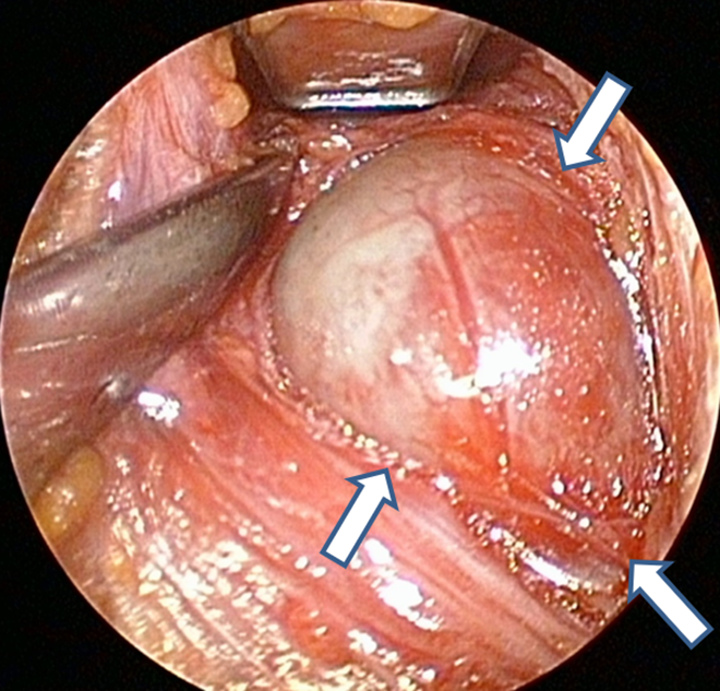
A 30-year-old female with right cervical swelling was referred to our department. Contrast-enhanced computed tomography (CT) showed the well-defined, oval-shaped cystic lesion (22×19×22 mm) with rim enhance was located anterior to the carotid vessels, anteromedially to the sternocleidomastoid muscle, and posterior to the submandibular gland (Figure 1). The lesion was diagnosed radiologically as branchial cyst. The patient underwent resection of the cyst under general anesthesia. A small skin incision was made, and the cyst was exposed by an anteromedial detachment of the sternocleidomastoid muscle (Figure 2). Needle aspiration of the cyst showed yellowish-brown fluid, and the cyst was removed completely. Pathological examination of the specimen showed the cyst had no epithelium and contained normal thyroid follicle cells with no signs of malignancy (Figure 3). Thus, it was confirmed that the lesion was a lateral ectopic thyroid cyst. In preoperative CT images, there was the thyroid in normal position. Blood tests revealed that thyroid hormone levels were normal. The postoperative course was uneventful, and there were no recurrences 6 years after surgery.
Computed tomography image. Computed tomography showed the well-defined, oval-shaped cystic lesion (arrow) with rim enhance was located anterior to the carotid vessels, anteromedially to the sternocleidomastoid muscle, and posterior to the submandibular gland.
Intraoperative view. After small skin incision, the cyst (arrows) was exposed by anteromedial detachment of the sternocleidomastoid muscle.
Pathology of the specimen. The cyst had no epithelium and contained normal thyroid follicle cells with no signs of malignancy.
An ectopic thyroid is rarely detected anomaly, and the prevalence of ectopic thyroid is approximately 1 in 100 000 to 300 000 people.5,9 Although 90% of ectopic thyroid is found in the area foramen cecum as lingual thyroid, 11 it may be found in any location along the pathway of migration, from the foramen cecum to the mediastinum. 11 The arrested descent of the lateral anlages during the fetal period causes ectopic thyroid tissue on the lateral neck region, between the submandibular space and proper location of the thyroid. 11 Approximately 1% to 3% of all ectopic thyroids are located in the lateral neck. 5 Of all lateral ectopic thyroids, 70% and 30% occur in submandibular region and carotid sheath region, respectively. 5 The vast majority of reported cases has occurred in women.5,7 Present case also was a young woman, and her lateral ectopic thyroid with cystic formation was located anterior to the carotid vessels, anteromedially to the sternocleidomastoid muscle, and posterior to the submandibular gland.
Ectopic thyroid as a lateral neck mass should be differentiated from metastatic thyroid cancer; other differential diagnoses include a submandibular tumor, branchial cyst, carotid body tumor, and lymphadenopathy of various etiologies.5,6 Some 76% of patients with true ectopic thyroid were reported as benign on cytopathogenic or histopathological examination (12%: malignancy, 12%; undetermined). 5 In the present case, ectopic thyroid was not a tumor but a cyst. To our knowledge, the present case was a first report with cystic formation of lateral ectopic thyroid.
The most common cystic neck lesion is branchial cyst, but metastatic lymph node may be misdiagnosed as a branchial cyst. According to Pietarinen-Runtti et al, 12 the incidence of unsuspected carcinoma in the cystic neck lesions initially diagnosed as branchial cyst by preoperative imaging appearances was 3.6% (squamous cell carcinoma: 3.1%, papillary carcinoma: 0.5%). Furthermore, several authors reported primary thyroid papillary carcinoma arising in ectopic thyroid tissue within branchial cyst.13,14 Although the initial clinical diagnosis was branchial cyst and final pathological diagnosis was an ectopic thyroid cyst in the present case, primary papillary carcinoma arising in ectopic thyroid tissue within branchial cyst or metastatic cystic degeneration of a cervical lymph node should always be considered as a potential differential diagnosis in patients with cystic lesion of the lateral neck.
Footnotes
Declaration of Conflicting Interests
The author(s) declared no potential conflicts of interest with respect to the research, authorship, and/or publication of this article.
Funding
The author(s) received no financial support for the research, authorship, and/or publication of this article.