
Abstract

A 66-year-old woman was referred to our hospital for evaluation, with a 3-month history of left facial and periorbital pain, headache, and purulent rhinorrhea. Although she was treated with medication at a local clinic, her condition did not improve. She reported no other health problems. Four months prior to her presentation, the woman had her left first molar extracted and replaced with a dental implant.
A nasal endoscopy identified a large amount of purulent discharge in the left middle meatus and nasopharynx. Computed tomography (CT) of the nose and paranasal sinuses showed soft-tissue densities in the left ethmoid, and in the maxillary sinuses, accompanied by a radiopaque mass in the ostium of the left maxillary sinus (Figure 1). The CT revealed a partial bone covering around the dental implant (Figure 1).
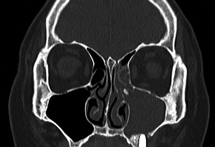
A, Coronal computed tomography (CT), showing soft-tissue densities in the left ethmoid and in the maxillary sinuses, accompanied by a radiopaque mass in the natural ostium of the left maxillary sinus. The CT reveals the dental implant with a partial bone covering around it.
Left endoscopic sinus surgery was performed under general anesthesia for removal of the foreign body, ventilation, and drainage of the left maxillary sinus. Immediately after an uncinectomy procedure, a significant volume of purulent discharge was drained. Following middle meatal antrostomy, a foreign body in the ostium of the left maxillary sinus was discovered (Figure 2A). The foreign body was a piece of bone, measuring 7 × 3 × 2 mm (Figure 2B). Subsequently, an anterior ethmoidectomy was performed, and the surgery was completed. The postoperative course was uneventful. One month postoperatively, complete resolution of the woman’s symptoms had been achieved.
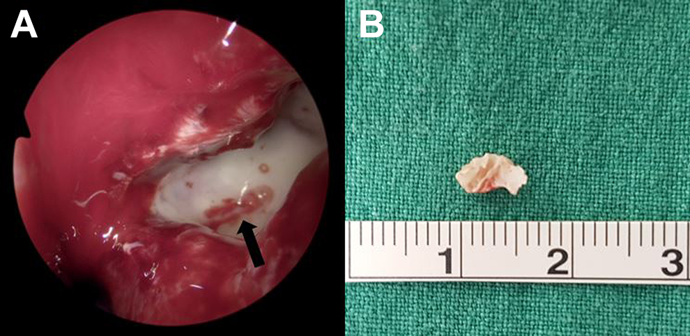
A, Endoscopic image showing a foreign body in the ostium of the left maxillary sinus. B, The foreign body is a piece of bone measuring 7 × 3 × 2 mm.
Earlier studies suggested that dental sinusitis made up approximately 10% of all sinusitis cases, 1 but recent articles suggest that it makes up 25% or more, of all chronic sinusitis cases. 2 Among the dental procedures, the placement of a dental implant into the maxilla is particularly difficult, owing to the close proximity to the maxillary sinus, and complications may arise from the surgery.
In this case, the piece of bone discovered in the maxillary sinus ostium may be related to the bone grafting material used in sinus elevation procedures or during the dental implant placement surgery. In the maxillary sinus, secretions move toward the ostium by a natural mucociliary action. 3 Similarly, a foreign body in the maxillary sinus will migrate toward the ostium by the natural mucociliary action. Once in the ostium, the displaced foreign body should be removed to prevent the occurrence of sinusitis. If the foreign body is small, it may pass through the maxillary sinus ostium. If the foreign body is too large to pass, obstruction will occur resulting in maxillary sinusitis. Once the foreign body is removed, the resolution of sinusitis should soon occur. Generally, if the foreign body is larger than 3 mm, endoscopic sinus surgery may be considered for its removal. We report an unusual case of bone grafting material, related to a dental implant procedure, causing ostial obstruction with associated sinusitis, and include the management of this associated condition.
Footnotes
Declaration of Conflicting Interests
The author(s) declared no potential conflicts of interest with respect to the research, authorship, and/or publication of this article.
Funding
The author(s) received no financial support for the research, authorship, and/or publication of this article.