
Abstract

A number of different crystalline structures (crystalloids) can be encountered in neoplastic and non-neoplastic salivary gland lesions, including tyrosine, oxalate, collagenous, intraluminal, and α-amylase crystalloids. While many crystalloids are nonspecific, α-amylase crystalloids have only been reported in benign lesions, and their identification may be helpful in distinguishing benign from malignant lesions. 1 Although these crystalloids are found in a variety of benign salivary gland tumors, they only rarely induce a granulomatous reaction, resulting in a condition known as “crystalloid granuloma” (CG). We present a case of CG in the parotid gland, misdiagnosed on cytopathology and subsequently surgically resected.
A 62-year-old male presented with a left parotid mass and complaints of sensation changes in his left cheek and preauricular area over the course of several weeks. His past medical history was unremarkable. This case report is exempt from institutional review board review.
Physical examination revealed a firm mass in the left parotid gland, numbness to light touch over the left cheek, and palpable bilateral nodules in the thyroid gland. An ultrasound-guided fine-needle aspiration (FNA) biopsy of the thyroid later showed the left-sided nodule was positive for papillary thyroid carcinoma, which was ultimately treated with a total thyroidectomy and central compartment neck dissection.
An MRI of the neck showed a 5.0-cm multilobular, well-defined, rim-enhancing lesion involving the left parotid gland superficial and deep lobe (Figure 1). This lesion was isointense on T1-weighted imaging, hyperintense on T2-weighted imaging, and showed restricted diffusion on diffusion-weighted imaging. The initial impression was a lymphatic malformation with recent hemorrhage, although a malignant or benign cystic neoplasm of the parotid gland was not excluded. An ultrasound-guided FNA of the parotid gland showed concretions of uncertain type, neutrophils, and histiocytes suggestive of an acute inflammatory response (Figure 2). Due to the unclear pathologic diagnosis and concern for a potential neoplasm, the patient was taken to the operating room for surgical excision.
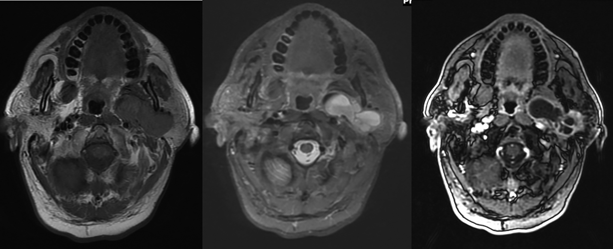
Magnetic resonance imaging (MRI) of the neck (axial T1, axial fat saturated T2, and post gadolinium fat-suppressed axial fast multiplanar spoiled gradient (FMPSPGR)) showed a 5.0 (transverse) × 2.7 (anteroposterior) × 2.4 (craniocaudal) cm multilobular, well-defined lesion involving the superficial and deep lobes of the left parotid gland.
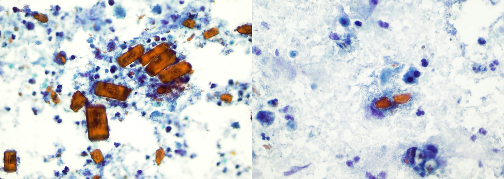
Microscopic examination of cytologic smears displayed a number of orange-stained extra- and intracellular crystalloids that were cuboidal and elongated. Crystals ranged in size, up to 50 microns.
A parotidectomy with facial nerve preservation was performed. Intraoperatively, the mass was thin walled and expressed thick, yellowish fluid during extirpation. Microscopically, the lesion was cystic with innumerable crystals and associated foreign-body granulomas. The deeply eosinophilic crystals, which varied in size and shape, were compatible with amylase crystals. A diagnosis of amylase CG in the parotid gland was rendered on final pathology.
Many crystalloids have been identified in both benign and malignant salivary gland lesions. The crystalloids in our case morphologically resembled the α-amylase crystalloids described in the literature. Amylase crystalloids have eosinophilic, variably shaped rectangular, polyhedral, rod-like, ovoid, and rhomboid structures (Figure 3 bottom). 1,2 Amylase crystalloids in the parotid gland are generally identified in sialadenitis, cystic lesions, and Warthin tumors. 3 Although other crystalloids may be seen in malignant salivary gland tumors, amylase crystalloids have only been reported in benign lesions. Typically, amylase-type crystalloids are nonbirefringent rectangular structures, ranging between 5 and 500 µm in size. 1 The detection of amylase crystalloids on FNA, coupled with a benign appearance on imaging, suggests that surgery may be avoided in patients with a higher surgical risk.
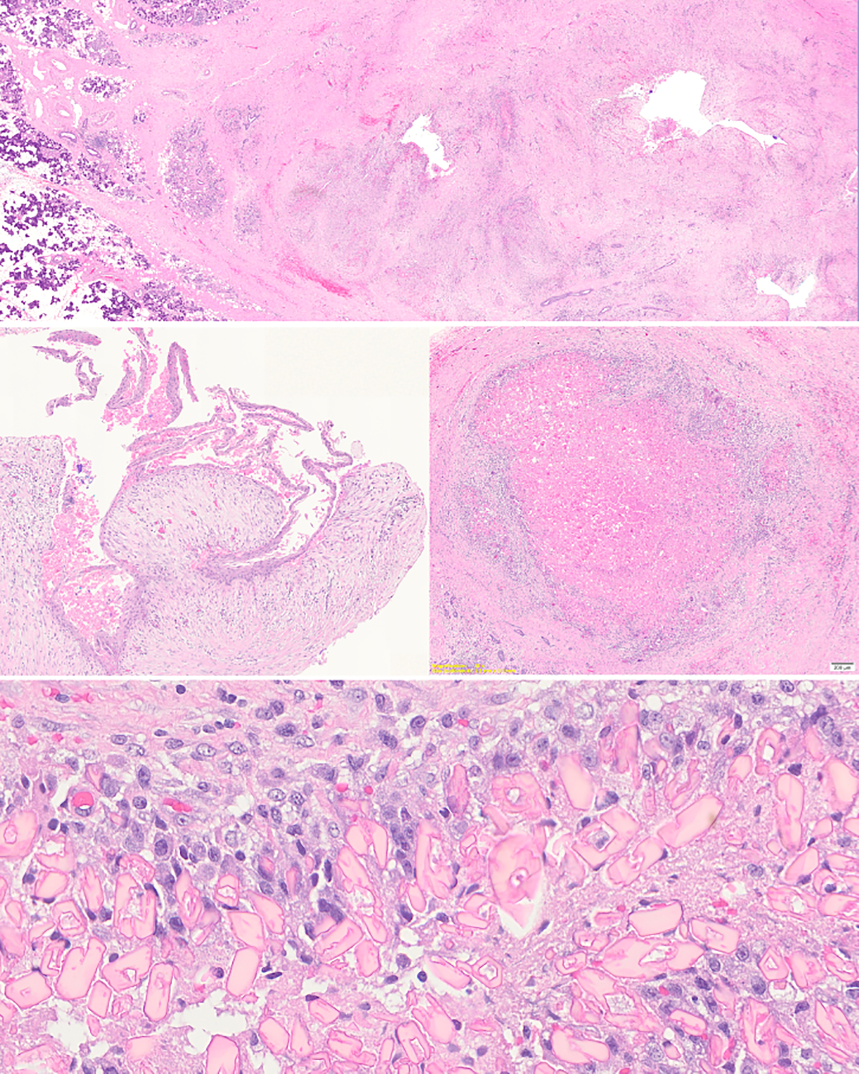
Top: Low-power magnification showed an intraparotid cystic process with dense solid inflammation. Middle left: Intermediate-power magnification showed a squamous-lined cyst. Middle right: Intermediate-power magnification showed an amylase crystalloid granuloma. Bottom: High-power magnification showed amylase crystalloids that were eosinophilic and variably shaped, including rectangular, polyhedral, rod-like, ovoid, and rhomboid crystalloids.
Over 50 cases of amylase crystalloids in the salivary gland have been reported in the literature, yet rarely do these crystalloids induce a florid granulomatous reaction to form a crystalloid granuloma as in the current case. 3 The mechanism for this type of crystal formation is not well understood. The leading hypothesis suggests that crystals arise from saturated saliva. 3 -5 It is thought that a cystic space is necessary for the formation of these crystalloids. When the cystic space is filled with excessive saliva, the cyst may rupture, inducing subsequent inflammatory and granulomatous reactions. The exact reasoning for the predilection of the parotid gland for CG is unclear but likely relates to the gland’s role as the main producer of α-amylase. 5 Conversely, sialolithiasis and sialadenitis are more common in the submandibular glands than in the parotid gland because the upward course of the submandibular duct is particularly conducive to the stasis of secretions. 4
We describe an unusual case of a 62-year-old man with crystalloid granuloma clinically resembling a salivary gland neoplasm. While CG clinically and radiographically resembles a neoplasm, the presence of amylase crystalloids suggests a benign process.
Footnotes
Declaration of Conflicting Interests
The author(s) declared the following potential conflicts of interest with respect to the research, authorship, and/or publication of this article: Dr. Urken is the Medical Advisor of the THANC Foundation.
Funding
The author(s) received no financial support for the research, authorship, and/or publication of this article.