



Abstract

A middle-aged male bluegrass singer with a 100 pack-year smoking history and history of vasculopathy was seen in referral for T1bN0M0 squamous cell carcinoma (SCC) primarily involving the anterior right true vocal fold. He was treated with radiation therapy (RT) to 63 Gy at an outside institution. He had decreased his smoking frequency but continued to smoke throughout his treatment. At his 2-month post-treatment visit, he reported odynophagia. His vocal folds were without erythema or lesions and fully mobile. At his 3-month post-treatment visit, he had continued throat pain with new arytenoid ulceration and vocal fold hypomobility but an adequate airway.
He presented 1 week later to his outside hospital for respiratory distress. A glidescope intubation and tracheotomy with subsequent suspension microlaryngoscopy and biopsy were performed. Pathology from posterior commissure biopsies revealed ulcerated squamous mucosa, dense submucosal fibrosis, and necrotic bone. He was treated for chondroradionecrosis (CRN) with 1 week of amoxicillin–clavulanic acid. At 4-month post-treatment, he reported improved throat pain after coughing a small, hard object out of his tracheotomy, which upon inspection was an intact, ossified arytenoid cartilage. Flexible laryngoscopy revealed absence of the left arytenoid with improvement in the appearance of the posterior laryngeal mucosa but bilateral immobility of the vocal folds. Several weeks later, he expectorated another small, hard object from his tracheotomy, his right arytenoid (Figure 1). He has remained without recurrence but is tracheostomy dependent due to posterior glottic stenosis and iatrogenic absence of his arytenoid cartilages (Figure 2).
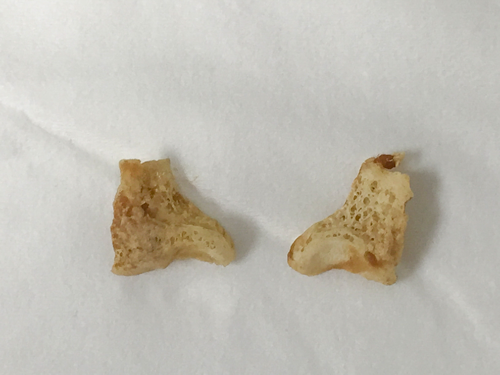
Bilateral arytenoid cartilages. Clinic photograph of intact bilateral arytenoid cartilage after expectoration.

Laryngoscopy following arytenoid expectoration. Clinic laryngoscopy demonstrating resolution of chondronecrosis, with absent arytenoid cartilage bilaterally.
Laryngeal CRN is a known complication of RT for early glottic SCC at a rate of 1% to 2%. 1 Reported risk factors for the development of laryngeal CRN include ongoing smoking, laryngeal trauma, and tumor cartilage invasion. 2 Typically, CRN manifests with a variety of presenting symptoms including weight loss, hoarseness, odynophagia, dysphagia, laryngeal edema, and upper airway obstruction. 2 Clinical features and imaging can be useful in diagnosis, but definitive diagnosis requires biopsy. Management includes voice rest, humidification, antibiotics, steroids, hyperbaric oxygen, supportive surgery (tracheotomy, gastrotomy tube placement), debridement, and laryngectomy. 2
Cartilage and bone can generally withstand large doses of RT without gross change in tissue. However, histopathologic studies suggest subclinical CRN can occur in rates as high as 27% of larynges previously irradiated for advanced stage laryngeal SCC.3,4 In previously irradiated larynges undergoing laryngectomy, the most common site of CRN was the arytenoid, with necrosis found in 51% of specimens. 4 Increased RT dose has been associated with higher rates of RT-related complications in early glottic SCC. 5 Although radiotherapy is often utilized as organ-sparing therapy for early glottic SCC, serious complications can occur especially in patients with vascular risk factors and continued tobacco use.
Footnotes
Acknowledgment
Presented at the Fall Voice Conference, October 2017; Washington DC
Declaration of Conflicting Interests
The author(s) declared no potential conflicts of interest with respect to the research, authorship, and/or publication of this article.
Funding
The author(s) received no financial support for the research, authorship, and/or publication of this article.