
Abstract
A 34-year-old man presented to our department of otorhinolaryngology with a diffuse facial swelling. He denied other systemic diseases, including congenital disease, and did not have habits of cigarette smoking, alcohol beverage drinking, or betel nut chewing. Tracing back the patient’s history, a mass over his left infra-auricular region was first noted about 1.5 years ago. The mass enlarged gradually accompanied with progressive facial swelling. On physical examination, a diffuse facial soft tissue swelling causing difficulties in eye opening and in examination of bilateral neck node status (Figure 1) was noted and a huge mass lesion could be palpated over the left lateral and posterior neck. The oral cavity was unremarkable, but medial bulging of the left oropharynx was prominent. No facial paralysis, no facial numbness, and no pus or blood from Stenson duct were noted. Left middle ear effusion was noted by otoscopy. Fiberscopy showed a nasopharyngeal mass with obstruction of left eustachian tube orifice and with downward extension over the left lateral pharyngeal wall. A computed tomography scan of the head and neck revealed increased soft tissue density over left face, nasopharynx, left oropharynx, left parapharyngeal space, and bilateral lymphadenopathy over neck levels I and V with compression of left internal jugular vein (Figure 2). Summing up the above findings, nasopharyngeal tumor was highly suspected. The nasopharyngeal biopsy confirmed a nonkeratinizing undifferentiated carcinoma. The Epstein-Barr virus (EBV) DNA load was 7474 copies/mL. Further tumor survey revealed bony metastasis of thoracic spine. The patient underwent induction chemotherapy and concurrent radiation chemotherapy thereafter.
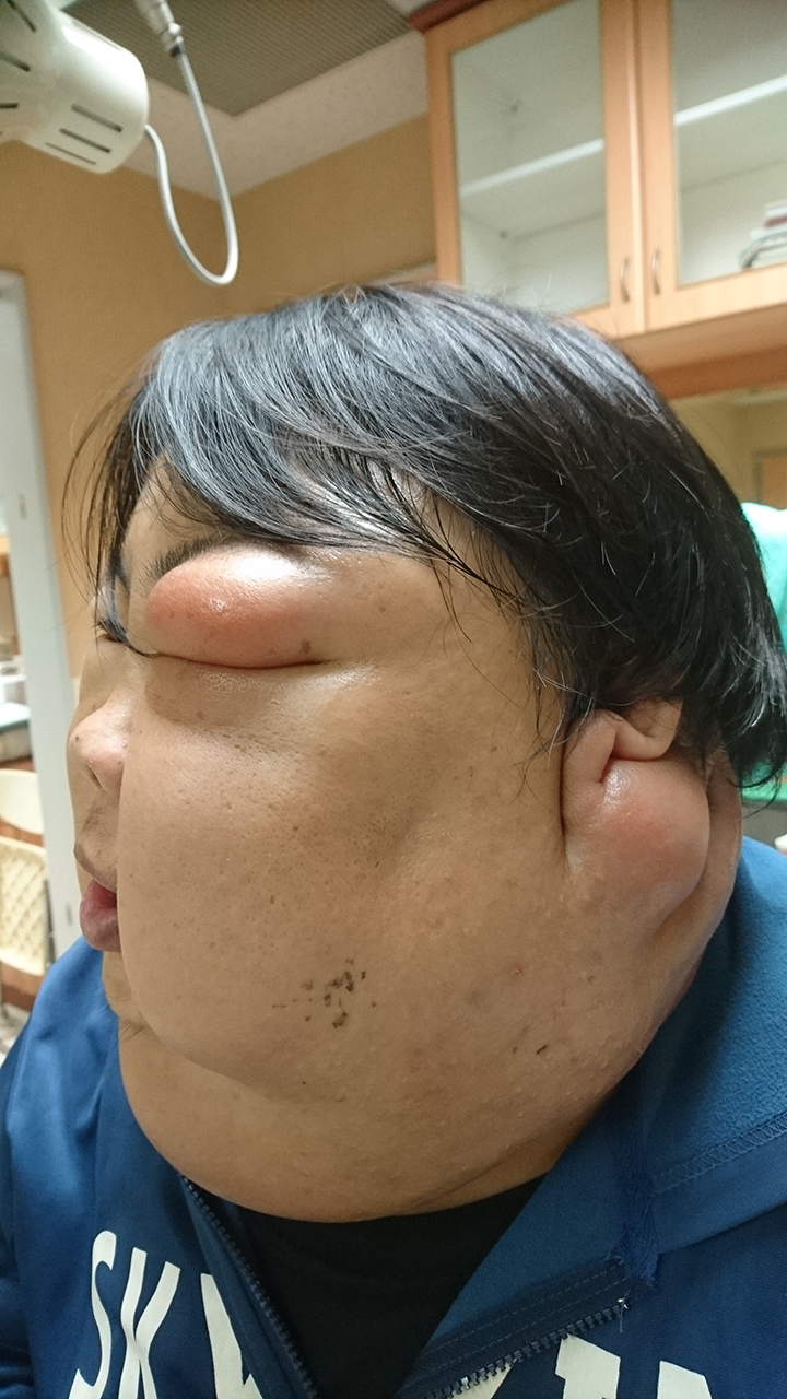
Oblique view of the patient at presentation showing a huge mass lesion over left posterolateral neck and a diffuse facial swelling.
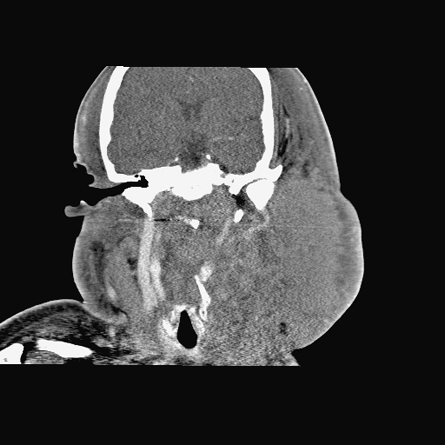
Coronal view of computed tomography scan showing bulging nasopharyngeal tumor, increased soft tissue density over left face, and neck with compression of left internal jugular vein.
The incidence of nasopharyngeal carcinoma (NPC) exhibited an evident geographical variation, which is endemic in southeast Asia, North Africa, Middle East, and Arctic, with a male preponderance. 1 The incidence rate of NPC for males in Taiwan is 7.7 to 8.8 per 100 000 person-years, 2- to 3-fold higher than that of females. 2 Clinical presentation of NPC is related to the extent of the primary and nodal diseases. The most frequent presenting symptom is painless neck masses (76%), followed by nasal symptoms in 73%, aural symptoms in 62%, and cranial nerve palsy in 20% of patients. 3
The clinical presentation of diffuse facial swelling is rare compared with most of the patients with NPC. This implies the presence of lymphedema, which occurs when the lymphatic load exceeds the transport capacity of the lymphatic system. The most common causes of head and neck lymphedema in cancer survivors include surgery, radiation, or blockage of the lymph nodes or vessels by the cancer. 4 In our case, the compression of internal jugular vein and possibly impaired lymphatic drainage by the very advanced lymphadenopathy may be the causes of diffuse facial swelling. The current case shows a distinct clinical presentation of NPC and the value of inspecting nasopharynx when the clinical presentations include neck masses.
Footnotes
Declaration of Conflicting Interests
The author(s) declared no potential conflicts of interest with respect to the research, authorship, and/or publication of this article.
Funding
The author(s) received no financial support for the research, authorship, and/or publication of this article.