
Abstract

Introduction
Teratomas are the most common neonatal tumor. They usually occur in the sacrococcygeal region. 1 Though rarer, neonatal teratomas of the head and neck cause significant mortality due to respiratory compromise. 2 Early identification and resection are critical for airway control and preventing malignant degeneration. 2–3
Case Report
An infant girl was diagnosed prenatally with a large nasopharyngeal and left neck mass. Magnetic resonance imaging (MRI) revealed both a fat-containing mass obstructing the nasopharyngeal airway and a cystic mass centered in the left parotid space. The cystic lesion extended to the left mid neck. It contained a solid component and was surrounded by soft tissue with homogenous contrast enhancement (Figure 1A and B). Computed tomography confirmed the presence of calcifications within the nasopharyngeal mass (Figure 2). The neck mass was resected by transcervical approach, and a tracheostomy was performed. Out of concern for potential fistula formation, the nasopharyngeal tumor was resected in a staged transoral approach 4 weeks later (Figure 3). The child’s tracheostomy was decannulated prior to discharge. She has demonstrated no recurrence after 7 months and has planned MRI surveillance for the near future.
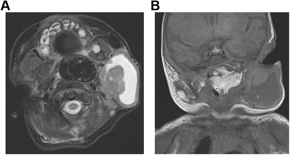
A, Noncontrast-enhanced T2-weighted axial image, with small arrow delineating nasopharyngeal component and long arrow delineating cervical disease. B, Noncontrast-enhanced T1-weighted coronal image, with small arrow delineating nasopharyngeal component, and long arrow delineating cervical disease.
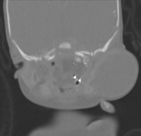
Noncontrast-enhanced coronal CT image, with arrows demonstrating calcification within the tumor along the left skull base.
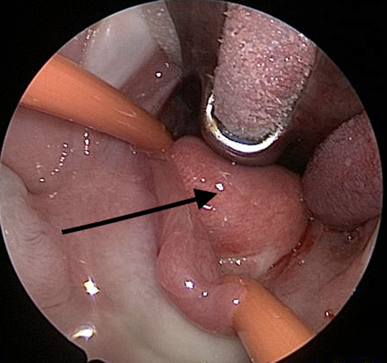
Transoral surgical view, at the beginning of nasopharyngeal tumor resection.
Discussion
Neonatal teratomas of the head and neck are rarely malignant. 4–5 They grow aggressively, however, and over half that go undiagnosed in the prenatal period result in infant death. 6 The appearance of encapsulated cystic, solid, or multiloculated masses is characteristic of teratomas. 7 Small foci of calcifications are also highly suggestive. Their anteromedial location distinguishes teratomas from other neck masses, such as lymphangiomas, hemangiomas, and bronchial cysts. 2 It is important to consider this entity in the differential diagnosis of all head and neck masses.