
Abstract

A 59-year-old woman was referred to the Department of Otolaryngology at the University Hospital for evaluation of cough, nasal discharge, and postnasal drip, which had progressively worsened over 6 months. Although she was treated with medication for headache at a local clinic, her condition did not improve.
Nasal endoscopy revealed a large amount of purulent nasal discharge in the left middle meatus and a small amount of purulent discharge in the right sphenoethmoidal recess. Coronal computed tomography scans showed opacities in the left frontal, anterior ethmoid, and maxillary sinuses, and an opacity in the right most posterior ethmoid sinus (Figure 1).
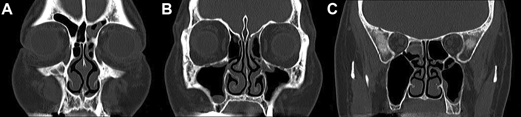
A and B, Coronal computed tomography (CT) scans showed opacities in the left frontal, anterior ethmoid, and maxillary sinuses. C, An opacity is shown in the right most posterior ethmoid sinus.
Under general anesthesia, a left middle meatal antrostomy and anterior ethmoidectomy were performed followed by a frontal sinusotomy. Due to an isolated most posterior ethmoid cell lesion, an endoscopic surgery of the right sinus was subsequently performed via the sphenoethmoidal recess (Figure 2A). The supreme meatus was obstructed by swollen mucosa and opened by resection of the lower half of the supreme turbinate (SpT). The most posterior ethmoid cell was found to contain several pieces of dark yellowish material (Figure 2B). These materials were removed by saline irrigation and suction of the sinus (Figure 2C). The results of the pathological examination were consistent with Aspergillus infection. The postoperative course was uneventful, and she experienced no further symptoms.
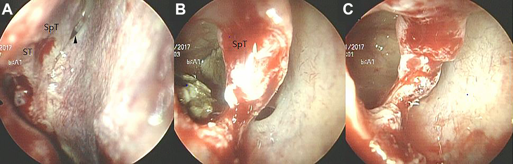
A, Endoscopy shows the structures, such as the superior, supreme turbinates, and sphenoid sinus ostium (arrowhead) in the left sphenoethmoidal recess. B, After resection of the lower half of supreme turbinate, the most posterior ethmoid cell can be seen to contain several pieces of dark yellowish material with a 30° endoscopy. C, These materials were removed by saline irrigation and suction of the sinus. ST indicates superior turbinate; SpT, supreme turbinate.
The site that is most commonly affected by aspergillosis is the maxillary sinus, followed by the ethmoid and sphenoid sinuses. Involvement of isolated posterior ethmoid cell is a very rare condition, and early diagnosis can often be difficult. Computed tomography scanning, with or without magnetic resonance image scanning, can aid in the diagnosis of the disease.
The nasal turbinates are important anatomical structures extending from the lateral nasal walls into the nasal cavity. The ethmoturbinal and maxilloturbinal structures are precursors of the nasal turbinates that appear between the eighth and tenth weeks of fetal life. 1 The maxilloturbinal gives rise to the inferior turbinate, while the ethmoturbinal forms the uncinate process, middle turbinate, superior turbinate (ST), and, when present, the SpT. 1
Orhan et al investigated the anatomical details of the ST to identify approaches to the ostium of the sphenoid sinus using 20 specimens of adult cadavers and an operating microscope. 2 In this study, a SpT was detected on the lateral wall of the nasal cavity in 60% of the cases. In approximately 42% of the specimens, the size of the SpT was found to be either equal to or larger than the ST. In such cases, Orhan et al suggested that the ostium of the sphenoid sinus could be difficult to identify during endoscopic sinus surgery.
When approaching the posterior ethmoid sinus, recognizing the ST and, when present, the SpT can be also very helpful. 3 The SpT has not been used as frequently as the ST as a landmark during endoscopic sphenoid and posterior ethmoid sinus surgery. Both the turbinates are consistent anatomic landmarks that allow safe entrance into the sinuses.
Footnotes
Declaration of Conflicting Interests
The author(s) declared no potential conflicts of interest with respect to the research, authorship, and/or publication of this article.
Funding
The author(s) received no financial support for the research, authorship, and/or publication of this article.