
Abstract
Congenital middle ear cholesteatoma is a common finding among pediatric patients, whereas congenital cholesteatoma between the layers of the tympanic membrane (TM) is a rare finding with limited information. Here, we present the case of a 4-year-old patient with intratympanic membrane congenital cholesteatoma (IMCC) who underwent IMCC removal using an endoscopic system.
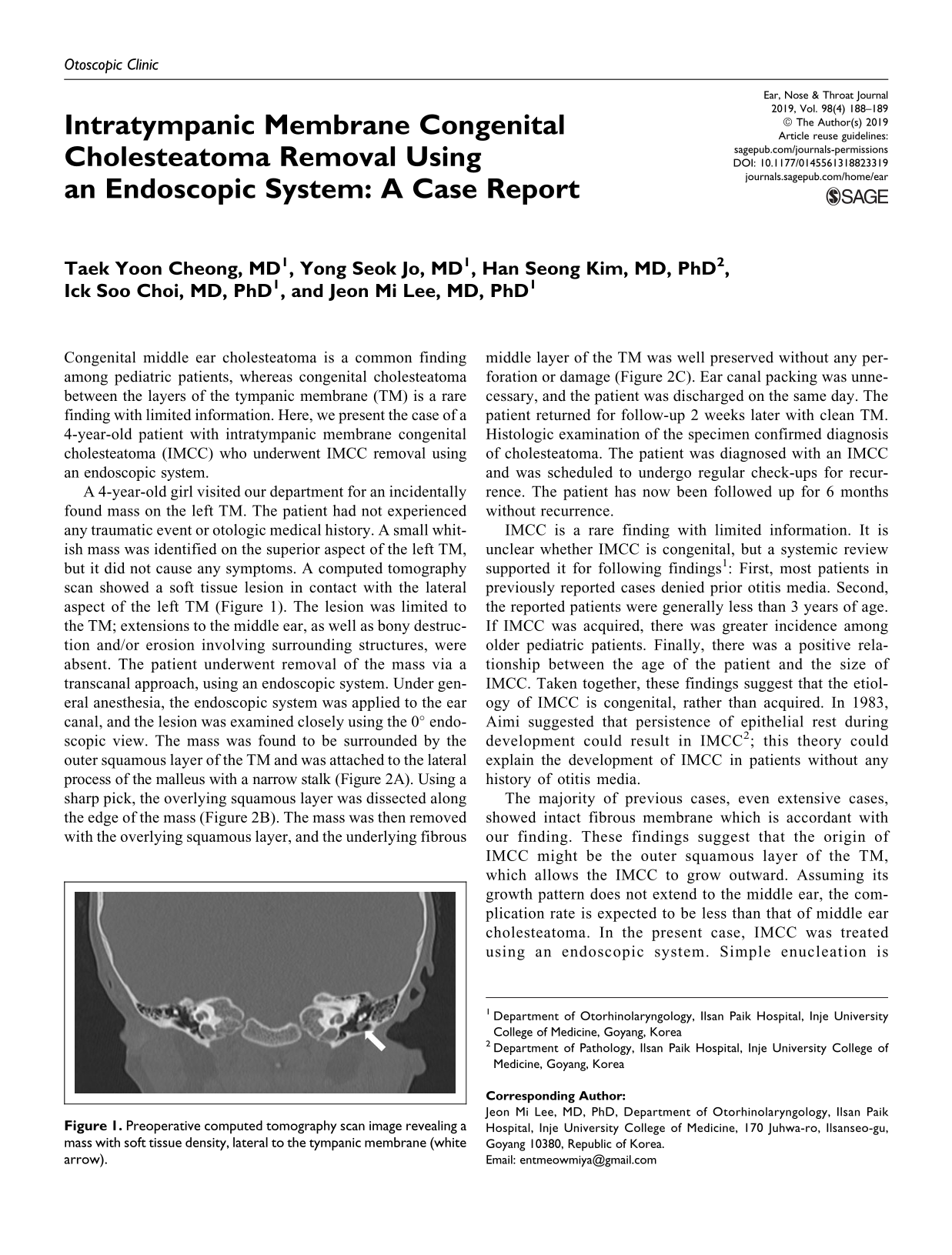
A 4-year-old girl visited our department for an incidentally found mass on the left TM. The patient had not experienced any traumatic event or otologic medical history. A small whitish mass was identified on the superior aspect of the left TM, but it did not cause any symptoms. A computed tomography scan showed a soft tissue lesion in contact with the lateral aspect of the left TM (Figure 1). The lesion was limited to the TM; extensions to the middle ear, as well as bony destruction and/or erosion involving surrounding structures, were absent. The patient underwent removal of the mass via a transcanal approach, using an endoscopic system. Under general anesthesia, the endoscopic system was applied to the ear canal, and the lesion was examined closely using the 0° endoscopic view. The mass was found to be surrounded by the outer squamous layer of the TM and was attached to the lateral process of the malleus with a narrow stalk (Figure 2A). Using a sharp pick, the overlying squamous layer was dissected along the edge of the mass (Figure 2B). The mass was then removed with the overlying squamous layer, and the underlying fibrous middle layer of the TM was well preserved without any perforation or damage (Figure 2C). Ear canal packing was unnecessary, and the patient was discharged on the same day. The patient returned for follow-up 2 weeks later with clean TM. Histologic examination of the specimen confirmed diagnosis of cholesteatoma. The patient was diagnosed with an IMCC and was scheduled to undergo regular check-ups for recurrence. The patient has now been followed up for 6 months without recurrence.
Preoperative computed tomography scan image revealing a mass with soft tissue density, lateral to the tympanic membrane (white arrow).
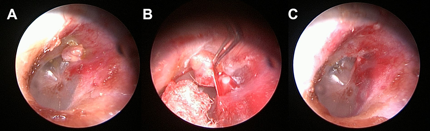
Intraoperative endoscopic images. A whitish mass abutted the lateral process of the malleus with a narrow stalk (A). The squamous layer was easily detached from the underlying fibrous layer (B). The mass and overlying squamous layer was removed, and the tympanic membrane was intact (C).
IMCC is a rare finding with limited information. It is unclear whether IMCC is congenital, but a systemic review supported it for following findings 1 : First, most patients in previously reported cases denied prior otitis media. Second, the reported patients were generally less than 3 years of age. If IMCC was acquired, there was greater incidence among older pediatric patients. Finally, there was a positive relationship between the age of the patient and the size of IMCC. Taken together, these findings suggest that the etiology of IMCC is congenital, rather than acquired. In 1983, Aimi suggested that persistence of epithelial rest during development could result in IMCC 2 ; this theory could explain the development of IMCC in patients without any history of otitis media.
The majority of previous cases, even extensive cases, showed intact fibrous membrane which is accordant with our finding. These findings suggest that the origin of IMCC might be the outer squamous layer of the TM, which allows the IMCC to grow outward. Assuming its growth pattern does not extend to the middle ear, the complication rate is expected to be less than that of middle ear cholesteatoma. In the present case, IMCC was treated using an endoscopic system. Simple enucleation is suggested as the first choice for treatment of IMCC since there is no statistical difference in recurrence or complications between the two surgical procedures, enucleation and complete excision with TM. 1 A sharp approach is required to detach the outer squamous layer from the middle fibrous layer for successful enucleation. The magnified clear vision provided by the endoscopic system enables operators to perform delicate procedures and remove lesions without damaging surrounding structures.
To our knowledge, this is the first case in the literature to remove IMCC with endoscopic system to report its favorable outcomes. It will be a good example of expanding the possibility of endoscopic system in treating various otologic diseases, with an understanding of this rare disease, IMCC.
Footnotes
Declaration of Conflicting Interests
The author(s) declared no potential conflicts of interest with respect to the research, authorship, and/or publication of this article.
Funding
The author(s) received no financial support for the research, authorship, and/or publication of this article.