
Abstract

A 3-year-old girl was brought to the otolaryngology clinic by her mother for evaluation of a left-sided anterior neck mass that had been present since birth. The size of the growth had not changed since birth, and the child did not complain of any symptoms related to it.
Findings on physical examination of the head and neck were unremarkable except for the 1 × 2-cm, nontender, firm, and mobile pedunculated subcutaneous mass located in the anterior neck just left of the midline at the level of the cricoid cartilage (figure 1). The consistency of the mass was suggestive of underlying cartilage. Computed tomography of the neck demonstrated a soft-tissue mass involving the skin and subcutaneous tissue.
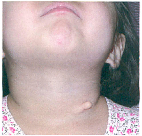
The pedunculated mass is seen at presentation.
The child was taken to the operating room for an excisional biopsy. The mass extended into the subcutaneous tissues and demonstrated a cartilaginous core. Histologically, the specimen exhibited a pedunculated squamous papilla with hair follicles and sebaceous glands. Within the core of the lesion were cartilage and surrounding fibroadipose tissue consistent with an accessory tragus (figure 2).
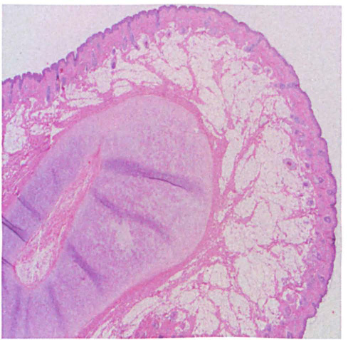
Histologic image of the cervical accessory tragus shows a pedunculated squamous papilloma with a central core of cartilage.
An accessory tragus is usually an isolated congenital defect. 1 It occurs as the result of a developmental anomaly of the first branchial arch, and it may be associated with other branchial cleft abnormalities. It manifests as a skin-colored nodule arising on or near the tragus (1) along an imaginary line drawn from the tragus to the angle of the mouth or (2) along the anterior margin of the sternocleidomastoid muscle.1,2 When these lesions occur, they are typically present at birth. Excisional biopsy is curative in most cases.