
Abstract

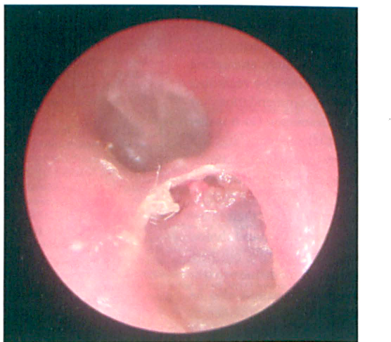
Otoscopic view of the left ear shows a skin-lined defect in the posterior bony external auditory canal.
A 34-year-old woman complained of left otalgia of 4 months’ duration. She denied otorrhea, facial nerve symptoms, or hearing loss. She had been treated previously with antibiotic/steroid drops without relief. She denied having ear surgery or trauma to her ear canal. On otoscopy, her left tympanic membrane was intact and mobile. However, there was a circular erosion of the posterior bony external auditory canal wall that was filled with squamous debris and granulation (figure). Computed tomography of the temporal bones confirmed erosion of the bony external canal.
The patient was taken to surgery where, through a postauricular incision, the eroded canal was examined. The defect extended into the mastoid to the level of the vertical segment of the facial nerve. The cavity was filled with granulation tissue and squamous debris. All disease was removed, the defect was smoothed with a diamond bur, and the canal was lined with temporalis fascia.
External canal cholesteatomas are the result of squamous epithelial deposition deep to the skin of the external canal. The disease can result from postsurgical implantation, radiation, or trauma. 1 Surgical treatment is recommended to prevent complications of further tympanic/mastoid bone erosion, such as facial nerve injury, ossicular erosion, and labyrinthine fistula.