



Abstract

A 75-year-old woman with Parkinson disease presented with complaints of a weak voice that had been worsening as her Parkinson disease progressed. She had been working with speech pathology with only minimal improvement in her vocal quality and volume.
On examination under fiberoptic laryngoscopy through the right nares, an incidental note was made of bilateral pulsating masses from the level of her larynx extending up into her nasopharynx (figure 1). Particularly on the right, which was the more prominent side, the mass was nearly in the midline. The pulsations were noted to be synchronous with the patient's heartbeat. High-resolution computed tomography (CT) confirmed the impression that these pulsating masses were in fact aberrantly positioned internal carotid arteries (figure 2).

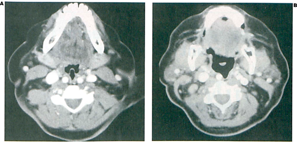
No further intervention was required, but this incidental finding did stress the due diligence required whenever one is performing procedures in the nasopharynx down to the hypopharynx (e.g., adenoidectomy, tonsillectomy). Failure to recognize midline pharyngeal pulsations indicating a medially displaced internal carotid artery could result in disastrous bleeding.
Regarding the patient's original complaint of a weak voice, further sessions with speech pathology were recommended. However, she did not pursue speech therapy.