
Abstract
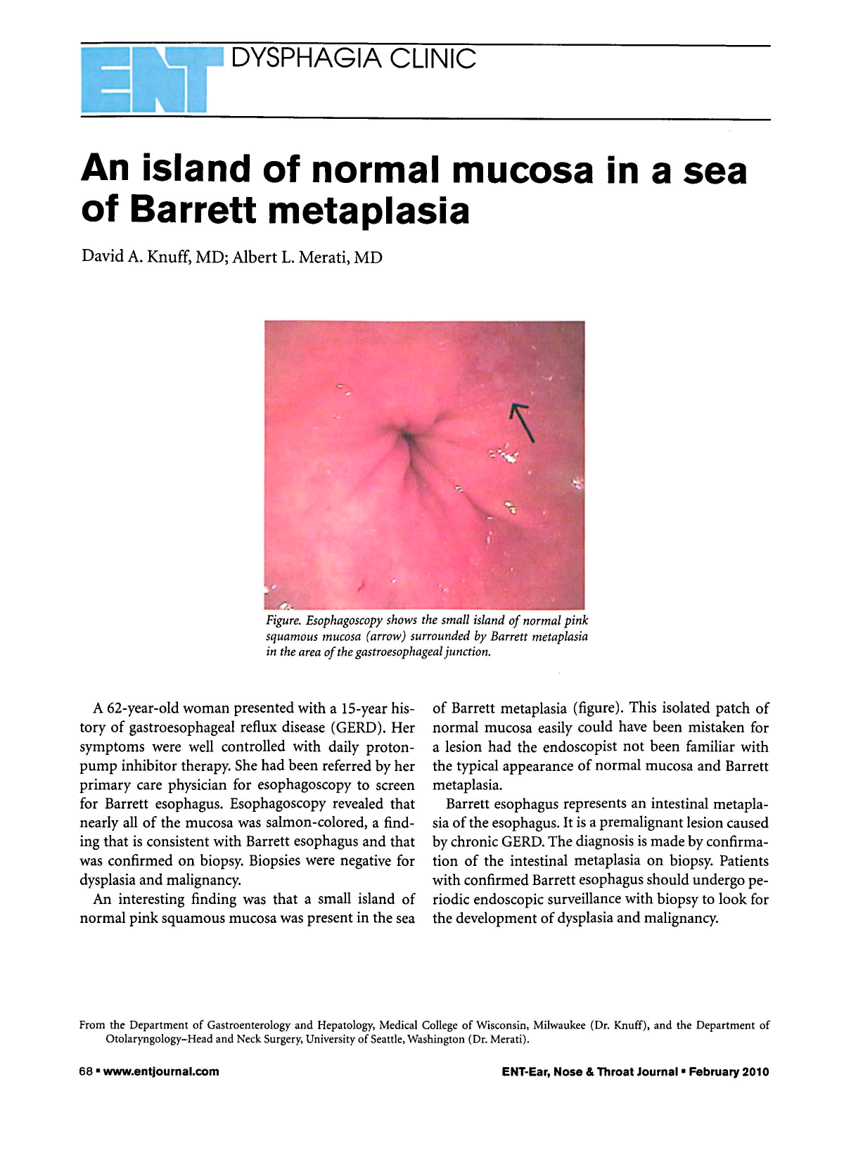
Figure. Esophagoscopy Shows the Small Island of Normal Pink Squamous Mucosa (Arrow) Surrounded by Barrett Metaplasia in the Area of the Gastroesophageal Junction.
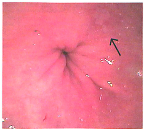
A 62-year-old woman presented with a 15-year history of gastroesophageal reflux disease (GERD). Her symptoms were well controlled with daily proton-pump inhibitor therapy. She had been referred by her primary care physician for esophagoscopy to screen for Barrett esophagus. Esophagoscopy revealed that nearly all of the mucosa was salmon-colored, a finding that is consistent with Barrett esophagus and that was confirmed on biopsy. Biopsies were negative for dysplasia and malignancy.
An interesting finding was that a small island of normal pink squamous mucosa was present in the sea of Barrett metaplasia (figure). This isolated patch of normal mucosa easily could have been mistaken for a lesion had the endoscopist not been familiar with the typical appearance of normal mucosa and Barrett metaplasia.
Barrett esophagus represents an intestinal metaplasia of the esophagus. It is a premalignant lesion caused by chronic GERD. The diagnosis is made by confirmation of the intestinal metaplasia on biopsy. Patients with confirmed Barrett esophagus should undergo periodic endoscopic surveillance with biopsy to look for the development of dysplasia and malignancy.