
Abstract
Camptothecin (CPT) is an important alkaloid used for anticancer treatment. It is mainly produced by two endangered and overharvested Camptotheca acuminata and Nothapodytes nimmoniana plants. Endophytic fungi are promising alternative sources for CPT production. In the present study, fungi residing within explants of Ixora chinensis were isolated and their CPT-producing capability of their endophytes was verified via thin-layer chromatography, high-performance liquid chromatography, liquid chromatography/high resolution mass spectrometry, and nuclear magnetic resonance analyses and compared with standards. In addition, MTT and sulforhodamine B assays were selected to test the anticancer effect. The endophytic fungi collection of 62 isolates were assigned to 11 genera, with four common genera (Diaporthe, Phyllosticta, Colletotrichum, and Phomopsis) and seven less common genera (Penicillium, Botryosphaeria, Fusarium, Pestalotiopsis, Aspergillus, and Didymella). Moreover, the anticancer activity of extracts was assessed against human lung carcinoma (A549). Among eight potential extracts, only Penicillium sp. I3R2 was found to be a source of CPT, while the remaining seven extracts have not been discovered potential secondary compounds. Thus, other prominent endophytic fungi might be potential candidates of phytochemicals with anticancer properties.
Introduction
Camptothecin (CPT) is an isoquinoline alkaloid first discovered in the stem bark of Camptotheca acuminate. 1 This bioactive compound has demonstrated its potential as an antitumor agent by inhibiting topoisomerase I interaction, ultimately leading to cell death.1–6 Currently, 43 plant species belonging to eight different families have been shown to be able to produce CPT. Nearly a half of CPT-producing plant species are members of the Icacinaceae family, and 28% belong to the Rubiaceae family. However, the primary commercial production of CPT still relies on Camptotheca acuminate and Nothapodytes nimmoniana. 7
An estimated 10 million cancer-related deaths were reported in 2020, 8 while patients had to face a significant financial burden ranging from $1500 to $22,000 per year for cancer treatments. 9 The cost is believed to continue rising due to the sheared budget for cancer drugs, new research, and clinical trials. 9 At present, approximately one ton of raw CPT material is required for the global market each year. 10 This demand brings C. acuminate and N. nimmoniana under threat due to overharvesting, driven by the need to fulfill the CPT requirement for the clinical market. It is crucial to explore diverse alternative sources of CPT to prevent these medicinal plants from extinction.
Besides medicinal plants, endophytic fungi are also a potential source of CPT. 11 In recent decades, many endophytes have been reported to have CPT-producing capabilities such as Fusarium solani strain ATLOY-8, 12 Meyerozyma sp. OmF3 and Talaromyces sp. OmF4, 13 Diaporthe sp. F18, 14 Aspergillus flavus ER, and Aspergillus terreus. 15 The CPT biosynthesis pathway in endophytes has not yet been reported; however, a recent study has provided evidence to support that some endophytes are CPT producers independent of their host plant. 16 These findings have addressed the constraints on CPT resources and cleared doubts about endophytic capabilities. Due to their rapid growth rate and adaptability for large-scale fermentation, their strains can be economically produced. Unfortunately, commercial CPT products from endophytes have not been successful, and endophytic fungi are inconsistent producers with lost or diminished CPT capability across generations, except for the Alternaria burnii NCIM1409 strain isolated from Nothapodytes nimmoniana. 16
Despite its widespread cultivation or natural growth in Southeast Asia and its frequent use used as a native medicinal plant, there is a lack of available publications about endophytes associated with Ixora chinensis plant.17,18 In addition, the flower extract of I. chinensis has been proven to completely inhibit specific tumors.19,20 Thus, with the aim of exploring alternative CPT resources, our research identified all endophytes from the Ixora chinensis plant and evaluated their CPT-producing potential.
Materials and methods
Sample collection
Ixora chinensis plants are wild-growing shrubs distributed in Bac Giang Province, Northern Vietnam (21.07° - 21.37°N, 105.53° - 107.02°E). Sample collection was performed during June 2020 at the flowering time of these plants. All parts of the plant (flower, leaf, stem, root) were collected from eight different healthy trees and deposited in separated zip lock bags and kept at 4°C before being brought to the laboratory for future studies at the National Key Laboratory of Gene Technology, Institute of Biotechnology (IBT)- Vietnam Academy of Science and Technology (VAST), Hanoi, Vietnam (Supplemental Figure S1).
Plant material surface sterilization
Shoot and root systems were separated before being washed under running water and then with deionized water to eliminate impurities and debris. Then, the shoot system was divided into three parts: stem, leaf, and flower. Each part underwent a similar surface sterilization process adapted according to a previous study, 2 in which ethanol and sodium hypochlorite were used to abolish the epiphytes. In detail, the plant materials were soaked in 75% ethanol for 3 min, followed by sodium hypochlorite for 3 min. These explants were cut into fragments of 2–3 cm and then soaked in 75% ethanol for one minute before being rinsed with sterile water three times for 3 min each time. Finally, plant fragments were cut into small segments measuring 5 mm (length) × 5 mm (width) for fungal endophyte isolation after being air-dried on sterilized filter paper by sterile flow.
Fungal endophyte isolation
Fungal endophytes were isolated according to a previously described protocol. 21 After sterilization, the treated explants were placed on potato dextrose agar (PDA) (TM media, No. M3D1LT01, India) Petri plates with the addition of antibacterial agents (penicillin 0.05% and streptomycin 0.1%) and cultivated at 28 ± 2°C for 20 days in the dark. The Petri dish was sealed using Parafilm (Bemis-PM966). To confirm the success of the sterilization process, 100 µL aliquots of water from the last washing step were plated on Luria–Bertani agar and PDA media for seven days under optimized conditions of 28 ± 2°C. The effectiveness of the surface sterilization protocol was presented by the absence of any observable colonies, indicating that epiphytic microbes were completely removed. 22 The fungal hyphae emerging from plant tissue were observed every day for 20 days of cultivation to check the growth of fungal endophytic colonies from small explants. The hyphae were subcultured onto a new PDA plate to attain the isolated fungal strains at their early stage. The purified colony was stored in glycerol at 50% (v/v) and deposited at −80°C for long-term storage. The endophytic strain was characterized by analyzing the morphologies of the colony, hyphae, mycelium, and reproduction features through observation and measurement under an optical microscope (Axiostar, Carl Zeiss, No. 45718). Fungal mycelium and spores were stained with lactophenol cotton blue to visualize their structures.23,24
Fungal DNA extraction and amplification
Genomic DNA of endophytic fungi isolated from I. chinensis was extracted by the AllPrep Fungal DNA/RNA/Protein Kit (Qiagen) according to the manufacturer's recommendation. The genomic DNA was used as a template for amplification of the internal transcribed spacer (ITS) region for taxonomic identification. The primer set (ITS4 forward primer-ITS5 reverse primer) designed by White and his coworkers was used to completely cover the ITS1-5.8S-ITS2 region. 25 The PCR reaction comprised 100 ng genomic DNA, 25 µL DreamTaq Green PCR Master Mix (2X), 0.5 µL forward primer, and 0.5 µL reverse primer. Sterile deionized water was added to bring the reaction mixture to a final volume of 50 µL. The PCR programs were set up as follows: preheating at 94°C for 3 min, 35 cycles of 94°C for 30 s, annealing at 58°C for 20 s, extension at 72°C for 1 min, and final extension at 72°C for 10 min. The resulting PCR products were then subjected to electrophoresis on a 1.5% agarose gel at 80 V for 50 min. Predicted DNA fragments were collected, purified by the GeneJET PCR Purification Kit (Thermo Fisher), and sequenced using the BigDye Terminator Cycle Sequencing Kit v 3.1 kit (Applied Biosystems) on the capillary system ABI Prism 3500 XL sequencer (Applied Biosystems) at the National Key Laboratory of Gene Technology (IBT-VAST, Hanoi, Vietnam).
Molecular phylogenetics of endophytic fungi
The acquired DNA sequences were qualified, and a consensus was constructed using BioEdit software version 7.0.5.3. 26 The consensus sequences were then compared to the available fungal sequences by using the online BLAST tool from the NCBI/GenBank database (National Center for Biotechnology Information -http://www.ncbi.nlm.nih.gov/BLAST/).
To determine the taxonomy of the fungal strains, ITS regions including 5.8S rDNA from isolates and reference strains were aligned by Clustal X software. 27 The neighbor-joining method was used to create a phylogenetic tree. 28 The percentage of replicate trees, displayed adjacent to the branches, represent the associated taxa determined through bootstrap analysis 29 with 1000 replicates in Mega X, and the sequences were uploaded to NCBI. 30
Fermentation and extraction of CPT
The liquid fermentation was prepared by using PDB media (pH 5.5) in two steps. First, the seed medium was incubated with 50 mL liquid medium without agar in a 250 mL flask at 28 ± 2°C on a rotary shaker at 160 r/min for three days to inoculate the endophytic fungal strains. Then, the seed liquid was transferred into the fermentation medium to a total of 250 mL under the same conditions for seven days. After liquid cultivation, the whole culture of each strain was transferred into a 250-mL Erlenmeyer flask and percolated with 100 mL of dichloromethane (DCM) and methanol (MeOH) with a 9:1 (v:v) ratio solvent mixture and sonication (33 MHz, Roop Telesonic, India) at room temperature for 60 min. The process was repeated twice for complete extraction. 31 After sonication, the extracts were filtered through 25 µm pored paper and then evaporated to dryness in vacuo (Buchi evaporator system R-300 Rotavapor, V-300 vacuum pumper, B-300 heating bath). To determine CPT content, the concentrate was weighed and transferred into polypropylene microcentrifuge tubes, mixed with high-performance liquid chromatography (HPLC) grade DCM/MeOH (3:1, v:v) at a concentration of 50 mg/mL, and vortexed for 30 s, followed by centrifugation at 11000 r/min for 2 min. The clean supernatants were applied directly onto HPLC 32 and Thin-layer chromatography (TLC) screening.
Sulforhodamine B and MTT assays
Sulforhodamine B and MTT assays were carried out to evaluate anticancer activities of the fermented extracts and derived compounds based on the optical density (OD) measured when the protein composition of the cells was stained with sulforhodamine B (SRB) and MTT - (3-(4,5-dimethylthiazol-2 - yl)- 2, 5 - diphenyltetrazolium), respectively. The human lung carcinoma A549 cell line was chosen for these experiments. Sulforhodamine B assay was performed according to the method of Skekan et al.,
33
starting with trypsinizing A549 cells to separate them, followed by cell counting in a counting chamber to adjust the density. Then, 190 µL of A549 cells were added to a 96-well plate for testing. The fermented extracts of 62 isolated endophytic fungi (samples-T) were dissolved in 100% DMSO to obtain a stock concentration of 20 mg/mL. The samples-T on a 96-well plate were diluted with cell culture medium (without FBS) into 4 ascending concentrations. 10 µL of diluted samples-T at various concentrations were introduced into the wells of the preprepared 96-well plate containing cells. Wells without SRB but supplemented with A549 cells (190 µL) and DMSO 1% (10 µL) were used as the no-growth control (day 0). After 1 h, cells in day 0 control wells were fixed with 20% trichloroacetic acid (TCA), then were incubated the plate at 37°C in a humidified incubator for 72 h. Subsequently, the samples-T and the day 0 were fixed with TCA for 1 h, stained with SRB for 30 min at 37°C, washed three times with acetic acid and then dried at room temperature. Add 10 mM unbuffered Tris base to dissolve the SRB, shake gently for 10 min and then read the OD results at 540 nm on an ELISA Plate Reader (Biotek). The percentage of inhibition was calculated using the following formula:


Thin-layer chromatography screening of fungal CPT
The presence of active metabolites in the extract was evaluated by TLC. Normal-phase TLC was performed on precoated aluminum plates (TLC silica gel 60 F254 Supelco), dried at room temperature for 30 min, and activated in an oven at 110°C for the next 30 min. The solvent system was DCM:MeOH (95:5). Approximately 2 µL of extract (50 mg/mL) was spotted on silica plates. The experimental plates were developed in a chromatographic chamber saturated with 5 mL of the solvent system. Plate development required approximately 5–7 min; plates were then visualized at 254 nm under a UV illuminator (Cleaver). Rf values of bands were recorded using a formula and were compared with a control of 2 µL CPT at 1 mg/mL concentration. 35
High-performance liquid chromatography analysis
Quantification of CPT was performed by following a previously described method. 36 Gradient analytical HPLC assays were performed on an Agilent 1100 instrument, and 10 µL of supernatant extract was loaded onto an octadecyl silane (ODS Agilent) (5 µm; Inertsil) column (150 × 4.6 mm). Acetonitrile:water (20:80 to 100:00 in 30 min) was eluted at a flow rate of 0.6 mL/min, and the alkaloids were detected at 366 nm by a UV detector (DAD). The peak areas corresponding to CPT were integrated by comparison with external standard calibration curves. 31 High-performance liquid chromatography assays of different extracts yielded chromatograms with a retention time of 17.7 min for CPT. Validation of the quantitative method was performed three times with samples. The results of the three injections from the same samples at the five concentrations (0.1–10 mg/mL) showed similar retention times. The standard deviation showed the accuracy of the quantitative method and the ability to determine the CPT content. The concentration of CPT in the cultured liquid was calculated from the same volume of fungal liquid culture.
High resolution mass spectrometry and nuclear magnetic resonance verification of endophytic fungi CPT production
Based on the screening results, potential samples, including the CPT compound, were verified by high resolution mass spectrometry (HRMS) analysis using an electrospray ionization source in positive mode. Extract samples were dissolve in DCM:MeOH (05:95, v/v) with the concentration 5 mg/mL. The system was set up at 300°C, and high-purity nitrogen was used to construct curtain gas (25 psi) chambers. The capillary voltage was constantly kept at 5500 V. The collision energy was set at 10 V, and the collision energy spread was zero. We used IDA mode to scan mass range from 300 to 400 (m/z). The sample was analyzed by an LC-HRMS system (J’phere column 4.6 × 150 mm, flow rate of 0.5 mL/min, and 60% ACN in water). The data were recorded on an Agilent QTOF 6530. Molecules of CPT were found using Agilent MassHunter Qualitative Analysis B.07.00.
For nuclear magnetic resonance (NMR) verification, a 70 L fermentation tank containing PBD media was prepared, and I3R2 was cultured for 7 days at 180 r/min and 28°C. CPT was extracted using the protocol mentioned above and purified via HPLC system (Agilent HPLC 1290 Infinity II) by collecting the fraction of the peak as CPT. Finally, 4 mg of purified CPT (90%) was dissolved in CDCl3 for 1H and 13C structural elucidation. 1D NMR spectra were recorded on a Bruker Avance 600 MHz spectrometer (Bruker Biospin, Rheinstetten, Germany) using TMS (tetramethylsilane) as an internal standard.
Preparation of the standard solution of CPT
A stock solution of CPT (Sigma Aldrich, CAS: 7689-04-4) was prepared by dissolving 5 mg of accurately weighed CPT in a DCM:MeOH mixture (3:1, v:v) and bringing the volume to 500 µL with methanol. From this stock solution, standard solutions of 0.1, 0.5, 1, 5, and 10 mg/mL were prepared by transferring aliquots of stock solution to a 1.5-mL microtube and adjusting the volume with a DCM:MeOH mixture (3:1, v:v). The calibration curve was calculated by injecting 10 µL of standard solutions of CPT into the column (Murthy 2019). All analysis experiments were carried out in triplicate. The peaks were detected at 366 nm, and the peak areas were recorded. Calibration curves of CPT were prepared by plotting peak area against concentration with a correlation coefficient of R2 = 0.9999 (Y = 778781X + 33712).
Results
Identification and colony characterization of endophytic fungi in I. chinensis
Fungal endophytes were isolated from explants of I. chinensis following the elimination of epiphytes through the surface sterilization process. Sixty-two isolates were obtained from distinct morphological analyses on PDA medium (Figure 1, Table S1).

Morphological characteristics of isolated endophytic fungi from Ixora chinensis. (A) Colony morphology of 62 isolated endophytes. (B) Typical colony types, the left side: is vegetative hyphae, the right side: is aerial hyphae; (B-1): the white cottony type; (B-2): the green powdery type; (B-3): the black spongy type; (C) Septate hyphae; C1: Phomopsis sp. I2T2; (C-2): Diaporthe sp. I5T3. (D) Conidiophore and conidial structure; D1: Pestalotiopsis sp. I1L3; (D-2, D-3): Colletotrichum sp. I1L2; (D-4): Penicillium sp. I3R2.
The colony morphological characteristics including aerial mycelia, vegetative mycelia, pigmentation, form, and the margin of the colony were first used to distinguish and identify the group of fungi. The results showed that 62 isolated fungal endophytes were assigned to four morphotypes, encompassing the green powdery, black spongy, white cottony groups, and a group comprising the remaining isolates (Figure 1). Microfeatures such as hyphal structures, conidiophores, conidia, and spore morphologies were combined with molecular identification to classify isolated endophytic fungi (Figure 1, Table S1). Hyphal structures showed a septate type with a difference in size, such as 36.3 ± 13 × 6.3 ± 2.2 µm for the I2T2 strain and 12.3 ± 1.6 × 5.3 ± 1.1 µm for the I5T3 strain (Figure 1). The reproduction of fungal endophytes on the PDA plate occurred by fragmentation, as in Pestalotiopsis sp. I1L3, or by producing spores from the tips of hyphae (e.g. Colletotrichum sp. I1L2) or from conidiospores (e.g. Penicillium sp. I3R2) (Figure 1D).
In addition, the endophytes were identified by ITS sequencing analysis. In detail, ITS consensus sequences containing ITS1, 5.8S and ITS2 regions were blasted against the NCBI database to obtain the closest matches. The results were compared with DNA sequences of types or epitypes and morphology characteristics of the specific taxon. The phylogenetic tree was also constructed using the ITS sequences of endophytic fungal strains (Figure 2F), and the data were matched with morphological results (Figure 1, Table S1). Finally, in the present study, 62 endophytic fungal strains isolated from I. chinensis were classified into 11 genera (Table S1). The results indicated that most species isolated from I. chinensis belong to four common genera: Diaporthe (18 strains), Phyllosticta (13 strains), Colletotrichum (11 strains), and Phomopsis (7 strains). For seven less-common genera, the colonization frequency of Penicillium was four strains, followed by Botryosphaeria (2 strains), Fusarium (2 strains), Pestalotiopsis (2 strains), Lasiodiplodia (1 strain), Aspergillus (1 strain), and Didymella (1 strain) (Table S1). The identified endophytes were consistent between morphological and ITS analyses and were deposited in glycerol (50% v/v) at −80°C at the National Key Laboratory of Gene Technology (IBT-VAST, Hanoi, Vietnam) for long-term storage. In addition, all strains were grown for the collection of crude extracts in the following studies.

Diversity and distribution of the endophytic community in Ixora chinensis explants (A–E) and phylogenetic tree (F).
Diversity and distribution of endophytic fungi from different parts of I. chinensis plants
The endophytic population from I. chinensis distributed unevenly among various plant tissue types (Figure 2, Table S1). Endophytes were the most abundant in stem, with 25 strains (40%), followed by leaf with 20 strains (32%). The flowers and roots hosted a minor number of fungal strains at 13% and 15%, respectively (Figure 2A). In addition, the distribution of specific endophytic species varied in different parts of the plant (Figure 2B–E). Diaporthe sp. exhibited abundances in root and stem tissues of 56% and 52%, respectively. Phyllosticta emerged only in leaf and flower parts. Several genera, such as Didymella sp. (12%), Pestalotiopsi sp. (10%), Aspergillus sp. (11%), and Lasiodiplodia sp. (4%), were unique to the flower, leaf, root, and stem parts, respectively.
Sulforhodamine B and MTT assays
Four extracts of endophytic fungi including I1L2, I1R7, I3R2, and I6T1 exhibited anticancer capability with inhibition greater than 50% at a tested concentration of 20 μg/mL in both SRB and MTT assays. However, some extracts only showed anticancer activity in the MTT assay (I3T3, I3T4, I6L2, I8T3). Among the positive strains, the extracts of I1R7, I1L2, and I6T1 indicated the strongest activity, with inhibition percentages of 90, 57, and 59.83%, respectively, in the SRB assay. Meanwhile, the inhibition percentages of these extracts were 100, 84.89, and 71.85% in the MTT assay, respectively (Table 1). Subsequently, the eight extracts were selected to determine the half maximal inhibitory concentration (IC50) of cytotoxicity on A549 human lung cancer cell line (Table 2).
Evaluation of the in vitro cytotoxicity of endophytic fungi.

IC50 values of the compounds against A549 (µg/mL).
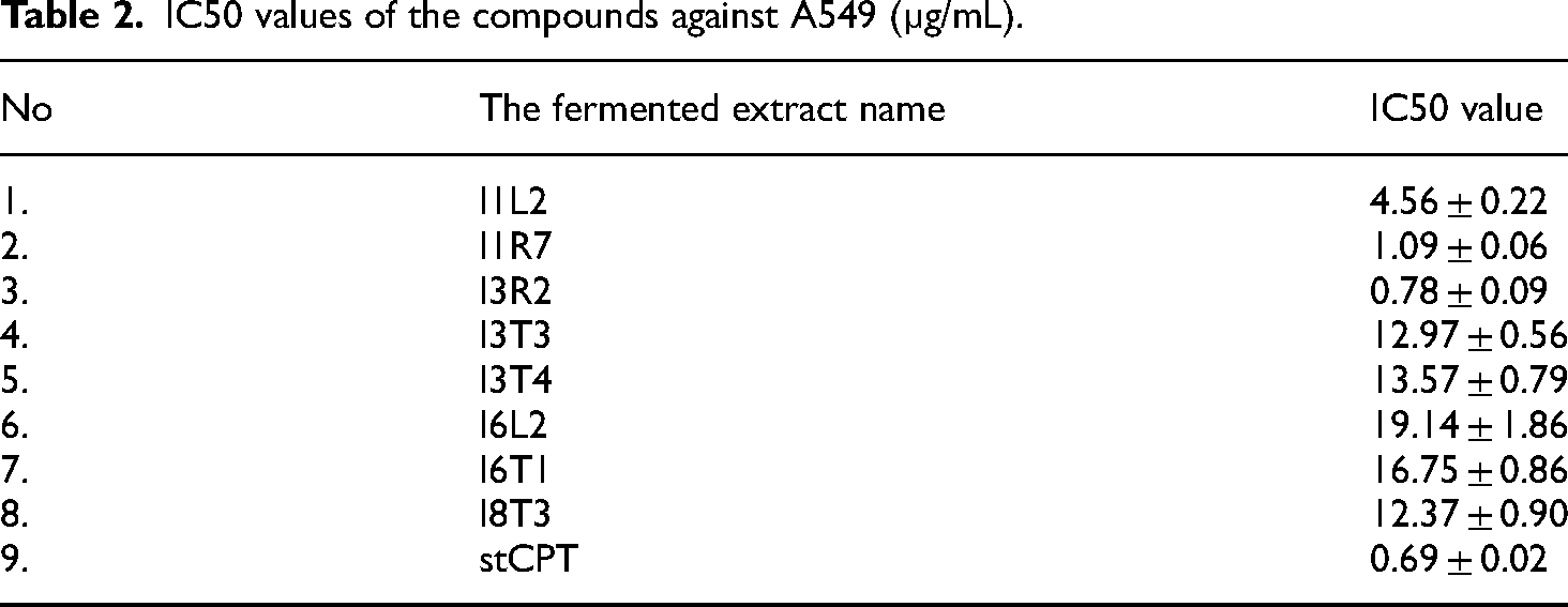
The above results have shown that the eight fermented extracts showed inhibitory activity against the tested cancer cell lines with IC50 values from 0.78–19.14 µg/mL. According to the standards of the US National Cancer Institute (NCI), the extract and the purified substance are considered to have good activity with IC50 ≤ 20 μg/mL and IC50 ≤ 5 μM, respectively. 37 The fermented extract derived from I3R2 strain displayed the highest cytotoxicity among eight samples with the IC50 value of 0.78 ± 0.09 μg/mL, and this half maximal inhibitory concentration was approximately with the one from standard CPT (0.69 ± 0.02 μg/mL).
Screening, quantification, and verification of CPT-producing endophytic strains
The crude extracts from the culture liquid of endophytic strains were screened to produce CPT using TLC and HPLC analyses. First, the CPT in the sample was characterized in comparison with standard CPT (Sigma Aldrich). Then, the presence of CPT in extracts was continuously analyzed by HPLC to detect and quantify CPT from fungal endophytes. A standard curve with a correlation coefficient R2 = 0.9999 allowed us to determine the exact CPT content in the extract of endophytes (Figure S2, Table S2). In the present study, a substance with a polarity like that of the CPT standard in the TLC experiment emerged in the extracts. The crude extracts were then subjected to HPLC to give a signal with the same retention time and UV spectrum as the standard CPT Rf of 17.7 min. The detailed results of TLC and HPLC were reported in supplemented Figures S3 and S4, respectively. These potential endophytic fungi were I1L1, I1L2, I1R1, I2T2, I3L2, I3R2, I3T2, I4H2, I5T3, I6T5, and I8T3. The potential CPT content in endophytic fungi ranged from 2.164 mg/L to 0.055 mg/L (Table 3).
The putative CPT concentration (mg/L) was determined from endophytic fungi via TLC and HPLC analysis.

N: not determined.
To confirm the presence of CPT in the extracts, the samples were analyzed by an LC-HRMS system. As a result, the presence of CPT in extracts of I8T3 and I3R2 was determined with molecular ions at m/z 349.1237 and 349.1242, respectively. These results were like that of standard CPT with a molecular ion at m/z 349.1228 (Figure 3). Base on the recovery yield from extract of I3R2 and I8T3, the strain I3R2 was applied in scale up cultivation for further NMR analysis. The ability of the I3R2 strain to produce CPT was reconfirmed by NMR analysis. CPT was isolated and verified with the chemical shifts of 1H and 13C NMR spectra (Figure S5, Figure S6). 1H NMR (600 MHz, CDCl3) d ppm 5.31 (2 H, s, H-5) 8.40 (1 H, s, H-7) 7.94 (1 H, d, J = 8.4 Hz, H-9) 7.66 (1 H, t, J = 7.2 Hz, H-10) 7.84 (1 H, t, J = 7.2 Hz, H-11) 8.24 (1 H, d, J = 9 Hz, H-12) 7.69 (1 H, s, H-14) 0.88 (3 H, t, J = 7.34 Hz, H-18) 1.90 (2 H, m, J = 7.2 Hz, H-19). 13C NMR (600 MHz CDCl3): 173.95 (C-21), 157.69 (C-17), 152.52 (C-2), 150.15 (C-15), 149.03 (C-13), 146.50 (C-3), 131.06 (C-7), 130.61 (C-11), 128.54 (C-6), 129.88 (C-12), 128.11 (C-9), 128.17 (C-8), 128.02 (C-10), 118.70 (C-16), 98.07 (C-14), 72.76 (C-20), 66.40 (C-17), 50.07 (C-5), 31.66 (C-19), 7.8 (C-18) (Table S3). The NMR spectral data of the isolated compound were in agreement with those of Sigma-grade CPT. 38 These data suggested that the I3R2 endophytic fungi from I. chinensis could produce CPT in the fermentation broth during its development.

Identification of CPT-producing endophytic fungi from the crude extract by HRMS analysis. Standard CPT (A), CPT yield from I3R2 (B) and I8T3 (C).
Discussion
Ixora chinensis is widely distributed in Southeast Asia and is considered as a promising candidate for its ability to inhibit tumors.17–20 However, little information has been reported related to this plant or endophytes associated with I. chinensis. This is the first study investigating the diverse fungal endophytes within I. chinensis, encompassing a total of 62 endophytic strains belonging to 11 genera (Figure 2, Table S1). The complex of Diaporthe and its Phomopsis anamorph (Diaporthe/Phomopsis) reported in this study were in line with previous research.39,40 The three frequent genera in our collection of isolates—Diaporthe, Phomopsis, and Colletotrichum—have also been identified as dominant genera in the endophytic fungal communities of other plants, such as Nothapodytes foetida, Icacinaceae, and Camptotheca acuminata.11,31,41 However, Trichoderma, which was commonly found in other plants, was not detected in this study.42,43 In addition, the results showed that endophytes were abundant in the leaves and stems. Pestalotiopsis, Phomopsis, and Didymella were found only in the leaves, while Aspergillus was observed only in the roots (Figure 2). These findings support the theory that variations in endophyte diversity arise from distinct geographic conditions and specific host plant species.42,44
Although eight extracts from endophytic fungi displayed anticancer activity, only two extracts were reported as candidates for CPT. Moreover, only the extract from I3R2 contained CPT, and the extract from I8T3 might be a CPT analog. The secondary metabolites in the six prominent extracts could be other active anticancer compounds. Further biochemical experiments are required to identify and classify bioactives. Thus, the observed cytotoxicity exhibited in this study can be originated from the compounds present in the fungal extracts. Moreover, many study have indicated that the extracts from endophytes are excellent producers of strong cytotoxic metabolites.45,46 According to the US NCI Plant Screening Program, a crude extract is generally considered to have in vitro cytotoxic activity if the IC50 values is <20 μg/mL. 37 The eight fermented extracts showed inhibitory activity against the tested cancer cell lines with IC50 values from 0.78 to 19.14 µg/mL.
The HPLC results revealed that 11 extracts might contain CPT. In this study, TLC and HPLC methods might impose certain limitations on the identification and quantification of compounds produced by endophytic fungi. Strain I2T2 exhibited a peak at a retention time and UV absorption spectrum like that of CPT, although the molecular mass peak of CPT (Figure 3) was not detected in the HRMS spectrum. This indicates that, in addition to HPLC, further research is required to confirm the presence of CPT. Only sample from I3R2 was identified as CPT by NMR analysis. This emphasizes the vital role of NMR in verifying the compounds present in the extracts.
In the present study, Penicillium sp. I3R2 was reported as a source of CPT, with a concentration of 0.05 mg/L from biomass. The yield was lower than that previously reported for Fusarium solani strain ATLOY-8 12 and Diaporthe caatingaensis MT192326. 47 However, its anticancer activity was more potent than others, such as the F. solani MTCC9667 strain from A. dimidiate 31 and P. chrysogenum. 15 The difference might depend on the enzyme system involved in the CPT biosynthetic pathway in screening CPT-producing endophytic fungi. In addition, the distinct culture medium caused variations in CPT-producing endophytic fungi.31,48 Optimized fermentation conditions activated biosynthesis-related genes for CPT biosynthesis, improved the concentration of bioactive compounds, and reduced self-toxicity during fungal metabolism.49,50 Diaporthe sp. F18 produced a 23-fold higher CPT content by using tryptophan in modified SDB medium. 14 In addition, the Box–Behnken design for Fusarium solani strain ATLOY-8 exhibited 1.4- and 1.2-fold increase in CPT production and biomass yield, respectively. 12 Therefore, subsequent studies might be designed to optimize culture conditions to increase the yield of CPT in fungal endophytes from I. chinensis.
Conclusion
This study has reported the diversity of endophytic fungi in I. chinensis and the potential CPT production of their endophytic members for the first time. Total 62 endophytic isolates were found in I. chinensis and classified into 11 genera based on morphological and ITS1-5.8S-ITS2 region analysis. Sulforhodamine B and MTT assays were chosen to evaluate anticancer activities of the fermented extracts. Although eight extracts from endophytic fungi exhibited anticancer activity, only the extract from I3R2 contained CPT, and the extract from I8T3 might be a CPT analog. Among them, the endophytic fungi I3R2 of Penicillium sp. exhibited a CPT yield of 0.055 mg/L when was cultured in PDB media at 30°C, pH 5.5 for seven days. The endophyte will be useful for studying to construct alternative sources of CPT for commercial manufacturing.
Supplemental Material
sj-docx-1-sci-10.1177_00368504241253675 - Supplemental material for Isolation, anticancer potency, and camptothecin—producing ability of endophytic fungi isolated from Ixora chinensis
Supplemental material, sj-docx-1-sci-10.1177_00368504241253675 for Isolation, anticancer potency, and camptothecin—producing ability of endophytic fungi isolated from Ixora chinensis by Thi Nhung Doan, Thi Dung Le, Ngoc Anh Ho, Thuong Thi Ho, Thi Thao Do, Ha Hoang, Mau Hung Nguyen, Thanh Mai Bui and Hoang Ha Chu in Science Progress
Supplemental Material
sj-docx-2-sci-10.1177_00368504241253675 - Supplemental material for Isolation, anticancer potency, and camptothecin—producing ability of endophytic fungi isolated from Ixora chinensis
Supplemental material, sj-docx-2-sci-10.1177_00368504241253675 for Isolation, anticancer potency, and camptothecin—producing ability of endophytic fungi isolated from Ixora chinensis by Thi Nhung Doan, Thi Dung Le, Ngoc Anh Ho, Thuong Thi Ho, Thi Thao Do, Ha Hoang, Mau Hung Nguyen, Thanh Mai Bui and Hoang Ha Chu in Science Progress
Supplemental Material
sj-docx-3-sci-10.1177_00368504241253675 - Supplemental material for Isolation, anticancer potency, and camptothecin—producing ability of endophytic fungi isolated from Ixora chinensis
Supplemental material, sj-docx-3-sci-10.1177_00368504241253675 for Isolation, anticancer potency, and camptothecin—producing ability of endophytic fungi isolated from Ixora chinensis by Thi Nhung Doan, Thi Dung Le, Ngoc Anh Ho, Thuong Thi Ho, Thi Thao Do, Ha Hoang, Mau Hung Nguyen, Thanh Mai Bui and Hoang Ha Chu in Science Progress
Supplemental Material
sj-docx-4-sci-10.1177_00368504241253675 - Supplemental material for Isolation, anticancer potency, and camptothecin—producing ability of endophytic fungi isolated from Ixora chinensis
Supplemental material, sj-docx-4-sci-10.1177_00368504241253675 for Isolation, anticancer potency, and camptothecin—producing ability of endophytic fungi isolated from Ixora chinensis by Thi Nhung Doan, Thi Dung Le, Ngoc Anh Ho, Thuong Thi Ho, Thi Thao Do, Ha Hoang, Mau Hung Nguyen, Thanh Mai Bui and Hoang Ha Chu in Science Progress
Supplemental Material
sj-docx-5-sci-10.1177_00368504241253675 - Supplemental material for Isolation, anticancer potency, and camptothecin—producing ability of endophytic fungi isolated from Ixora chinensis
Supplemental material, sj-docx-5-sci-10.1177_00368504241253675 for Isolation, anticancer potency, and camptothecin—producing ability of endophytic fungi isolated from Ixora chinensis by Thi Nhung Doan, Thi Dung Le, Ngoc Anh Ho, Thuong Thi Ho, Thi Thao Do, Ha Hoang, Mau Hung Nguyen, Thanh Mai Bui and Hoang Ha Chu in Science Progress
Footnotes
Acknowledgments
This study was supported by the project of the Vietnam Academy of Science & Technology (VAST): “Research for the function of camptothecin composition or other substances with cancer activities in labor lever from endophytic fungi on medical plant” (project no. TĐCNSH.03/20-22).
Availability of data and materials
Declaration of conflicting interests
The author(s) declared no potential conflicts of interest with respect to the research, authorship, and/or publication of this article.
Funding
The author(s) received no financial support for the research, authorship, and/or publication of this article.
Supplemental material
Supplemental material for this article is available online.
Author biographies
Thi Nhung Doan was a Master of Biotechnology at Graduate University of Science and Technology. She is working at National Key Laboratory of Gene Technology, Institute of Biotechnology (IBT), Vietnam Academy of Science & Technology (VAST), Hanoi, Vietnam. Nhung had published several scientific papers on a number of topics related to metagenomic, microorganism genomic, antibiotic, chloroplast, secondary metabolites, human ancient DNA. Currently, she is interested in machine learning and metabolomic.
Thi Dung Le graduated master's degree in plant biotechnology from Sejong University, South Korea. She has published several papers on microbiology, genetic technology, and DNA sequencing. Currently, she is a PhD student at the University of Science and Technology of Hanoi, and works at Center for DNA Identification, Institute of Biotechnology, Vietnam Academy of Science and Technology.
Ngoc Anh Ho is a researcher at the Institute of Biotechnology (IBT), Vietnam Academy of Science and Technology. In 2019, he earned his Doctor of Philosophy degree in Biomodulation from Myong Ji University, Korea. He recently worked in the Environmental Bioremediation Laboratory at IBT. He has published numerous scientific papers on various topics, including the isolation and purification of compounds from natural resources, as well as the application of NMR and MS spectroscopy for structure elucidation. His current research interests focus on evaluating the biological activity of native Vietnamese plants and their purified compounds.
Thuong Thi Ho graduated PhD in Biotechnology at the Graduate University of Science and Technology in 2023. Currently, she is a researcher at Applied DNA Technology, Institute of Biotechnology, Vietnam Academy of Science and Technology. She has had 10 years of research experience in the construction, expression, characterization, and evaluation of the immunogenicity of plant-based proteins such as Hemagglutinin protein (A/H5N1, A/H7N9), Spike protein (PEDV), GP5-M protein (PRRSV). She has published several scientific papers on various topics related to vaccines, plant biotechnology, medicine, and nanomaterials.
Thi Thao Do received her PhD degrees in Cellular Biology at the Institute of Biotechnology, Vietnam Academy of Science and Technology in 2007. She was appointed as an associate professor in 2016 and senior researcher in 2017. She has been carrying out research on developing and applying the in vitro and in vivo bioassays for drug discovery. She has worked on thousands of herbal and traditional medicinal plants' extracts, fractions and pure compounds in order to look for active agents of which possess anticancer, antioxidant, antidiabetic, hepatoprotective, antimalarial, anti-inflammatory activities, etc. Several promising compounds were developed as active ingredients in supplemental drug products or alternative drugs for clinical treatments (VINDOXIM), VDK, KHUONG-THAO-DAN, etc. Now, she is working at Bioassay Group, Institute of Biotechnology (IBT), Vietnam Academy of Science & Technology (VAST), Hanoi, Vietnam.
Ha Hoang holds a PhD in Microbiology from Vrije Universiteit Amsterdam. At the present, he is working at Department of application and development technology, Vietnam Academy of Science and Technology (VAST). His publication related to endophytic fungi, plant genomic, human genetics and sequencing technology.
Mau Hung Nguyen hold a master's degree of practical biology at Thai Nguyen University. At the present, he is working at National Key Laboratory of Gene Technology, Institute of Biotechnology (IBT), Vietnam Academy of Science & Technology (VAST), Hanoi, Vietnam. His publication is related to human genetics and microbiology.
Thanh Mai Bui is a Master of Microbiology at Thai Nguyen University. Now, she is working at Center for DNA Identification, Institute of Biotechnology (IBT), Vietnam Academy of Science & Technology (VAST), Hanoi, Vietnam. Her publications involve microbiology and plants.
Hoang Ha Chu is a professor of gene technology at Vietnam Academy of Science & Technology (VAST). He is working at National Key Laboratory of Gene Technology, Institute of Biotechnology (IBT), Vietnam Academy of Science & Technology (VAST), Hanoi, Vietnam. He had published several scientific papers on various topics, including genome editing, bioactivities, immunology, ancient DNA, stem cells, metagenomics, and plant physiology.
References
Supplementary Material
Please find the following supplemental material available below.
For Open Access articles published under a Creative Commons License, all supplemental material carries the same license as the article it is associated with.
For non-Open Access articles published, all supplemental material carries a non-exclusive license, and permission requests for re-use of supplemental material or any part of supplemental material shall be sent directly to the copyright owner as specified in the copyright notice associated with the article.