
Abstract
Rodents are the most widely used species for scientific purposes. A critical pre-requisite of their use, based on utilitarian ethical reasoning, is the provision of a humane death when necessary for scientific or welfare grounds. Focussing on the welfare challenges presented by current methods, we critically evaluate the literature, consider emerging methodologies that may have potential for refinement and highlight knowledge gaps for future research. The evidence supports the conclusion that scientists and laboratory personnel should seek to avoid killing laboratory rodents by exposing them to carbon dioxide (CO2), unless exploiting its high-throughput advantage. We suggest that stakeholders and policymakers should advocate for the removal of CO2 from existing guidelines, instead making its use conditionally acceptable with justification for additional rationale for its application. With regards to physical methods such as cervical dislocation, decapitation and concussion, major welfare concerns are based on potential inaccuracy in application and their susceptibility to high failure rates. There is a need for independent quality-controlled training programmes to facilitate optimal success rates and the development of specialist tools to improve outcomes and reliability. Furthermore, we highlight questions surrounding the inconsistent inclusion criteria and acceptability of physical methods in international regulation and/or guidance, demonstrating a lack of cohesion across countries and lack of a comprehensive ‘gold standard’ methodology. We encourage better review of new data and championing of open access scientific resources to advocate for best practice and enable significant changes to policy and legislation to improve the welfare of laboratory rodents at killing.
Why consider killing methods for use in laboratory rodents?
Rodents remain the most widely used species for scientific research due to their small size, low cost, rapid sexual maturity and scope for genetic manipulation. Despite significant interest in the replacement of animals for scientific purposes,1,2 the numbers involved are still large, and growing, primarily due to advances in molecular genetics. 3 In 2019, global animal use for scientific research was estimated to be 192.1 million annually. 4 In Europe (and Norway), approximately 6.4 million mice and rats were used in 2018 (most recent published data), accounting for around 62% of the animals reported across member states of the European Union (EU). 5 Therefore, global numbers are likely to be substantial, potentially exceeding 100 million rats and mice annually.
Regulation requires animals to be killed humanely upon completion of the work (i.e. at the end of the experiment or breeding programme) and when humane endpoints are reached.6–9 This necessitates the killing of millions of laboratory rodents each year. Ethical harms relating to the harm of death notwithstanding, 10 the scale of this activity makes welfare at the time of killing an important issue. Arguably, identification and use of methods that offer a death with minimal suffering is a moral imperative, and crucial to support the justifiability of animal-based research based on utilitarian harm/benefit ethical reasoning.
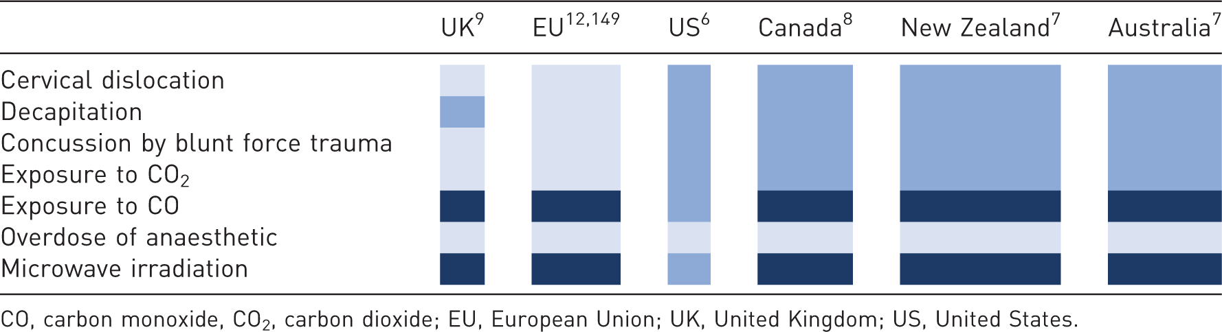
There are diverse international requirements for killing methods for laboratory rodents (Table 1), which include: overdose of anaesthetic (with various inhalant or injectable agents), concussion by blunt force trauma, cervical dislocation, decapitation, exposure to carbon dioxide (CO2) or carbon monoxide (CO) and microwave irradiation. If animals are unconscious (e.g. anaesthetised), methods such as exsanguination, air embolism and injection of potassium chloride or ethanol are permitted. 11 Across member states of the EU and in the United Kingdom (UK), killing laboratory rodents is regulated by law; however, in the United States (US), Canada, Australia and New Zealand this is not the case.6–8 Instead, approaches are mandated through national guidelines and local policy rather than law, and methods are classified according to whether they are considered capable of providing a ‘humane’ death. Thus, they may be described as acceptable, conditionally acceptable (humane only when done correctly and appropriately and only when operator health and safety concerns are mitigated) or unacceptable. Although not intended to be exhaustive, Table 1 highlights the international significant differences in requirements for rodents at the time of killing. These represent the potential for misalignment of animal care standards and killing practices which could present numerous challenges to animal welfare including unnecessary suffering at the time of killing.
Overview of killing methods from published euthanasia guidelines for adult laboratory rodents across scientifically advanced countries. Light blue represents methods permitted or recommended for use, dark blue represents methods where additional permissions are required, navy blue represents methods not permitted or recommended.

The aim of this review is to critically evaluate the literature to identify the welfare challenges presented by current methods used to kill adult laboratory rodents. Attention is given only to those methods used across scientifically advanced countries that have published and/or evidence-based guidance. Consideration will be given only to primary methods that have the potential to affect the animals’ conscious experience prior to loss of consciousness. In examining various methods available and in current use, this review will highlight welfare costs and benefits, comment on reliability and the challenges posed to operator health and safety. Finally, we describe emerging methodologies that may have potential for refinement and highlight knowledge gaps for prioritisation of future research efforts.
Relevant definitions and terminology
The term ‘killing’ refers to any intentional act or process that results in the death of an animal. 12 Euthanasia is considered to mean a ‘good death’ and therefore one without pain and suffering and, in some contexts, refers to a death that is in the animal’s interests (i.e. to end pain and suffering).13–15 The use of euthanasia throughout guidance and regulation protecting laboratory animals implies that approved killing methods are humane and may falsely alleviate potential public concern. 16 As such, because there is considerable uncertainty about the welfare consequences of existing methods used to kill laboratory rodents, the universal term ‘killing’ will be used rather than ‘euthanasia’ throughout this review. A large component of the extent of suffering at killing depends upon the time to loss of consciousness. 17 Loss of consciousness can be defined as the transition from a state of conscious awareness, where the animal is aware of their surroundings and is responsive to external stimuli, to unconsciousness, which occurs when the awareness of self and surroundings are lost, becoming unresponsive to external and internal stimuli.17–19 The majority of rodents are conscious during handling and the application of killing methods and are therefore capable of experiencing negative states (e.g. pain and fear) until they lose consciousness.12,14,15
Assessing welfare at killing
Concern for animal welfare during killing stems from animals’ ability to experience potentially negative sensations occurring during the conscious phase of the process. To fully determine the welfare consequences of a killing method, it is important to determine (a) the time taken for the animal to be rendered unconsciousness inclusive of pre-handling time and kill method application and (b) the presence and character of potentially negative experiences during the conscious phase.
Behaviourally, it is possible to determine the transition from consciousness to unconsciousness by assessing the gradual loss of reflexes and goal-directed behaviours and ultimately, motor coordination. Generally, the cessation of movement and onset of recumbency is considered to be a marker of unconsciousness in animals, which is typically measured in rodents by their inability to effectively right their posture, referred to as loss of righting reflex or loss of posture.20,21 We can also measure changes in behavioural output, through the transition from goal directed behaviour (e.g. motivated escape behaviours)22,23 to the presence of spontaneous behaviours such as jumping, gasping etc., through to the presence of tonic and clonic convulsions in the unconscious phase.24–26 Neurophysiologically, we can measure brain activity to infer the likely degree of consciousness. Recordings of the electroencephalogram (EEG) provide a method for determining changes in global brain state,27–29 and appear to be common across vertebrates,30,31 where changes in signal amplitude and frequency reflect the transition between consciousness and unconsciousness.32–35 Unconsciousness is typically characterised by the presence of high amplitude, low frequency activity in the EEG signal (so-called slow wave activity), which, upon brain death, diminishes so that only low-level residual noise is present with no meaningful EEG signal (isoelectric).34,36–38
The presence of negative experiences can be assessed using both behavioural and physiological measures. Behaviourally, species-specific ethograms are employed, identifying behaviours associated with panic, fear, stress and/or anxiety, which have been extensively studied and validated. 3 For example, the administration of an anxiolytic results in reduced frequency of fear behaviour (i.e. escape attempts). 39 Physiological indicators include changes in the circulatory system and respiratory parameters, for example, increases are associated with the presence of negative emotions (e.g. stress and/or fear), and provide some indication of suffering at time of killing.40,41 However an unavoidable caveat to both behavioural and physiological measures is the difficulty in disentangling them from the dying process, given the obvious direct consequences of the method applied on both the cardiac and respiratory systems,17,34,36 as well as cognitive function.42,43 Therefore, although useful, the available welfare measures are not without their limitations, which must be considered carefully during study design and during the subsequent interpretation of their findings.
Scientific, ethical and practical considerations for personnel
In addition to animal welfare consequences, each killing method is associated with important, scientific, ethical and practical implications, which the operator must carefully consider in evaluation, selection and application. Importantly, the method by which an animal is killed has the potential to affect scientific outcomes.6,44 For example, the administration of chemical compounds may have unwanted toxicological side effects for downstream molecular targets or unwanted pathological consequences in specific organs.6,44 Further, the emotional wellbeing of the operator must be considered. The action of killing an animal is potentially distressing,45,46 which, combined with the direct, non-aesthetic nature of some of the physical methods (e.g. cervical dislocation, decapitation, blunt force trauma) can have negative emotional consequences. This likely explains why some people are less comfortable with the application of physical methods compared with non-contact approaches. 47 Furthermore, the safety and wellbeing of operators must be considered. Some methods pose greater risk to operator safety than others, for example anaesthetic agents that have specific health and safety implications. 48 The success and welfare implications of some methods also relies heavily on the competence of staff, presenting serious risks with the quality of training and assessment. Thus, the goal of achieving an optimal killing method is multi-faceted, representing a balance between animal welfare outcomes, reliability, scientific integrity, operator safety and wellbeing of personnel.
Cervical dislocation
Cervical dislocation involves the separation of the cervical vertebrae resulting in lethal trauma to the spinal cord. It is considered to induce rapid unconsciousness due to concussion and damage to the brain and/or cerebral ischemia.49–52 Typically, in rodents, it involves placing the finger (manual) or an instrument (mechanical) behind the base of the skull whilst pulling the tail firmly to achieve rapid separation of the high cervical vertebrae. As a physical method it is often considered aesthetically unpleasant, 47 but it remains a common choice due to several advantages, including absence of toxicity associated with the administration of chemical compounds affecting scientific outcomes, rapid application and a lack of requirement for specialist equipment. However, there remains very little scientific evidence showing that cervical dislocation produces a reliable and/or a humane death. 53 Assumptions that cervical dislocation offers a humane death arise from data extrapolated from the decapitation of rats,54–58 and data surrounding the specific welfare impacts of cervical dislocation remain sparse in rodents.53,59 This is particularly concerning given that cervical dislocation is very different from decapitation, with a different mode of action, and of course mice are not small rats. The few studies undertaken raise major concerns about the accuracy and efficacy of the dislocation and hence the success and reliability of the method.53,59,60 For mice, due to their small size, failure to apply dislocation accurately poses a serious risk, as reliably severing the cervical and not thoracic region of the spine can prove difficult.53,59,60 Indeed, studies have reported that 20–25% of mice exhibited thoracic rather than cervical fractures,59,60 and that 9.6% of examined mice exhibited no cervical dislocation at all. 59 This is concerning because it is important to ensure high cervical spine dislocation to ensure rapid concussion, neurogenic shock, loss of consciousness and death.53,61–63 This was confirmed by Carbone et al., 53 who demonstrated that midthoracic dislocation alone did not induce respiratory arrest and death (100% failure rate). This is in line with previous work confirming inaccuracy of dislocation location when using three cervical targeting dislocation techniques (78% had thoracic and/or lumbar lesions), demonstrating an overall failure rate of 21%.59,60 There is, however, evidence to suggest that correctly performed cervical dislocation can induce rapid loss of consciousness and cortical function. 59 Cartner, Barlow and Ness (2007) evaluated EEG amplitude and visually evoked potentials (VEPs) following cervical dislocation and decapitation in mice. Brain activity significantly decreased 5–10 s following cervical dislocation and 10–20 s following decapitation, leading to the conclusion that if cervical dislocation is done correctly, it is possible that it can result in quicker loss of cortical activity than decapitation.53,59,64,65 This is likely due to the concussive effect of cervical dislocation due to extensive widespread trauma to the spinal cord and brain stem evoking massive depolarisation of neurons and neurogenic shock.61,62,65
Work to date concurs that cervical dislocation is particularly susceptible to a high failure rate and that proper technique is crucial for an effective and high welfare method for killing laboratory rodents.53,59,60 Work from agriculture highlights the benefit of a tool to improve dislocation when dispatching poultry on farm.66–68 Therefore, more research is urgently needed to explore innovative method refinements to help standardise the technique, with the potential use of aids/tools to improve accuracy in order to ensure cervical rather than thoracic dislocation in laboratory rodents.
Decapitation
Decapitation of conscious laboratory rodents has been controversial following findings reported by Mikeska and colleagues, 64 who showed that brain activity was sustained for around 14 s after decapitation. Isoelectric activity, a marker commonly used to determine brain death, occurred after around 27 s. Furthermore, they suggested that the high frequency EEG signals observed were indicative of discomfort, pain and negative affective responses to decapitation. However, this claim remains highly controversial and unsubstantiated, and the assertion that such high frequency signals reflect pain and discomfort has been heavily criticised.54,56,58 One strong counterargument comes from findings showing that high frequency EEG signals are also present whilst under general anaesthesia,33,69,70 as well as during rapid eye movement (REM) sleep. 27 Moreover, it has also been argued that, following decapitation, rapid blood loss would result in hypoxia rendering the decapitated head unconscious in less than 2.7 s, 55 and that lack of blood supply would be unable to support ongoing brain activity. 64 In subsequent EEG work, it took 17 s following decapitation for the EEG signal to become isoelectric, with the power of the frequency bands expressing cognitive activity (13–100 Hz) decreasing exponentially to less than 50% of baseline power, representative of an unconscious state, after 3.7 s. 58 This result was corroborated in 1992 by loss of consciousness reported in 3–6 s, 56 as well as disputed by a later study demonstrating that brain activity (EEG and VEP assessment) was sustained for relatively long periods following decapitation (15–20 s). 59 EEG and VEP assessment are useful indicators but have limitations to their use. VEP assessment requires the application of a series of visual stimuli and as such provides a binary (yes or no) response to the presence of a visually evoked potential at designated discrete time points. Therefore, it does not provide a continuous response and does not necessarily correlate with the degree of consciousness as VEP signals in the visual cortex have been recorded despite being under desflurane anaesthesia. 17 Further, for gradual killing methods, the transition between awake to unconsciousness represents a continuum. Therefore, the discrete nature of VEP makes recording the exact point of loss of consciousness implausible. At present it remains unclear which EEG patterns are representative of consciousness and not responsiveness (vigilance), 71 and, therefore, their use for determining the welfare at time of killing are limited. 72
Although decapitation is highly effective in terms of inducing a non-recovery state, controversy surrounding its ability to elicit pain and the duration that the brain remains conscious are still strongly debated and remain under investigation.58,73 Until conclusively proven, a conservative approach is to use upper durations for loss of consciousness to form conclusions about the techniques ability to induce a rapid and humane death. Currently, evidence suggests longer latencies to loss of consciousness than correctly performed cervical dislocation.
Concussion of the brain by blunt force trauma
Concussion is a physical method that involves applying a severe blow to the skull with sufficient force to produce haemorrhage and depression of the central nervous system (CNS), rendering the animal unconscious instantaneously by concussion that disrupts normal brain function.74,75 Rapid acceleration of the head causes the brain to impact on the inside of the skull, disrupting electrical activity due to changes in intra-cranial pressure along with potential irreversible damage to blood vessels and nervous tissue. 76 Like cervical dislocation, this method is highly reliant on the ability of the human operator to achieve a correctly targeted blow and is therefore susceptible to human error and potentially high failure rates. 77 If the operator does not deliver sufficient force or strikes the incorrect anatomical location, then the strike is highly likely to induce pain and suffering if the animal remains conscious. It is important to highlight that this method is internationally considered a stun only, and must be immediately followed by another killing method such as cervical dislocation.6,8,9 This is because concussion is reversible, such that the animal could regain consciousness and recover.
Perhaps surprisingly, blunt force trauma has not been studied specifically for laboratory rodents, but some evidence can be extrapolated from other species. Most farm animals are stunned prior to exsanguination for slaughter, which is usually achieved by physical, electrical or gas approaches.74,75 Concussive stunning methods typically used for poultry provide the most meaningful comparison. In poultry, physical concussion is typically achieved by specialist hand held devices such as non-penetrating captive bolt guns which must be followed immediately by exsanguination or dislocation, 76 and have been assessed extensively for animal welfare impacts using both physiological and behavioural responses.30,78–80 Turkeys stunned using three different concussive non-penetrating captive bolt guns showed a 94% success rate, with the turkeys rendered unconscious within 10 s, demonstrated by the significant reduction in total power of the EEG (<84%), followed shortly by isoelectric activity. 78 However, two birds (6%) had EEG activity continuing for up to 60 s after the blow and demonstrated rhythmic respiration, neck tension and nictitating membrane reflexes, showing they were not concussed or killed immediately by these tools. Failure was proposed to be due to a combination of incorrect positioning of the instrument by the operator but also malfunction of the equipment itself, highlighting the importance of accuracy and correct functioning and maintenance of equipment. At present, there is no specialist equipment commercially available to deliver a consistent concussive blow for use in laboratory rodents, although a single recent study assessed the performance of a penetrating bolt gun in guinea pigs. 81 Blunt force trauma therefore requires scientific validation in laboratory rodents to confirm the specific techniques and forces required to induce immediate unconsciousness, to safely underpin their continued use in a laboratory setting. It is crucial that future work focuses on investigating failure rates, operator variability and species differences in relation to bodyweight and anatomy, in addition to methodological factors such as determining the actual force applied by existing techniques. Furthermore, it may be beneficial to investigate whether the use of specialist tools could be adapted for use in rodents and offer advantages for achieving a consistent and effective concussive blow. It is possible that, if done correctly, with sufficient and consistent force, using specialist tools to mitigate against operator variability, then concussion could provide a high welfare method of killing, as outlined in previous work in poultry.82,83
Exposure to CO2 gas in a rising concentration
Exposure to a rising concentration of CO2 is the most commonly used technique for killing laboratory rodents.13,84 Some systems are fully automated and enable the animals to be killed in their home cage along with their cage mates, which offers several advantages over physical methods, such as its high-throughput and non-contact nature, elimination of stress associated with handling, isolation and restraint, as well as minimising the impact of operator error.85–87
Inhalation of CO2 has wide-ranging effects on the respiratory, circulatory and nervous systems. At low concentrations (5–35%) it causes hyperventilation, bradycardia and hypertension and results in increased activity of the hypothalamic-pituitary-adrenal (HPA) axis via activation of glucocorticoid receptors.86,88–90 At higher concentrations, hyperventilation is followed by depression and failure of the circulatory and respiratory systems, resulting in an anaesthetised state before death due to neuronal acidification, reduced intracellular pH and hypoxia. 86 Although used commonly in laboratory and agricultural contexts, killing animals by exposing them to CO2 is a source of growing welfare concern.13,72,77,84 Specific potential welfare insults arise from the capacity of CO2 to induce negative sensations and experiences such as pain, fear, anxiety and respiratory distress, breathlessness (dyspnoea) and air hunger, all of which have been reviewed elsewhere and will be expanded upon here.13,72,84,91,92 The discussion below will focus on a brief summation of the literature with regards to pain, negative affective states such as fear and anxiety and consideration with regards to severity versus duration for methodological factors such as fill method and flow rate.
Evidence of pain
The suggestion that exposure to CO2 is painful for animals arose initially from work conducted on humans that reported concentrations of around 50% CO2 as painful and capable of inducing distress.88,93,94 Subjects judged increasing concentrations of CO2 progressively more noxious, from ‘highly unpleasant’ at 50% to ‘painful’ at 100% CO2. 93 The pain associated with exposure to CO2 is likely due to the formation of carbonic acid when gaseous CO2 comes into contact with moist tissues and mucous membranes, specifically within the nasal and ocular epithelia.93–96 These concerns are easily extrapolated to animals exposed to CO2 via identical mechanisms.93,95,96
Rodents have similar nociceptors in the mucous membranes and at comparable densities that respond to CO2 at similar concentrations as in humans.93–97 Therefore, it seems reasonable to assume that concentrations above 50% are also painful for laboratory rodents and, according to Leach, 99 in humans and rats, most nociceptors are activated at a concentration of 40% CO2. In addition, pain researchers have for many years used exposure to CO2 to induce pain in laboratory rodents and therefore it is an accepted noxious stimulus at concentrations above 25%.95,96 However, the rate of exposure can determine whether the animal loses consciousness prior to activation of nociceptive activity, making it a crucial factor in evaluating the welfare impact of this methodology.
In laboratory rodents, there is evidence that exposure to CO2 induces rapid loss of consciousness at concentrations above 40% and cessation of life occurs above 70%. 99 This is why guidelines recommend that animals are exposed to a rising concentration of CO2 (i.e. 20% of the chamber volume per minute) rather than exposed to a pre-filled chamber in order to mitigate against exposure to high CO2 concentrations prior to loss of consciousness.6,8,9
Although most of the evidence demonstrating that CO2 exposure induces pain is associated with exposure to high concentrations, some evidence has suggested that low levels of CO2 are also capable of inducing pain. Increased neural firing in the medullary dorsal horn (a pain-sensing area) has been demonstrated upon exposure to 25% CO2, with activity increasing linearly with increasing CO2. 95 Therefore, there is potentially overlap between anaesthetic and nociceptive concentrations such that pain could be experienced before loss of consciousness during gradual fill application.
Evidence of fear and/or anxiety
When an animal is exposed to a stressful situation, a series of well-understood signalling events are activated, preparing the body for a ‘fight or flight’ response. Acute stress results in activation of both the sympathetic-adrenal-medullary system (SAM) and the HPA axis, and the release of a number of neurotransmitters and hormones (e.g. norepinephrine and corticosterone). 100 This results in autonomic physiological responses, including increased heart rate and blood pressure, in addition to measurable changes in the hormones and neurotransmitters themselves. Exposure to CO2 induces a stress response as indicated by initial increases in heart rate and blood pressure,89,101 before bradycardia is observed as the anaesthetic properties of CO2 occur. 86 Changes in tissue histology, decreased blood pH indicative of acidosis and increases in plasma corticosterone are also observed,93,102–104 indicative of a possible stress response. Although these measures are useful, they cannot necessarily indicate the animal’s subjective experience and are difficult to interpret for two primary reasons. First, these responses lack emotional valence, whereby both positive and negative events can result in the same physiological outcome, e.g. increased heart rate in both exciting and stressful situations. 105 Second, it is often difficult to disentangle the physiological consequences of stress from the killing process because dying has obvious consequences for several physiological systems, such as depression of respiratory and cardiac responses and activation of key reflexes.
To circumvent these issues, a large number of studies have measured changes in behaviour upon exposure to CO2.22,25,98,99,106–110 Spontaneous behaviour provides important information with regards to how exposure to CO2 affects normal behavioural repertoire, and the presence of concerning behaviours (e.g. escape attempts) can inform us of the animal’s likely experience. Spontaneous behaviours such as increased locomotion, jumping, rearing, gasping, defecation and urination, escape behaviours and seizures have all been reported when animals are exposed to CO2.37,98,99,106 However, although informative, it remains difficult to infer the animals experience objectively, and, crucially, it cannot be easily determined whether certain behaviours are elicited during the conscious or unconscious phase of CO2 exposure.
Application of behavioural paradigms that are focussed on goal directed (active) rather than spontaneous (passive) behaviours may provide a route to better understand the experience of rodents when exposed to CO2. These include approach-avoidance and aversion-avoidance paradigms. They differ from one another in terms of their motivations for either a food reward (approach-avoidance) or their motivation to avoid a known aversive stimulus such as bright light (aversion-avoidance), and both aim to provide a way of measuring the degree of aversion expressed by an animal when exposed to a situation.22,25,98,108–114
The approach-avoidance paradigm has been used widely and involves the animal choosing to remain in a chamber in order to obtain a food reward or choosing to forego the reward in order to leave the chamber.25,98,99,109,112–114 By contrast, aversion avoidance testing involves the animal choosing to leave the environment in order to access a separate aversive environment, such as exposure to a brightly lit compartment.110,111,113 The premise of these tests is that they induce a motivational conflict, and therefore the stronger the motivation to escape the greater the aversion is deemed to be. 112 The majority of studies employing these paradigms have shown that rodents will actively avoid exposure to CO2,98,99 even if this means spending time in an environment they find aversive,22,110,111 or foregoing a food reward whilst under food-deprived conditions.22,25,108–110,112,114 In approach-avoidance tests, the strength of aversion can only be measured if the incentive of the reward is known, and therefore use of an appropriate reward is crucial to the interpretation of findings.112,114 Another important consideration is the strength of the animals’ feeding motivation and the fact that this can be manipulated by the experimenter makes it an attractive choice. It is hypothesised that if severely food-deprived rats are not willing to tolerate the stimulus (CO2 exposure), then their aversion to the stimulus must be strong. However, Kirkden et al. highlighted that the relationship between food deprivation and motivation may be more complex. 112 They found that the animal’s willingness to remain in the chamber filling with CO2 did not increase with increasing food deprivation, and past a certain deprivation level (7–7.5 h), the animals motivation to remain was found to decrease despite increasing hunger. 112 Thus, although this paradigm still proves a useful tool to determine an animal’s aversion to a given stimulus, caution must be applied when making inferences about the strength of that aversion. Aversion-avoidance testing is not without its limitations in this regard; the latency to exit the chamber can be influenced by a number of factors, including onset of ataxia due to the anaesthetic effect of CO2. 115 Although these paradigms potentially provide important information with regards to an animal’s motivation to avoid CO2, careful interpretation and design of studies are crucial to their use.
A major factor considered to cause distress and feelings of panic and/or fear upon exposure to CO2 is the sensation of breathlessness, referred to as dyspnoea, with the most debilitating component of dyspnoea referred to as ‘air hunger’.88,91,116,117 Dyspnoea is known to be an unpleasant sensation and highly distressing in humans, where people have reported the sensation of not being able to get a full breath when exposed to concentrations of CO2 above 8%.88,116 Air hunger describes the conscious appreciation of an urge to breathe and, in humans, is associated with anxiety, frustration and fear. 117 Therefore, it is plausible that air breathing mammals too can experience feelings of dyspnoea, raising a significant welfare concern during CO2 exposure, even at relatively low concentrations. 91 However, there is a lack of research on the presence and magnitude of this phenomenon in non-human animals and is an extremely difficult phenomenon to measure.116,117
Severity versus duration: prefill versus gradual exposure
Current guidelines (EU, UK, USA, Australia and New Zealand) recommend that rodents must be gradually exposed to CO2 in a rising concentration rather than placed in chambers that have been prefilled with the gas.6,8,9,118–120 Gradual fill is intended to result in a slowly increasing concentration of CO2 until the animal loses consciousness, to mitigate against the exposure to high concentrations associated with pain,6,8,9 but notably this approach would not prevent dyspnoea and air hunger.
When considering the term ‘gradual exposure’, there is considerable ambiguity in the classification of what constitutes ‘gradual’. Several studies have focussed on determining what flow rates might offer the best approach when killing laboratory rodents using CO2. Flow rate affects the speed at which an animal loses consciousness (faster flow rates mean that animals often lose consciousness quicker than slower flow rates), 121 and the duration of the period where suffering is a possibility. This effect is thought to be mediated by the rate of change in pH in the cerebrospinal fluid that underlies loss of consciousness. 25 Therefore, if concern for negative sensations such as dyspnoea and air hunger were not upheld to limit suffering due to pain, a balance must be struck between the time to loss of consciousness and the concentration of CO2 at which this occurs.
Whether or not employing a gradual fill does in fact avoid fear, anxiety and pain in rodents has been the focus of several studies that have employed interpretation of spontaneous behaviour and preference testing.25,26,87,89,101,114,122–124 Findings have been contradictory, with some studies reporting no signs of behavioural distress upon gradual CO2 exposure,87,89,122 while others have reported signs of distress and/or dyspnoea.25,26,41,101,123,124 Contradictory findings are likely mediated by different methodological factors such as fill location (top versus bottom fill), in addition to different definitions of behavioural distress. A variety of flow rates have also been investigated. Niel et al. investigated flow rates ranging between 3% to 27% of the chamber volume per minute and found that rats left the chamber at all flow rates, and no rat remained in the chamber until loss of consciousness. 25 This indicates that even the most gradual flow rate investigated (3%) was aversive, which is at odds with current recommendations that exceed such exposure.6–9
A significant body of evidence supports the conclusion that exposure to CO2 is aversive for laboratory rodents. Whether slower or faster flow rates of CO2 are more aversive remains controversial and therefore the suitability of current guideline recommendations is unclear. Focus should be on the trade-off between duration and severity; specifically, whether longer durations with lower severity are optimal compared with shorter durations with higher severity. However, the ability to quantify an animal’s degree of aversion remains difficult. An obvious and unavoidable consequence of exposing animals to CO2, is that the animal succumbs to the agents’ chemical properties and quickly loses its ability to show its level of aversion. At this time, it is possible that the animal remains fully conscious, experiencing negative sensations but unable to respond accordingly. Given the limited scope for refinement with CO2, future research should focus on developing alternative high-throughput and non-contact methodologies capable of providing laboratory rodents with a higher welfare death. One methodology that may offer promise is hypobaric hypoxia achieved by gradual decompression. A recent study evaluating the pathological and behavioural consequences of gradual decompression in anaesthetised laboratory mice demonstrated the methods ability to elicit a non-recovery state (100% kill success) with minimal pathological consequences. 125 However, given that the animals remained anaesthetised during exposure, the full welfare consequences remain to be elucidated.
Exposure to CO
Fatal hypoxia is induced by CO, as it binds irreversibly to iron in haemoglobin, blocking the uptake of oxygen by erythrocytes. 85 CO is colourless, tasteless and lacks odour, so insidious exposure is highly dangerous and a serious risk to human health and safety. Little research has focussed on whether the exposure of laboratory rodents to CO provides a humane methodology for killing purposes and research from other species including pigs, cats and dogs reported agitation during the conscious phase.126–128 Only one study has determined the aversion to CO induced hypoxia in laboratory rats at various flow rates (3–7%). 23 The study found that most rats chose to avoid CO; however, 1 rat out of 21 remained in the test cage until recumbency at the highest flow rate and 1 rat became recumbent immediately after exiting the chamber following exposure to a 6% flow rate. In a follow-up study, latency to recumbency for the same three flow rates (without the possibility for escape) was investigated, demonstrating that latency was shorter when exposed to CO compared with CO2 and inhalant anaesthetic gases such as isoflurane.84,89,129 However, all rats exposed to CO showed behavioural signs of aversion and all rats exhibited convulsions after recumbency. Whether the animals were unconscious whilst recumbent was not determined given the lack of neurophysiological data; however, convulsions usually occur after loss of consciousness. 23 Given the lack of evidence supporting its benefit over CO2 for rodent welfare, combined with its high risk to human health and safety, CO remains a method that is rightly avoided on safety grounds.
Overdose of an anaesthetic
Anaesthetic agents used for killing laboratory rodents fall into two classes: barbiturates or halogenated anaesthetic agents. Barbiturates, most commonly sodium pentobarbital, are injectable agents and are typically administered at large doses via either intraperitoneal or intravenous routes.6,8 By comparison, halogenated anaesthetic agents such as halothane, enflurane, isoflurane and sevoflurane are all inhalational agents and, when administered in high concentrations in oxygen, lead to overdose and death. The welfare concerns associated with anaesthetic agents relate to the chemical properties of the compounds themselves and the requirement for large doses and thus their ability to induce pain and discomfort.6,130,131
The most common anaesthetic agents used to kill laboratory rodents are barbiturates, typically, sodium pentobarbital administered intraperitoneally. 130 Its mechanism of action involves depressing the central nervous system by acting on GABAA receptors, ultimately leading to loss of consciousness as assessed by loss of the righting reflex within approximately 104–140 s, 131 and respiratory and cardiovascular depression and cessation within approximately 283–485 s depending upon the dose.130–135 Pentobarbital is highly alkaline (pH ∼10), leading to the suggestion that its administration into the peritoneal cavity is likely to be associated with discomfort and pain. 136 Indeed, increased neuronal expression of c-fos-like immunoreactivity in the spinal dorsal horn has been demonstrated with intraperitoneal injection of sodium pentobarbital, suggesting greater nociceptive activation compared with rats that also received a local anaesthetic. 136 As such, administration of local anaesthetics may offer some amelioration of irritation and pain. Lidocaine or bupivacaine decreased abdominal writhing in rats without affecting the latency to induce unconsciousness and without causing unwanted effects on scientific outcomes such as in immunohistochemical assays.130,136
In the only study to date focussing on assessing the behaviours of laboratory mice post injection with sodium pentobarbital, Dutton et al. concluded that laboratory mice showed no behavioural signs of pain when sodium pentobarbital was injected subcutaneously into the hind paw. 137 However, this study focused only on specific behaviours considered to be indicative of pain in laboratory mice (i.e. abdominal writhing), rather than a full behavioural ethogram following injection, and it is possible that some pain-related behaviours were not identified. A full behavioural assessment should be conducted alongside control animals that are administered analgesics/local anaesthetics before making welfare inferences.
An additional concern relates to the potential experience of distress and discomfort by the animal due to a combination and accumulation of procedures: handling, restraint and needle puncture. For laboratory mice, restraint is usually achieved by scruffing, whereas for rats, two people are usually required, with the rat restrained using two hands (one over the shoulders and one holding the rear legs) and a second operator performing the injection, both of which have been evidenced as stressful.138–140 Therefore, when assessing the welfare impact of injectable agents, researchers should incorporate the stress associated with handling and restraint of the animal.
Inhalational anaesthetic agents are non-contact and so mitigate against some of these issues; however, there are concerns related to their possible aversive properties. A number of studies have focussed on whether various anaesthetic gases (e.g. enflurane, halothane, sevoflurane, etc.) offer a humane death, especially compared with exposure to CO2.13,85,98,99,109,111,112,121,129,141 However, findings from various studies are not in agreement; with some suggesting that these inhalational agents offer a possible refinement over CO2, with animals losing consciousness rather than foregoing a food reward, 129 or escaping to a brightly lit aversive compartment,113,129 whereas others have demonstrated that, as with CO2, rodents will actively avoid a chamber filling with halogenated anaesthetic agents.98,99,109,113,121,129,141 Compared with CO2, studies have shown that mice exposed to isoflurane and sevoflurane exhibited greater vocalisations, stress induced grooming and activation of neuroendocrine responses resulting in elevated adrenaline, noradrenaline, ACTH and corticosterone plasma concentrations.121,141 Furthermore, the degree of aversion has been found to increase upon repeated exposure,111,112,129,142 whereby both rats and mice will actively avoid the agent if already exposed to it once before, which is of particular concern given the likelihood of pre-exposure through general anaesthesia for scientific procedures.
It is possible that rats may benefit from the administration of local anaesthetics alongside sodium pentobarbital, although further investigation is needed for laboratory mice. Evidence demonstrating that the use of inhalational anaesthetic agents offers a possible refinement over CO2 remain controversial. This is likely due to differences in aversion testing protocols and/or potential differences in anaesthetic and oxygen flow rates. Future research must explore different flow rates to assess whether slower rates of induction result in lower levels of aversion. Although some evidence suggests this is possible, greater aversion upon re-exposure remains a significant concern for welfare.
Microwave irradiation
Focused beam microwave irradiation used for killing purposes is a relatively new method and is permitted for laboratory rodents only in the US. 143 It involves rapidly and remotely heating the brain by application of a high energy beam, halting brain enzyme activity and inducing loss of consciousness due to diathermal syncope.6,144,145 The approach was first used in neurobiology studies for fixing the brain and its metabolites, whilst maintaining anatomic integrity of the tissue;146–148 however, it has recently gained traction for the use in the commercial stunning of cattle. 144 Presently, one of the greatest limitations to its use is the high cost of the equipment. Although welfare has not been directly assessed when using this methodology, it has been suggested that the animal loses consciousness and is killed rapidly (approximately 600–900 ms for rats and 100–330 ms for mice. 146 However, the full welfare consequences of the technique are yet to be fully explored, including factors such as pre-handling and restraint. Further research should focus on determining the latency to loss of consciousness, along with other negative welfare consequences as well as operational risks (e.g. failure rate).
Conclusions
This review critically evaluates the welfare outcomes associated with currently available methodologies used to kill laboratory rodents. The range of considerations involved demonstrate that careful consideration of a range of factors is important when selecting an appropriate methodology that protects both animal welfare and scientific integrity. The available evidence suggests that researchers and laboratory personnel should seek to avoid killing laboratory rodents by exposing them to CO2, given the plentiful evidence of aversion, even at low concentrations and flow rates. Substantial questions surrounding this technique’s ability to provide a humane death persist, calling into question its approved status and extensive use worldwide. Future work should be focussed on the development of a humane high-throughput alternative. Until then, the use of CO2 may be warranted only as a high-throughput methodology, especially if the alternative was to employ physical methods, which rely on the success of human operators and may not achieve reliable successive kills. One methodology that may offer promise is hypobaric hypoxia achieved by gradual decompression; 125 however, further work is needed in conscious animals to elucidate the welfare consequences before recommendation and consideration in legislation can commence. At present, policies, guidelines and/or legislation do not include classification of circumstances. Until an alternative is fully validated, we suggest policymakers and stakeholders should advocate for the removal of CO2 from existing permitted methods of humane euthanasia, unless institutions provide additional rationale for its use (e.g. exploiting its high-throughput nature).
Although the evidence base surrounding physical methods is somewhat lacking in comparison with CO2, existing work does encourage the possibility that some of these provide a fast death with minimal negative experiences. However, successful cervical dislocation, blunt force trauma or decapitation rely on the technique being performed correctly by the operator. A major limitation with physical methods especially is the accuracy of the technique, leading to a high error rate in their application. This supports the need for standardised and independently quality-controlled training programmes to facilitate optimal success rates, in addition to new research focussed on developing aids and/or specialist tools to help improve their uptake, accuracy and success rate. The evidence presented in this review leads to questions around the inconsistent inclusion criteria and acceptability of cervical dislocation, blunt force trauma and decapitation in regulation and/or guidance. Perhaps the reasons are purely humancentric, since decapitation poses more risk to operator health and safety via direct injury and exposure to pathogens. It is also an emotive and understood term by the general public. These issues probably represent a case where potential benefits to animal welfare are outweighed by the risks to human safety and poor public acceptance.
More generally, research is also urgently needed to allow improved assessment of time to loss of consciousness (including the development of novel methods to accurately assess this) in laboratory rodents if we are to bring about meaningful changes to existing guidelines and policies. Furthermore, methodologies demonstrating negative affective states should be validated using species-specific ethograms and appropriate analgesic controls, as well as recognising method-specific differences in responses (e.g. recumbency ≠ unconscious).
Finally, there needs to be better outreach and dissemination of research findings, specifically with regards to advances focussed on making refinements to existing methods. Better review of new data and championing of open access scientific resources is crucial if we are to advocate best practice and have this reflected in policy and legislation. It is evident that there is lack of cohesion across countries and lack of a comprehensive ‘gold standard’ methodology. The inclusion of conditionally acceptable methods in guidelines across the US, Canada, Australia and New Zealand presents significant concern for animal welfare given their increased potential of providing an inhumane death if performed inadequately. We recommend that industry organisations, stakeholders and governments take a collaborative approach to evaluate and disseminate refinements to ensure all laboratory rodents are killed in the most humane manner currently available (Table 2).
Summary for commonly used killing methods for laboratory rodents according to physical or chemical properties. Rapidity: ++ very fast, + fast, − slow; Efficacy: ++ very effective, + effective, − not effective.
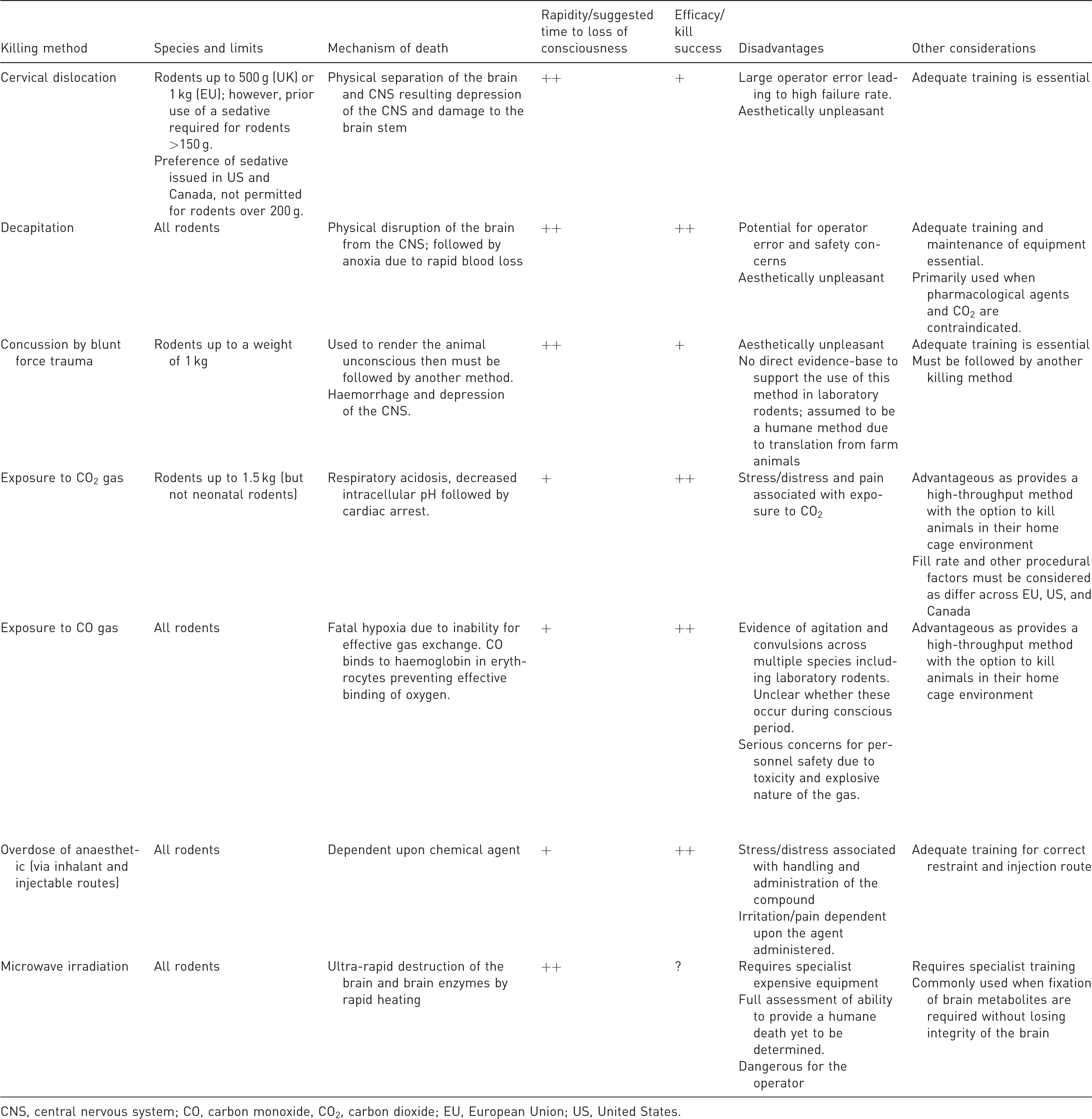
CNS, central nervous system; CO, carbon monoxide, CO2, carbon dioxide; EU, European Union; US, United States.
Footnotes
Acknowledgements
The authors would like to thank Ross Muers for his constructive comments on the manuscript. We would also like to thank the Biotechnology and Biological Sciences Research Council (BBSRC) for funding the research project (BB/S007210/1). The Roslin Institute is funded by a BBSRC Institute Strategic Program Grant BB/P013759/1. For the purpose of open access, the author has applied a Creative Commons Attribution (CC BY) licence to any Author Accepted Manuscript version arising from this submission.
Author contributions
JMC drafted the manuscript with JEM and DEFM making comments and edits in both substance and style.
Data availability statement
Data sharing is not applicable to this article as no new data were created or analysed in this study.
Declaration of Conflicting Interests
The author(s) declared no potential conflicts of interest with respect to the research, authorship, and/or publication of this article.
Ethical Statement
This publication did not require ethical board approval because it did not contain new data from human or animal trials.
Funding
The author(s) received no financial support for the research, authorship, and/or publication of this article.