
Abstract
The use of sheep in experiments is widespread and is increasing worldwide, and so is the need to develop species-specific anaesthetic techniques to ensure animal safety. Previous studies have mentioned several protocols involving the administration of alpha-2 adrenergic agonists in sheep; however, assessment of the efficacy and safety of these infusion techniques is still relatively new. Thus, the aim of the present study is to assess the effectiveness of detomidine constant rate infusion (CRI) in sheep by measuring the cardiovascular and respiratory parameters, blood gas variables and sedation scores. Eight adult female Santa Inês sheep received 20 µg/kg of detomidine hydrochloride intravenously as a bolus loading dose, followed by an infusion rate of 60 µg/kg/h. The heart rates and respiratory rates changed continuously during the CRI period. No arrhythmias were observed. The reduction in arterial partial pressure of oxygen (PaO2) was not significant, but one animal showed signs of hypoxaemia (minimum PaO2 of 66.9 mmHg). The arterial partial pressure of carbon dioxide (PaCO2) increased, but the animals did not become hypercapnic. The bicarbonate (HCO3−), pH and base excess (BE) tended towards metabolic alkalosis. The cardiac output (CO), stroke volume (SV), cardiac index (CI) and ejection fraction (EF%) showed no significant changes. The fractional shortening (FS%) decreased slightly, starting at T45min. Sedation scores varied between 3 (0/10) after sedation and during recovery and 7 (0/10) during CRI. We concluded that administering detomidine at an infusion rate of 60 µg/kg/h in Santa Inês sheep is a simple technique that produces satisfactory sedation for minimally invasive procedures.
The sheep is a convenient and popular large animal model for biomedical research. The need to determine species-specific sedation protocols has increased significantly, as most drugs used in sheep are often selected based on their effectiveness in other species. Ideal protocols for chemical restraint and sedation should facilitate animal handling, systemic analgesia and rapid recovery. 1 The development of adequate sedative and analgesic protocols should focus on simplifying techniques, on causing no detrimental side-effects, and on minimizing general anaesthesia requirements for surgical procedures, thus reducing associated complications.2,3
Alpha-2 adrenergic agonists are potent sedative drugs that provide muscle relaxation and analgesia and are widely used in sheep.1–5 Although this class of drugs is widely used, dose-dependent cardiopulmonary effects, such as decreased cardiac output (CO), bradyarrhythmia, peripheral vasoconstriction, biphasic arterial blood pressure response, respiratory depression, hypoxaemia and acute pulmonary oedema, have been observed.6–11 Also, individual, breed or strain variability can influence the presence or absence of such effects. 7
Furthermore, ruminants are known to be more sensitive to the effects of alpha-2 adrenergic agonists, when compared with other species, such as horses.1,3 Detomidine is more alpha-2-specific than xylazine, which means that a lower dose is required to achieve a desirable sedation with fewer side-effects. 12 Administration routes for detomidine include intramuscular (IM) and intravenous (IV) as a bolus or constant rate infusion (CRI). 13 Cardiopulmonary effects have a faster onset in IV routes than IM routes, which tend to be more reliable.1,2,5,14
In other species, such as horses, the administration of IV detomidine as a bolus3,5,7,15 or CRI15–18 is well established, and has been proven to cause excellent sedation without significant cardiopulmonary depression and ataxia.3,16,19 For standing chemical restraint, infusion rates varying from 5 to 40 µg/kg/h have been proven to be effective for prolonged sedation, with the caveat that they do not provide sufficient analgesia for surgical procedures.16–18 Other studies have compared steady state infusion with IV bolus and concluded that the plasma concentration of detomidine is directly proportional to adequate sedation and cardiovascular responses. 15
In sheep, a xylazine infusion regimen provided an effective and predictable steady state analgesia without the variability observed with bolus administration. 8 In pregnant ewes and preterm foetal sheep, the infusion of dexmedetomidine produced satisfactory results for sedation and pain control, with minimal foetal response. 10 These studies did not evaluate the occurrence of hypoxaemia or other respiratory effects. However, the effects of detomidine CRI are yet to be reported. It is believed that this technique can also provide prolonged and good quality sedation with minimal side-effects. 15 Moreover, there is not enough information regarding the haemodynamic parameters, sedation scores or infusion doses for sheep. Thus, the present study assesses the effectiveness and safety of detomidine CRI in sheep through the evaluation of cardiovascular and respiratory parameters, blood gas variables and sedation scores.
Materials and methods
Animals
The experimental protocol was approved by the Ethics Committee for Animal Research of Goiás Federal University (UFG), Brazil. Eight clinically healthy adult female Santa Inês sheep (Fazenda Anicuns, Anapolis, Brazil), weighing 34.98 ± 6.32 kg (mean ± standard deviation [SD]), were used. The animals were kept indoors, fed with alfalfa hay and commercial food (Nutroeste Nutricao Animal, Goiania, Brazil) twice daily, and provided with water ad libitum.
Experimental preparation
The animals were housed in group pens in the research facility with enough time to acclimatize to the environment. Feed was withheld for 18 h and water for 4 h prior to the experimental procedures. For drug administration, a 16 G intravascular catheter (Angiocath; Becton-Dickinson Infusion Therapy Systems Inc, Sandy, UT, USA) was inserted into the left jugular vein. For invasive blood pressure assessment and blood gas analysis, a 22 G intravascular catheter (Angiocath) was inserted into the auricular artery and kept patent with a heparin lock.
Drug treatment
Detomidine hydrochloride 20 µg/kg (Dormiun V; Agener União–Saúde Animal, Sao Paulo, Brazil) was given intravenously as a bolus loading dose followed by an infusion rate of 60 µg/kg/h. The interval between the bolus dose and the onset of infusion was 10 min, after which the sedation scores were assessed. The sheep were placed in right lateral recumbency, and the infusion was administered using an automatic syringe pump (DigiPump SR8x; Digicare Animal Health, Boynton Beach, FL, USA). During the experimental anaesthetic procedure, the animals were spontaneously breathing room air, and 0.9% saline was administered at a maintenance rate of 5 mL/kg/h. The detomidine CRI period was 60 min.
Assessments
Heart rate and rhythm; respiratory rate; mean, systolic and diastolic blood pressures; and peripheral oxygen saturation (SpO2) were evaluated using a multi-parameter monitor (Dixtal DX2010; Dixtal Biomédica Indústria e Comércio Ltda, Manaus AM, Brazil). The arterial partial pressure of oxygen (PaO2), arterial partial pressure of carbon dioxide (PaCO2), bicarbonate (HCO3−), base excess (BE), and pH were evaluated using a blood gas and electrolyte analyser (Cobas b 121; Hoffmann-La Roche Ltd, Basel, Switzerland). Sedation was assessed by a single experienced observer, based on the Kästner et al. (2003) 20 study, which assigned scores from 0 to 10, where 0 represented standing, with alert and normal behaviour, and 10 represented lateral recumbency, with no movements.
In addition, the cardiovascular function qualitative analysis was performed using Doppler echocardiography in two-dimensional mode through the right parasternal window. The obtained images allowed the measurement of interventricular septal thickness in systole (IVSs) and diastole (IVSd); left ventricular diameter in systole (LVIDs) and diastole (LVIDd); and left ventricular free wall thickness in systole (LVFWs) and diastole (LVFWd); followed by calculation of left ventricular fractional shortening (FS%) and ejection fraction (EF%). The cardiac index (CI) was calculated based on CO, using the following formula: CO/body surface area (BSA), where BSA = weight0,667/10. To determine CO, images were obtained from the right ventricular outflow tract, in which the annulus diameter of the pulmonary valve was measured, and pulmonary artery flow was subsequently recorded to determine the heart rate. The images were obtained using an ultrasound machine (Logic-e ultrasound – BT12; GE Healthcare, Little Chalfont, UK).
All parameters were evaluated before the experimental procedure (TBASAL), and the baseline values were determined as the arithmetic means of three consecutive measurements. All parameters were then evaluated at the following times: at the start of detomidine CRI (10 min after loading the bolus) (T0), every 15 min during infusion (T15, T30, T45, T60) and 30 min after the end of CRI (TREC). At all evaluation times, the sheep were observed for indications of sedation, including increases in salivation and degree of ptosis, and changes in responsiveness to external non-painful stimuli, such as noise. All voluntary attempts to move were recorded.
Data analysis
The data were summarized and expressed as the mean ± SD. Statistical analyses were performed by SigmaPlot for Windows (version 11.0). The Shapiro–Wilk test was used to verify a normal data distribution. Parametric values were analysed by one-way repeated measures analysis of variance (ANOVA), followed by Tukey’s test. Friedman’s test was chosen for non-parametric data. Values of P < 0.05 were considered to be significant.
Results
The sedation scores (median and quartiles) recorded during detomidine CRI are shown in Figure 1. The mean values and SDs of clinical parameters evaluated during the experimental procedure are summarized in Table 1.
Sedation scores (median ± quartiles) in response to 20 µg/kg of intravenous detomidine bolus followed by a continuous infusion of 60 µg/kg/h. TBASAL: time of bolus injection; T0: time when infusion started; TREC: time at recovery. Clinical parameters in response to 20 µg/kg bolus of intravenous detomidine followed by continuous administration of 60 µg/kg/h in domestic sheep (n = 8). HR: heart rate; RR: respiratory rate; SpO2: peripheral oxygen saturation; SBP: systolic blood pressure; DBP: diastolic blood pressure; MAP: mean arterial pressure; T℃: temperature (degrees Celsius); CI: cardiac index; EF%: ejection fraction; FS%: fractional shortening; SV: stroke volume; PaO2: arterial partial pressure of oxygen; PaCO2: arterial partial pressure of carbon dioxide; HCO3−: bicarbonate; BE: base excess; TBASAL: time of bolus injection; T0: time when infusion started; TREC: time at recovery. Values are given as mean ± standard deviation (*P < 0.05).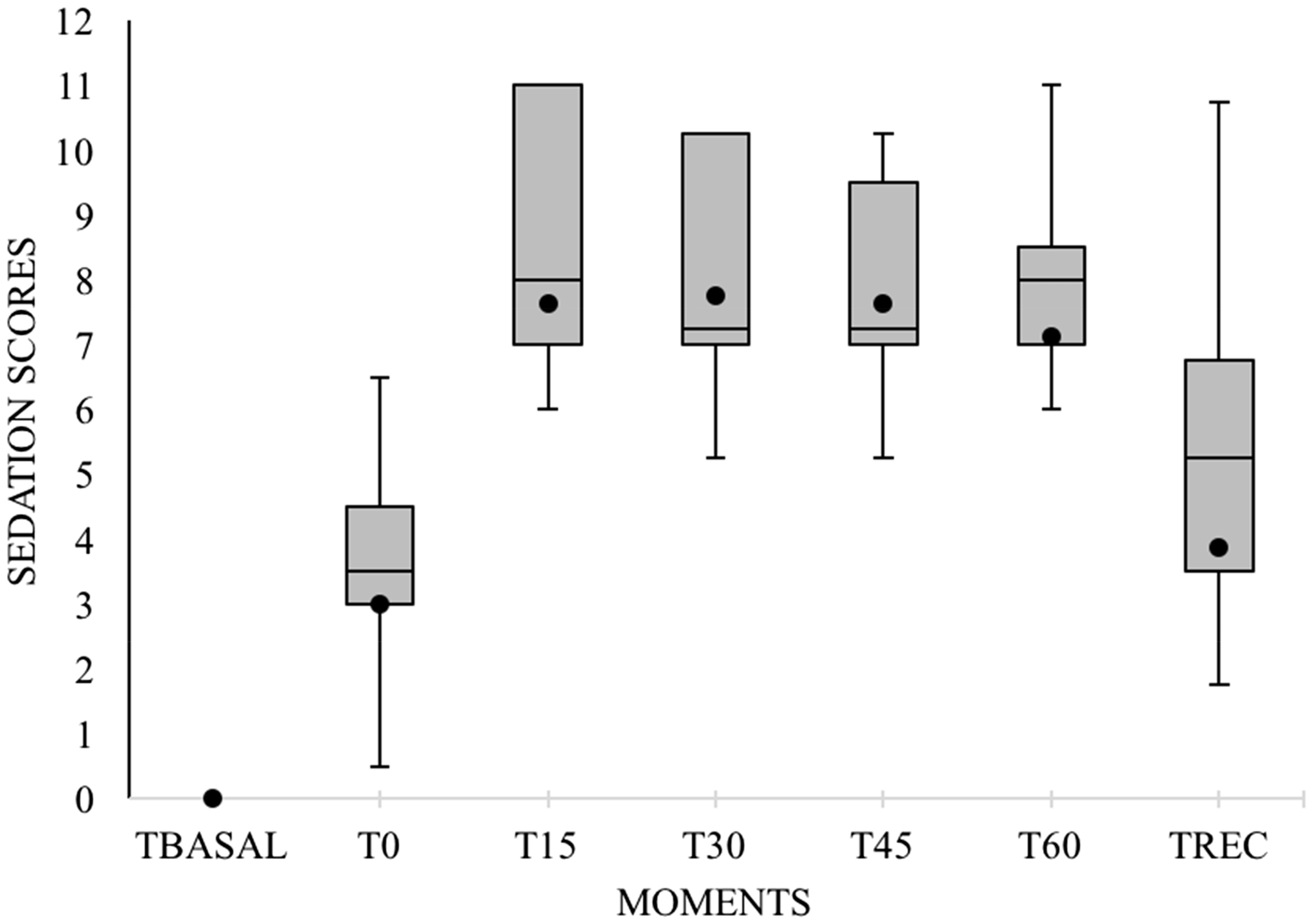
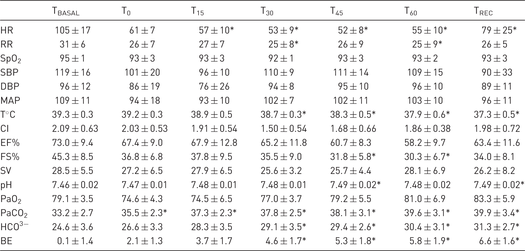
The heart rate decreased continuously, starting at T15, and the sheep became bradycardic (heart rate below 80 beats/min) 21 30 min after infusion started, but no arrhythmias were observed. The respiratory rate decreased at T30, but remained within normal reference values for the species. The values for both these variables returned to baseline at recovery (TREC). Blood pressure also remained within normal reference values during the 90 min evaluation period; neither hypertension (systolic, diastolic and mean blood pressure above 130, 90 and 110 mmHg, respectively) 22 nor hypotension (systolic, diastolic and mean blood pressure below 90, 60 and 70 mmHg, respectively) 22 was observed. The body temperature decreased gradually, and the lowest values were obtained after 45 min of infusion.
Results from the arterial blood gas analysis showed no significant changes in PaO2 mean values. One animal showed a significant decrease in PaO2 at T30, compared with baseline (TBASAL = 73.3 mmHg; T30 = 68.9 mmHg). Another animal remained significantly hypoxaemic (PaO2 below 80 mmHg) 23 during the whole infusion period (TBASAL = 81.7 mmHg; T0 = 69.6 mmHg; T60 = 67.4 mmHg). Both returned to values similar to baseline at T45 and TREC, respectively, while still in lateral recumbency. A significant increase in the PaCO2 mean values occurred gradually in all animals. The HCO3−, pH and BE values increased, starting at T30.
The cardiovascular function variables remained constant across the experimental period. Parameters such as CI, EF% and stroke volume (SV), were not altered significantly, compared with the baseline values. However, FS% showed a significant decrease at T45 and T60.
Regarding the sedation scores, the animals received a grade of 3 (0/10) at T0, and this increased to 7 (0/10) at T15. Throughout the infusion period (T30, T45, T60), the sedation scores remained constant (7/10). At TREC, during the anaesthetic recovery period, the animals’ average grade was 3, similar to T0.
Discussion
Despite previous reports of complications after the administration of alpha-2 adrenergic agonists in sheep, neither arrhythmias nor breathing pattern changes were observed in the present study. However, decreases in heart and respiratory rates were also expected, due to the inhibition of sympathetic tone caused by presynaptic norepinephrine reuptake. Bradycardia is a common side-effect, which was confirmed by our results.6,24 In previous studies, similar results for heart rates were reported with IV administration of detomidine 30 µg/kg in sheep7 and 10 µg/kg in cattle. 14 Contrary to our results, Celly et al. reported a significant increase in the respiratory rate after detomidine administration, starting 2 min after the bolus and lasting up to 30 min. In the present study, no assessments were conducted in the first 10 min after the loading dose; however, no significant changes were observed at T0 and T15, compared with baseline. At T30 and T60, the respiratory rate decreased significantly.
A biphasic arterial blood pressure response was also expected after the administration of alpha-2 adrenergic agonist.7,14,25,26 Due to the initial stimulation of alpha-1 adrenergic receptors, this blood pressure response is characterized by transient hypertension followed by hypotension 1 and is responsible for peripheral vasoconstriction. 6 No significant changes in the mean blood pressure values were observed during the experimental period. However, because evaluation began only 10 min after the detomidine loading dose, it is possible that the transient hypertensive peak was missed.
In the present study, no significant changes in PaO2 mean values were observed. On the other hand, a significant increase in PaCO2 was observed, probably due to a decrease in ventilation which led to a CO2 build-up. Alpha-2 adrenergic agonists have been reported to induce a variable degree of hypoxaemia in sheep after IV administration.7,9,11,20 So, despite the results obtained, it is worth considering individual, breed and strain variabilities. Two animals (2/8) showed significant reductions in PaO2 values, which confirm the effects of alpha-2 adrenergic agonists on oxygenation. Our results showed a reduced occurrence of hypoxaemia, but it is not possible to infer that these results would be similar under other conditions such as body position or breed selection. Thus, the establishment of oxygen therapy is strongly recommended, depending on the individual response. Hypoxaemia can be attributed either to hypoventilation or the presence of pulmonary oedema; however due to the limitations of our study, we could not determine why only two animals showed severe hypoxaemia (PaO2 below 70 mmHg). 23
The variables HCO3−, pH and BE increased gradually, and values obtained for pH and HCO3− in sheep were similar to those observed in horses in response to a detomidine infusion rate of 20 µg/kg/h. 15 Although the values also remained within the normal reference values for the species, there was a tendency toward metabolic alkalosis. Thus, based on blood gas analysis, it can be inferred that continuous detomidine administration may further lead to metabolic alkalosis as a compensation for respiratory acidosis. 7
Cardiovascular function parameters remained constant across the experimental period; however, FS% values had decreased at two of the evaluation stages (T45 and T60). A decrease in FS% alone does not directly interfere with left ventricular systolic function as it can be influenced by preload and afterload, which were not evaluated in this study. Furthermore, the standard parameter for systolic function evaluation is EF%, which remained constant. 27 Alpha-2 adrenergic agonists are expected to decrease the CI values,24,28,29 mainly due to a decreased heart rate.24,28 Although a significant decrease in heart rate took place, there were no changes in CI values, unlike the results of previous studies.24,30 The present results were obtained by measuring CO using echocardiography; and although reliable values were obtained from aortic and pulmonary arterial flow in other species,27,31,32 these values have been proven to be highly correlated with the thermodilution technique, 33 and high variability had previously been observed in sheep. 28
In this study sedation scores varying between 3 (0/10) after IV detomidine bolus and 7 (0/10) during CRI were considered to be satisfactory for performing simple procedures that were minimally invasive and not painful. During CRI, the sheep remained in lateral recumbency and were unresponsive to noise, however after the touching stimuli, they assumed sternal recumbency but were still unable to support their heads. External stimuli were not painful and consisted of encouraging the sheep to change position by touching them. Similar depths of sedation were obtained after IV administration of 50 µg/kg detomidine in goats 2 and 40 µg/kg in sheep. However, the sedation patterns were more variable, achieving optimal sedation periods with subsequent decreases, unlike those observed with CRI. 5 In addition, the animals showed prolonged recovery times, between 60 5 and 120 min, 2 which were also different from the values obtained in the present study. The observed recovery can be explained by the previously proven correlation between sedation and drug plasma concentration. 20
We can conclude that detomidine infusion at a rate of 60 µg/kg/h in Santa Inês sheep is a simple technique that can produce satisfactory sedation to facilitate handling and be used for minimally invasive procedures. The sedation produces no significant cardiorespiratory effects and provides rapid and high quality recovery. Despite these promising results, it is worth considering individual variability, and it is also worth evaluating the need to establish oxygen therapy in the event of hypoxaemia.
Footnotes
Acknowledgements
We thank Programa de Pós-Graduação em Ciência Animal from the Federal University of Goiás (UFG) for the award of a Master’s scholarship grant, and the Veterinary Teaching Hospital where the experiment was conducted.
Declaration of Conflicting Interests
The author(s) declared no potential conflicts of interest with respect to the research, authorship, and/or publication of this article.
Funding
The author(s) disclosed receipt of the following financial support for the research, authorship, and/or publication of this article: A Master’s scholarship grant was awarded by the Programa de Pós-Graduação em Ciência Animal from the Federal University of Goiás (UFG).