
Abstract
We used immunofluorescence staining (monoclonal antibody N45.1) with cytological imprinting to study changes in the intranuclear distribution of 8-hydroxy-2'-deoxyguanosine in renal cells of male Wistar rats after oxidative stress by ferric nitrilotriacetate. In the control proximal tubule cells, small spherical signals were uniformly distributed throughout the nuclei. Under oxidative stress, immunofluorescence intensity was increased, especially near nuclear membrane. In cells with nuclear shrinkage or deformity, intense, diffuse signals throughout the nuclei were observed. Our results suggest that specific nuclear sites are vulnerable to oxidative DNA damage and that diffuse intense signals precede cell death after oxidative stress.
Keywords
8-H
This study was undertaken to examine the intranuclear distribution of 8-OHdG and to confirm the specificity of the antibody. To this end, a prototype free radical-induced renal tubule injury model mediated by Fe-NTA (Toyokuni et al. 1994,1997; Toyokuni 1996) was used. Cytological imprinting was applied to normal testis and to normal or oxidatively stressed kidney 3 hr after IP administration of 15 mg Fe/kg Fe-NTA to 5-week-old male Wistar rats (Shizuoka Laboratory Animal Center; Shizuoka, Japan). Renal nuclear levels of 8-OHdG were 1.01 ± 0.18 and 2.60 ± 0.28 (per 105 dG; means ± SE; n =3; unpaired t-test, p<0.01), respectively, by the HPLC and ECD method (Toyokuni et al. 1997). The cells imprinted on silane-coated glass slides were fixed with 95% ethanol, then exposed to microwave radiation in 10 mM citrate buffer (pH 6.0) for antigen retrieval, subjected to the avidin-biotin complex method (10 μg/ml MAb N45.1; streptoavidin-FITC; nuclear counterstaining by propidium iodide), and observed with a confocal laser scanning microscopy at optical slices of 1 μm (Fluoview; Olympus). Addition of 100 μM 8-OHdG (Wako; Osaka, Japan) in the primary antibody reaction completely eliminated the immunofluorescent signal.
During prophase or prometaphase, normal spermatogonia showed 8-OHdG signals solely on the chromosomes, confirming the specificity of MAb N45.1 (Figure 1D). Because the major targets in the kidney for oxidative stress by Fe-NTA are proximal tubule (PT) cells (Toyokuni et al. 1994), we concentrated on those cells at interphase. By Papanicolaou staining, nuclei of the control PT cells presented fine chromatin with either no or one nucleolus (Figure 1A), whereas more than half of the nuclei of PT cells after Fe-NTA administration showed chromatin aggregation, nuclear shrinkage (Figure 1B), and sometimes even vacuoles and deformity (Figure 1C). Unexpectedly, nuclei of PT cells in the untreated normal rats showed small, uniformly dispersed spherical patterns of immunofluorescence (Figure 1E). Some of the signals were extremely large, but their association with nuclear function and structure requires further investigation. Under oxidative stress, the spherical signals became more intense and larger, especially near the nuclear membrane in the PT cells, which showed little morphological change from controls (Figure 1F), and intense diffuse signals eventually covered the entire nucleus in the PT cells showing nuclear deformity and which were expected to become necrotized (Figure 1G). In the latter cells, DNA integrity and chromatin structure may have been destroyed. Our results suggest that there are specific vulnerable sites for oxidative damage in the nuclei and that intense diffuse nuclear 8-OHdG signals identify cells destined for death. This method may be useful for evaluation of oxidative DNA damage in a number of cytological preparations.
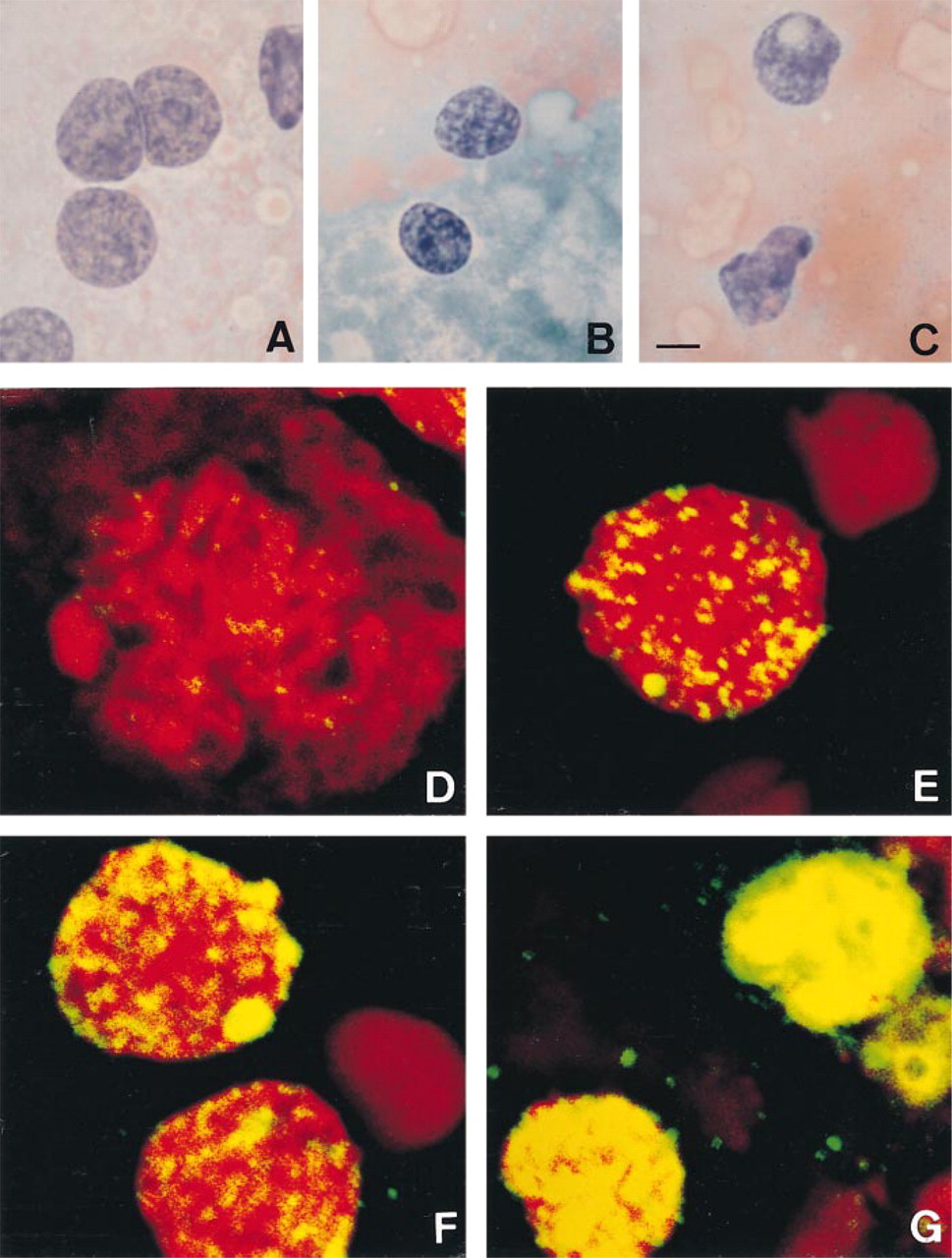
Cytology (A-C) and immunocytochemistry (D-G; 8-hydroxy-2'-deoxyguanosine) of touch preparations from rat kidney (A-C,E-G) and testis (D). (A,E) Control renal proximal tubule cells; (B,C,F,G) renal proximal tubule cells after oxidative stress by ferric nitrilotriacetate; (D) control spermatogonium at prophase. See text for details. Bar in C: A-C = 5 μm; D-G = 2.5 μm.
Footnotes
Acknowledgments
Supported in part by a Grant-in-Aid for scientific research from the Ministry of Education, Science, Sports and Culture, Japan, by a grant from the Japanese Owners' Association, and by the Program for Promotion of Basic Research Activities for Innovative Biosciences.
We thank James E. Strickland (NCI, NIH; Bethesda, MD) for critical reading of the manuscript.