
Abstract
We developed a sensitive chemiluminescence in situ hybridization assay for detection of human papillomavirus (HPV) DNA for objective and semiquantitative evaluation of the results. The hybridization reaction was performed using either digoxigenin-, biotin-, or fluorescein-labeled probes, visualized with alkaline phosphatase as the revealing enzyme and a highly sensitive 1,2 dioxetane phosphate as chemiluminescent substrate. The light emitted from the hybridized probes was detected, analyzed, and measured using a high-performance, low light-level imaging luminograph connected to an optical microscope and to a personal computer for quantification of the photon fluxes and for image analysis. The system operated in consecutive steps: First, hybridized specimens were recorded in transmitted light. Then the net luminescent signal was recorded, and then an overlay of the two images provided by the transmitted light and by the luminescent signal allowed the spatial distribution of the target DNA to be localized, measured, and evaluated. Biopsy specimens from different pathological conditions associated with HPV, which had previously been proved positive for HPV DNA with the polymerase chain reaction (PCR), were analysed. The chemiluminescence in situ hybridization proved sensitive and specific with digoxigenin-, biotin-, or fluorescein-labeled probes, and provided an objective evaluation of the results. The results obtained with chemiluminescence in situ hybridization were also compared with results obtained with in situ hybridization with colorimetric detection, with good concordance of the data. Chemiluminescence in situ hybridization therefore offers the possibility of detecting HPV DNA with great sensitivity in biopsy specimens. Moreover, the images of the samples, stored in the computer, are a permanent record of the reaction and can also be sent for evaluation or comparison to other laboratories using computer networks.
H
Recently, chemiluminescent substrates have been proposed as a more sensitive alternative to colorimetric substrates in various analytical techniques in which small amounts of analytes or enzymes have to be detected (Martin et al. 1995; Lorimier et al. 1993; Zerbini et al. 1993; Holtze et al. 1992; Musiani et al. 1991a,b). Because the signal intensity is proportional to enzyme activity or concentration, precise and accurate quantitative analysis can be achieved. Moreover, using newly synthesized dioxetane derivates as chemiluminescent substrates for alkaline phosphatase, we detected as low as 10 fg of viral DNA in dot-blot hybridization format, with the chemiluminescent substrates proving about 10- to 50-fold more sensitive than the colorimetric ones (Girotti et al. 1995). Continuing improvements in chemiluminescent substrate efficiency have been matched by new developments in photon imaging instrumentation, such as high performance luminographs based on a CCD videocamera or a high dynamic range pick-up tube (Saticon) combined with a video amplifier (Girotti et al. 1996; Roda et al. 1996). These instruments not only allow quantification of emitted light at the single photon level but also permit localization of the chemiluminescent emission on a target surface. Moreover, the connection of the luminograph to an optical microscope localizes the light emission inside tissues or cells. All these data prompted us to develop a sensitive chemiluminescence in situ hybridization assay for detection of HPV DNA in biopsy specimens that could give objective and semiquantitative results. In our study we explored the use of highly sensitive chemiluminescent substrates and a high-performance luminograph connected to a light microscope for in situ detection of HPV DNA, using differently labeled probes visualized with alkaline phosphatase as the revealing enzyme.
Materials and Methods
Samples
Paraffin-embedded biopsy specimens from one sample of vulvar condyloma acuminatum, one sample of anal condyloma acuminatum, two samples of cutaneous Bowen's disease, one sample of a plantar common wart, and one sample from a squamous cell carcinoma, which had been previously diagnosed as positive for HPV by PCR (Tosti et al. 1994; Manos et al. 1989), were analyzed in the study.
As negative controls, paraffin-embedded biopsy specimens from one sample of normal skin, two samples of normal squamous ectocervical epithelium, and two samples of laryngeal lesions, which had been previously proved negative for HPV by PCR and by in situ hybridization with colorimetric detection, were also analyzed.
Five μm-thick sections were cut from paraffin-embedded tissue blocks and were placed on silanated slides. Paraffin-embedded tissue sections were dewaxed by two 5-min incubations in xylene and then washed in absolute ethanol for 5 min (Musiani et al. 1990; Gentilomi et al. 1993).
The CaSki cervical carcinoma cell line containing 500-600 integrated copies of the HPV16 DNA sequence in each cell was used as a positive control. HeLa cells, which are known to contain as few as 10-50 integrated copies of HPV 18 DNA in each cell, were used for assessment of the sensitivity of the assay. Cells smeared on silanated glass slides were fixed with 4% paraformaldehyde in PBS for 30 min (Gentilomi et al. 1992). After fixation, cells were washed three times in PBS for 10 min each and then dehydrated with ethanol washes (95% and 100%) for 2 min each. Biopsy specimens and cells were then air-dried and stored at 4C until use.
Papillomavirus Probes
Three different commercial omniprobes for HPV detection were employed; (a) a fluorescein-labeled cloned probe detected by rabbit anti-fluorescein Fab fragments conjugated with alkaline phosphatase (Dako; Glostrup, Denmark); (b) a digoxigenin-labeled cloned probe detected by anti-digoxigenin Fab fragments conjugated with alkaline phosphatase (Kreatech; Amsterdam, The Netherlands); and (c) a biotinylated DNA cloned probe detected by streptavidin conjugated with alkaline phosphatase (Biohit; Helsinki, Finland).
In Situ Hybridization Reaction
Biopsy specimens were incubated for 20 min with 500 μg/ml of proteinase K in prewarmed (37C) PBS. Cell smears were digested with 1 μg/ml of proteinase K in prewarmed PBS. Biopsy specimens and cell smears were dehydrated in 95% ethanol for 1 min, followed by absolute ethanol for 1 min, and then air-dried.
Samples and controls were hybridized separately with the three HPV omniprobes prediluted in their own hybridization solution (ready to use). In brief, 20 μl of each hybridization mixture containing the labeled probe was added to the slides. Each slide was then covered with a glass coverslip and the edges were sealed with nailpolish to prevent loss of the mixture during denaturation and hybridization. Probe and target DNA were denatured simultaneously by placing the slides for 6 min on a prewarmed (95C) heating block. The hybridization was then performed by incubating the samples at 37C overnight in a humidified chamber. After hybridization, the coverslips were carefully removed and the slides were washed under appropriately stringent conditions: (a) The slides hybridized with the fluorescein-labeled probe were washed twice for 5 min at room temperature (RT) in Trisbuffered saline (TBS), once for 30 min at 48C in stringent wash solution (1.5 mM sodium citrate, 15 mM NaCl, 2 mM MgCl2, 0.1% Triton X-100, 0.05% BSA, 0.3 mM NaN3), and once for 2 min at RT in TBS. (b) The slides hybridized with the digoxigenin-labeled probe were washed three times for 5 min at 37C in washing buffer (50% formamide in 2 X SSC. (c) The slides hybridized with biotinylated DNA probe were washed once for 3 min at RT in 2 X SSC, twice for 5 min at 37C in 2 X SSC, and once for 3 min at RT.
Probe Detection
Each hybridized labeled probe was detected with the method recommended by the manufacturer, as follows: (a) The samples hybridized with fluorescein-labeled probe were incubated for 30 min at 37C with alkaline phosphatase-conjugated Fab fragments to fluorescein isothiocyanate (Dako). (b) The samples hybridized with digoxigen-labeled probe were incubated for 30 min at 37C with alkaline phosphatase-conjugated anti-digoxigenin Fab fragments (Kreatech). (c) The slides hybridized with biotinylated DNA probe were incubated for 30 min at 37C with alkaline phosphatase conjugated to streptavidin (Biohit).
After incubation, samples were washed twice for 3 min in TBS and once for 2 min in distilled water.
Chemiluminescent Detection
Chemiluminescent detection of alkaline phosphatase was performed treating the samples with undiluted adamantil-1,2-dioxetane phenyl phosphate substrate (CDP Star) (Tropix; Bedford MA). After an optimized incubation of 30 min at RT the solution was removed and the luminescent signal from the hybrid formation was detected and analyzed with a system consisting of the Luminograph LB-980 (EG&G Berthold; Bad Wilbad, Germany), which is a high-performance, low light level imaging apparatus, with a high dynamic range pickup tube (Saticon) combined with a video amplifier, connected to a model BH-2 light microscope (Olympus Optical; Tokyo, Japan), and to a personal computer for image analysis. The microscope was enclosed in a dark box to prevent contact with external light. The system operated in consecutive steps. First, biopsy specimens and cell smears were recorded in transmitted light. Then the luminescent signal from the hybrid formation was measured. After computer processing of the luminescent signal with pseudo-colors corresponding to the light intensity, an overlay of the two images on the screen provided by the transmitted light and by the luminescent signal allowed the spatial distribution of the target analyte to be localized and evaluated.
Evaluation of Chemiluminescent Signal
Digital images of the light emission from samples were optimized at 22-sec intervals integration time for 1 min total accumulation time. The light emission from each cell was quantified by defining a fixed area and summing the total number of photon fluxes from within this area.
Control negative biopsy specimens provided the threshold background levels. The optimized calculation of threshold values was performed by analyzing 10 microscopic fields from negative reference biopsies and calculating the average values (±SD) of the background light emission (expressed as photons/sec/area). The average value of the background signal plus fivefold its SD was considered the threshold value above which the chemiluminescent signal resulting from the hybridized HPV DNA could be considered as positive. The net light signal of the sample was obtained automatically on the screen after subtraction of the threshold values using appropriate software. Corrections for instrumental background and flat field variations were automatically performed by the LB-980 apparatus.
Colorimetric Detection
After two washes for 3 min each in TBS and one wash for 2 min in distilled water, the samples were incubated with alkaline phosphatase substrate solution [45 μl of Solution A (75 mg/ml of nitroblue tetrazolium salt in 70% dimethyl formamide) and 35 μl of Solution B (75 mg/ml of 5-bromo-4-chloro, 3-indolyl phosphate, toluidine salt in dimethyl formamide) plus 920 μl of substrate buffer (100 mM Tris-HC1, 100 mM NaCl, 50 mM MgCl2, pH 9.5)] for 15-30 min.
The development of a dark-blue precipitate at the enzyme site in positive cells was monitored by microscopic examination.
Results
In the analysis of the six biopsy specimens from different HPV-associated diseases that had previously proved positive for HPV DNA by PCR, the chemiluminescent in situ hybridization gave clearly positive signals without any background (Figure 1), using either fluorescein-labeled, digoxigenin-labeled, or biotinylated probes in all the samples. In one sample (cutaneous Bowen's disease), the chemiluminescence in situ hybridization clearly documented the fact that the viral HPV genome can persist at low copy number in undifferentiated basal stem cells of the epithelium and at higher copy number in more differentiated superficial cells, giving a semiquantitative result of the reaction. In fact, increasing values in light emission from basal to superficial cell layers were noticed after chemiluminescence hybridization, and the increase of emitted signal, proportional to the quantity of HPV genomes in the single cells, was more evident after the computer processing of the signals with pseudo-colors (Figure 2).
As negative controls, five biopsy specimens that had previously proved negative for HPV DNA by PCR and by in situ hybridization with colorimetric detection proved completely negative by in situ chemiluminescence hybridization with the three different labeled probes.
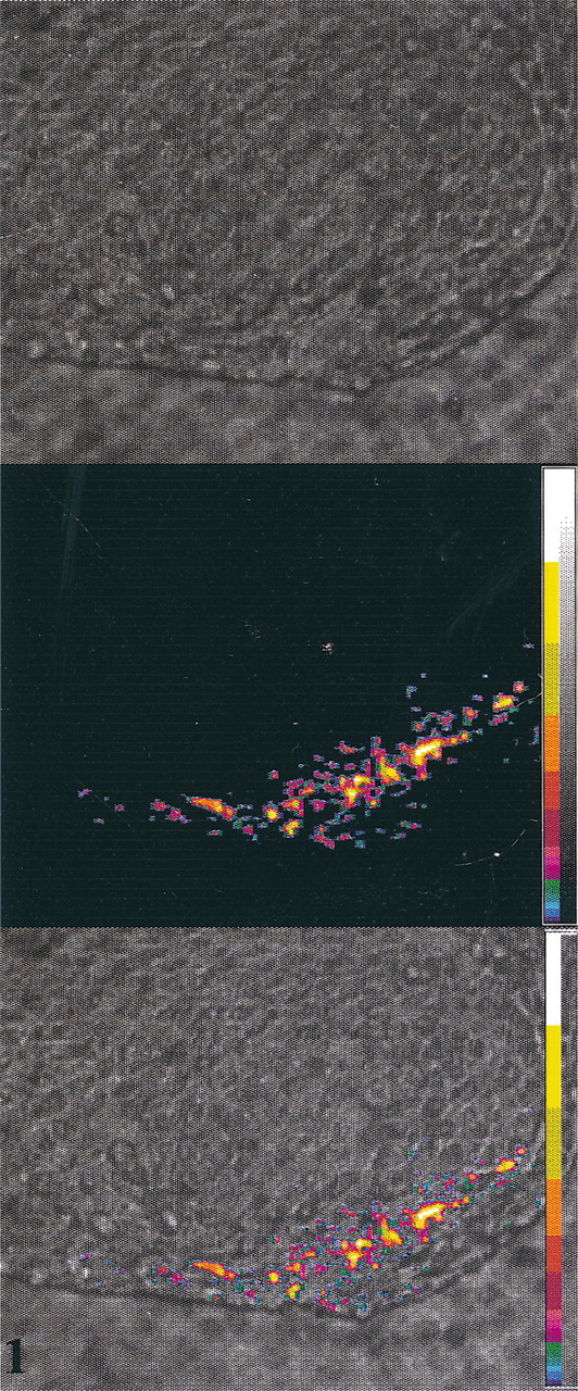
Chemiluminescence in situ hybridization using digoxigenin labeled probe in a sample of vulvar condiloma acuminatum. From top to bottom: live image; luminescent signal processed with pseudo-colors; overlay of the live image and luminescent image.
To assess the reproducibility of the chemiluminescence assay, positive and negative samples were analyzed in triplicate in different runs, and the results were concordant with the expected data.
The specificity of the chemiluminescent signal was also confirmed by the following control experiments. (a) HPV-positive CaSki cells were used as positive controls for each run, and they proved positive by chemiluminescence in situ hybridization with the three different HPV probes. (b) No chemiluminescent signal was observed when HPV-positive biopsy specimens were treated with the HPV-labeled probes and with the chemiluminescent substrate, omitting the incubation with alkaline phosphatase-conjugated detector systems. (c) No luminescent signal was detectable when HPV-positive specimens were hybridized with plasmid DNA probes (fluorescein-labeled, digoxigenin-labeled, and biotinylated) followed by treatment with detector systems conjugated with alkaline phosphatase and with the chemiluminescent substrate. (d) Reference HPV-negative skin specimens proved completely negative after hybridization with the HPV-labeled probes and treatment with detector systems conjugated with alkaline phosphatase, followed by the chemiluminescent substrate.
The data obtained with chemiluminescence in situ hybridization were also compared with data obtained with in situ hybridization and colorimetric detection, performed in the same run, with the same batch of probes and using consecutively cut tissue sections. Of the six positive samples examined, five samples proved positive at colorimetric detection with all three different labeled probes. The sample of vulvar condyloma acuminatum, on the other hand, proved negative with the fluorescein-labeled and biotinylated probe, whereas with the digoxigenin-labeled probe only a few cells with a weak dark-blue product were noticed. The control samples that had proved negative with chemiluminescence in situ hybridization were confirmed negative with in situ hybridization and colorimetric detection.
To compare the two methods and to assess the reproducibility of results, we performed hybridization using digoxigenin-labeled probes on CaSki cells (containing 400-600 copies of HPV 16 DNA), and on HeLa cells (containing 10-50 copies of HPV 18 DNA), and we compared the results with chemiluminescent and colorimetric detection. The mean values of emitted photons/sec/cell after analysis of 100 CaSki cells and 100 HeLa cells were 8.44 × 10² and 1.15 × 10², respectively. Single values were considered after subtraction of the threshold values obtained after the analysis of 100 CaSki cells and 100 HeLa cells previously hybridized with the negative control probe pACYC 184 DNA. Colorimetric detection was positive for CaSki cells but not for HeLa cells. CaSki and HeLa cells were analyzed in triplicate, in different runs, and on different days, and the results were consistent with previous data.
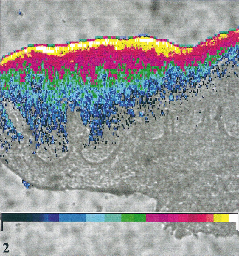
Chemiluminescence in situ hybridization revealing increasing concentrations of HPV DNA from basal to superficial epithelial cell layers in a sample of cutaneous Bowen's disease.
Discussion
In recent years there has been a growing interest in the application of in situ hybridization for the diagnosis of viral diseases, especially for those viruses, such as papillomaviruses, that cannot be diagnosed by isolation procedures.
In this study we developed a chemiluminescence in situ hybridization assay for the search of HPV DNA, which combined the sensitivity of alkaline phosphatase chemiluminescent substrates and the spatial morphological resolution of in situ hybridization, using a high-performance, low light level imaging luminograph, connected to a light microscope and to a personal computer for quantitative image analysis. In our assay we used digoxigenin-labeled, fluorescein-labeled and biotinylated probes, and sensitive results were achieved with all the probes tested. The use of alkaline phosphatase as the detectable enzyme avoided many of the disadvantages of horseradish peroxidase, such as interference from endogenous peroxidase activity in cells and the need to use cell pretreatment to inhibit the enzyme. The chemiluminescent detection of the alkaline phosphatase was performed using the 1,2 dioxetane substrate CDP-star, which represents one of the most sensitive detection systems, being able to reveal as few as 1.6 zeptomoles of the enzyme. Moreover, CDP-star has glowing kinetics with a steady-state emission, which permits easier handling and analysis of the samples (Beck and Koster 1990). Our chemiluminescent in situ hybridization assay used a very sensitive chemiluminescent substrate and signal acquisition was performed after removal of the liquid film of the substrate over the section. This permitted a sensitive, sharp topographic localization of the signal, because mobility of the luminescent product within the liquid film was avoided and the path and scatter of the photons were as restricted as possible. Moreover the use of an image superposition function avoided the problems that may arise from the sequential analysis of the tissue structure and the chemiluminescent signal, which are obtained on the screen in turn (Lorimier et al. 1993).
A sharp topographic distribution of the probe within the cell was achieved because the LB-980 instrumentation with the conventional setup has a spatial resolution of 240 μm, i.e., one pixel corresponds to this value, but once connected to the microscope and using the X40 and X100 lenses, the pixel size is reduced to 1.1 μm and 0.41 μm, respectively (Roda et al. 1996). The threshold values for cell samples were optimized considering the average value of the background signal plus fivefold its SD, because in the chemiluminescent reaction revealed by the microscope-luminograph assembly, the background is relatively high but with a low fluctuation (Bernroider 1994). With our assay, because the positive signal was considered above threshold values, an objective evaluation of the results could be achieved without any training at the microscope to read the slides and so doubts about positive or negative results were minimized. Chemiluminescence in situ hybridization offers a permanent record of the reactions, as all the images of the samples are stored in the computer and these images can be printed or sent for an evaluation in other laboratories, using floppy disks or other computer networks.
With the chemiluminescence hybridization assay, the use of the luminograph gave a semiquantitative analysis of the presence of HPV DNA in infected cells. Results on smears of CaSki and HeLa cells demonstrated that the luminescent signal changed in proportion to the known numbers of copies per cell. Because of differences in fixation, the sensitivity of the assay on formalin-fixed material cannot be inferred from these results, but the cell line results do demonstrate signal changes in response to copy number and the reproducibility of signal in repeat assays. Because the sensitivity and objectivity of the chemiluminescent detection, sample evaluation can be facilitated, especially in the routine use of in situ hybridization with colorimetric detection for diagnosis of HPV infections. In fact, it has been shown that for positive results there is a wide variation in the percentage of labeled cells detected, owing to several factors that include the type of viral infection (productive or non-productive) and the number of copies of viral genome and its physical state (episomic or integrated) in each cell. Chemiluminescence in situ hybridization revealing the distribution and level of amplification of HPV genetic information may also prove promising for study of the molecular aspects of HPV infection, with wide applications for both diagnostic and research purposes.