
Abstract
This study targeted the development of a novel microarray tool to allow rapid determination of the expression levels of 58 different tyrosine kinase (tk) genes in small tumor samples. The goals were to define a reference probe for multi-sample comparison and to investigate the variability and reproducibility of the image acquisition and RT-PCR procedures. The small number of tk genes on our arrays enabled us to define a reference probe by artificially mixing all genes on the arrays. Such a probe provided contrast reference for comparative hybridization of control and sample DNA and enabled cross-comparison of more than two samples against one another. Comparison of signals generated from multiple scanning eliminated the concern of photo bleaching and scanner intrinsic noise. Tests performed with breast, thyroid, and prostate cancer samples yielded distinctive patterns and suggest the feasibility of our approach. Repeated experiments indicated reproducibility of such arrays. Up- or downregulated genes identified by this rapid screening are now being investigated with techniques such as in situ hybridization. (J Histochem Cytochem 49:1057–1058, 2001)
O
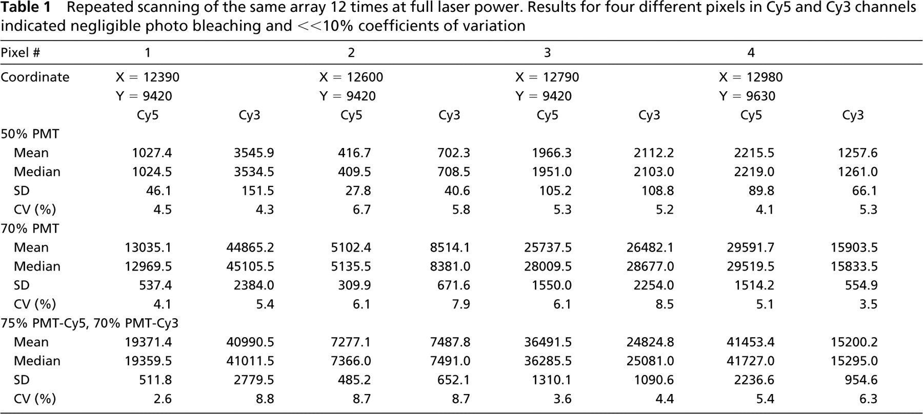
To test the extent of the scanner's intrinsic noise, slides were scanned 12 times repeatedly at full laser power. Four separate pixels belonging to four intensity groups were selected and their mean, median, SD, and CV were determined (Table 1). Results indicated an average of 5.0% and 6.0% CV for Cy5 and Cy3 channels, respectively. Noises were negligible and did not correlate with PMT voltages; the deviations of measured intensities were within experimental errors (≪10%). Repeated scanning up to 24 times did not decrease the fluorescence intensity, eliminating the concern of photo bleaching.
Repeated scanning of the same array 12 times at full laser power. Results for four different pixels in Cy5 and Cy3 channels indicated negligible photo bleaching and ≪10% coefficients of variation
Two properties regarding the RT-PCR amplification were examined: Do the amplified probes represent the true abundance of each transcript in cells? Does the probe saturate the arrays? First, we mixed five tk genes in three ratios (1, 20, and 400) and used them as PCR templates. Hybridizations of such PCR product to a small five-tk gene array showed a “compressed” ratio of three- to fourfold despite a 400-fold input template ratio. Nevertheless, the relative order of abundance of the five tk templates was conserved. Saturation has likely occurred in PCR, which may have also depleted some tk primers due to their high degeneracy. Second, we compared single-probe hybridizations at various concentrations. Four to six hundred ng of PCR amplified products typically were used for random prime labeling. The labeled probe was purified and different dilutions were applied to the arrays. It was determined that one tenth of the labeled probe per channel provided optimal signals for analysis without saturating the arrays.
Repeated hybridizations have identified distinctive patterns among thyroid, breast, and prostate cancer cell lines (Figure 1). Although the panel of 58 tk genes may not represent all the tk expressed in any single cancer tissue, we have reason to believe they represent over 50% of human tk genes expressed in a particular tissue type (Robinson et al. 1996). These distinctive expression patterns, as unveiled by microarray, become molecular fingerprints of tk gene expression for each tumor.
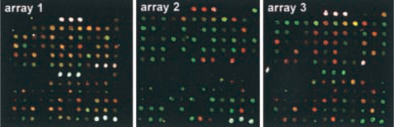
Multiple hybridizations confirmed distinctive patterns for three tumor cell lines. (
Footnotes
Acknowledgments
Supported by a grant from the Director, Office of Science, Office of Biological and Environmental Research of the US Department of Energy under Contract DE-AC03–76SF00098, by a fellowship from the Cancer Research Foundation of America (to HBH), and by grants from the Breast Cancer Research Programs, US Army Medical Research and Material Command, US Department of Defense (BC98–0937, DC991395). All experiments involving human cells or cell lines were approved by the UC Berkeley and the UCSF Human Subject Use Institutional Review Board.