
Abstract
The in situ end-labeling (ISEL) method demonstrates DNA fragmentation, commonly regarded as a marker of apoptosis. We investigated by the ISEL procedure a series of 52 thyroid lesions, including 24 lesions of mitochondrion-rich oxyphilic cells, both benign and malignant, and 28 non-oxyphilic control tumors. A high percentage of nuclear ISEL staining (approximating to 100% in most cases) was observed in the vast majority of oxyphilic cells from both adenomas and carcinomas, in the absence of morphological apoptotic changes and with no immunocytochemical evidence of caspase activation. This pattern of DNA fragmentation was not observed in non-oxyphilic lesions and was confirmed in total extracted DNA. Moreover, a peculiar cytoplasmic staining was also observed in oxyphilic cells from both benign and malignant lesions, probably related to abnormal fragmentation of mitochondrial DNA. Similar staining patterns were detected in oxyphilic cell tumors of other organs (parathyroids, salivary glands, and kidneys). These findings are consistent with an extensive DNA fragmentation peculiar to oxyphilic cells, which is not directly related to apoptosis and whose origin and biological significance are presently unknown. (J Histochem Cytochem 49:1003–1011, 2001)
O
In thyroid pathology, the issue of OC lesions is controversial. Some authors and also the WHO classification (Hedinger et al. 1988) categorize OC (Hurthle cell) tumors within the spectrum of follicular tumors on the basis of their architectural features rather than on the presence or degree of oxyphilic transformation. Other authors believe that OC tumors have to be kept separate for histopathological, behavioral, and possibly molecular and etiopathogenetic features (Watson et al. 1984; Carcangiu et al. 1991; Rosai et al. 1992).
Great attention has been paid in recent years to combining morphological and genetic characteristics in thyroid tumors, and molecular studies shed light on the role of various oncogenes in different subsets of thyroid tumors (Fusco et al. 1987; Suarez et al. 1990). Molecular analysis has also shed some light on the origin and significance of oxyphilic cells and related tumors (Canzian et al. 1998). A peculiar loss of genetic material in 10q in oxyphilic thyroid tumors and renal oncocytomas (Tallini et al. 1994), as well as monosomy of chromosome 2 in a subset of Hurthle cell tumors (Tallini et al. 1999), has been described. More interestingly, several molecular defects in mitochondrial DNA or alterations in mitochondrial metabolism have been detected in oxyphilic cells and related tumors, including overexpression of regulatory proteins (Faure–Vigny et al. 1996), mutations and/or deletions of mitochondrial DNA, and cytochrome C oxidase deficiency (Muller–Hocker 1992; Maximo et al. 1998; Muller–Hocker et al. 1998).
Studies focused on apoptosis and on the expression of apoptosis-related proteins in thyroid neoplasms reported a low apoptotic index (AI) in all tumor categories thus far investigated (Branet et al. 1996; Brocker et al. 1996; Basolo et al. 1997; Kikuchi et al. 1997; Moore et al. 1998; Soda et al. 1999; Yoshida et al. 1999; Sreelekha et al. 2000). Moreover, an inversely proportional alteration between p53 and bcl-2 expression was found to characterize poorly and undifferentiated carcinomas. To date, no large study on OC thyroid tumors has been reported. Recently, a morphological investigation on apoptosis-related changes failed to demonstrate significant differences between benign and malignant OC thyroid tumors (Lazzi et al. 1999). Interestingly, Muller–Hocker (1999) recently reported the overexpression of p53 in a high percentage of both OC adenomas and carcinomas of the thyroid, which was inversely related to bcl-2 immunostaining. The same expression pattern was previously reported in a series of poorly differentiated oxyphilic carcinomas (Papotti et al. 1996). In addition, in OC tumors we detected a peculiar pattern of overexpression of the E2F-1 transcriptor factor, which is alternatively involved in cell cycle progression and apoptosis according to the p53 function (unpublished observation).
One of the most widely used techniques employed to correlate the morphological features of apoptotic nuclei with specific biochemical changes (DNA fragmentation) is the ISEL method, which detects DNA strand breaks as revealed by labeled nucleotides incorporated by terminal deoxynucleotidyl transferase (TdT) (Gold et al. 1993; Wijsman et al. 1993) However, it must be pointed out that this method lacks specificity for apoptosis because labeling due to random DNA fragmentation may occur in necrotic cells (Yasuda et al. 2000) and labeling may also be seen in single cells committed to apoptosis but not yet showing its morphological features (Ansari et al. 1993; Eastman 1993; Gorczyca et al. 1993).
The aim of this study was to investigate in detail apoptotic phenomena in oxyphilic and non-oxyphilic lesions of the thyroid. Tissues were analyzed not only for the occurrence of DNA fragmentation using the ISEL technique but also for the presence of immunocytochemically detectable caspase, a cytoplasmic enzyme whose activation plays a key role in the proteolytic cascade leading to apoptotic cell death.
Materials and Methods
Case Selection
A total of 52 cases of thyroid lesions were selected from the files of the Department of Pathology, University of Torino, between 1993 and 2000. It comprised 24 OC lesions, including five hyperplastic goiters with oxyphilic features, seven oncocytic adenomas, three papillary carcinomas with oncocytic features, five oncocytic carcinomas and four poorly differentiated oncocytic carcinomas, and 28 nonoxyphilic tumors, including five follicular adenomas, 11 papillary carcinomas, three well-differentiated follicular carcinomas, six poorly differentiated (insular) carcinomas, and three anaplastic carcinomas. In two cases, material from pre-operative fine needle aspiration biopsies was also available. Control cases included five cases of normal thyroid parenchyma and 10 cases of OC tumors developed in the kidney (three “oncocytomas”), in the salivary gland (three Warthin's tumors and one “oncocytoma”), in the pituitary (two oxyphilic adenomas), and in the parathyroid gland (one oxyphilic adenoma). Representative paraffin blocks were available from all cases for conventional histological examination, immunohistochemistry, and ISEL procedures.
Immunohistochemistry
Serial sections were collected on poly-
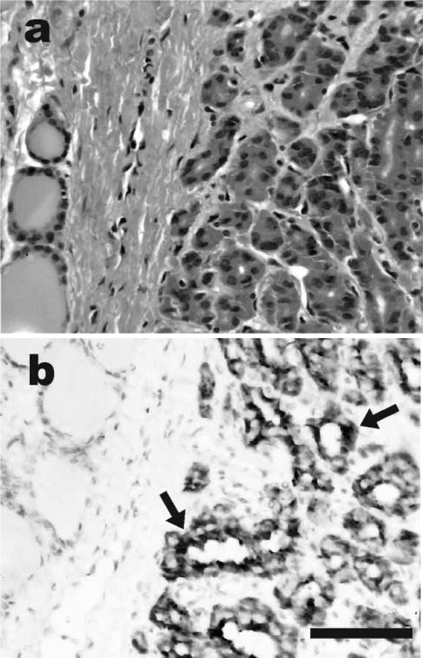
Oxyphilic thyroid adenoma. (
In Situ End-Labeling (ISEL) Technique
Deparaffinized sections were digested in a mixture containing 0–2.5 μg/ml proteinase K (Sigma; St Louis, MO) for 15 min at room temperature, optimizing protease concentration for every case (Labat-Moleur et al. 1998). Alternative pretreatment procedures were based on microwave oven heating (three 5-min cycles at 750 W). After quenching of endogenous peroxidase activity in 4% H2O2, sections were incubated in terminal deoxynucleotidyl transferase (TdT) buffer (25 mmol/liter Tris-HCl, 200 mmol/liter sodium cacodylate, 5 mmol/liter cobalt chloride) (Boehringer-Mannheim; Mannheim, Germany) for 5 min and then in TdT solution (20 U of TdT enzyme and 1 nmol of fluorescein-11-dUTP, in 100 μl of TdT buffer) (Boehringer) for 2 hr at 37C. After washing in 2 × SSC and in a solution containing 1 × Tris-buffered saline (TBS), 0.3% Triton, and 2% BSA, the reaction was revealed incubating with anti-fluorescein sheep peroxidase-conjugated antibody (Boehringer-Mannheim) for 30 min at 37C and then in diaminobenzidine solution. Specificity controls included the alternative omission of the fluoresceinated UTP, of the TdT enzyme, or of the anti-fluorescein antibody.
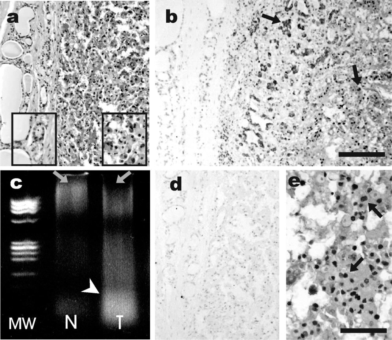
Oxyphilic thyroid adenoma. Comparing DNA extracted from normal peritumoral tissue (
DNA Fragmentation
Selected cases of paraffin-embedded material of both thyroidal and non-thyroidal tumors were microdissected under stereomicroscopic assistance to separate OCs from normal cells from the same paraffin block. The material obtained was processed for isolation of DNA after conventional phenol/chloroform extraction and ethanol precipitation. Ten μg of total DNA was loaded in 3% agarose gel containing ethidium bromide, run for 40 min at 100 V, and then visualized under UV light.
Results
Immunohistochemistry
All OC lesions had variable expression of mitochondrial markers (antimitochondrial antigen and biotin) in 70–100% of tumor cells (Figure 1). Among oxyphilic tumors, the proliferative index was <1% in goiters, 7% in adenomas, and from 3–5% in carcinomas both differentiated (including papillary) and poorly differentiated. The mean proliferative index of non-oxyphilic tumors was generally lower than that of the oxyphilic counterparts. Caspase activation was confined to single sparse tumor cells and to lymphoid cells in chronic thyroiditis foci.
Nuclear ISEL Staining
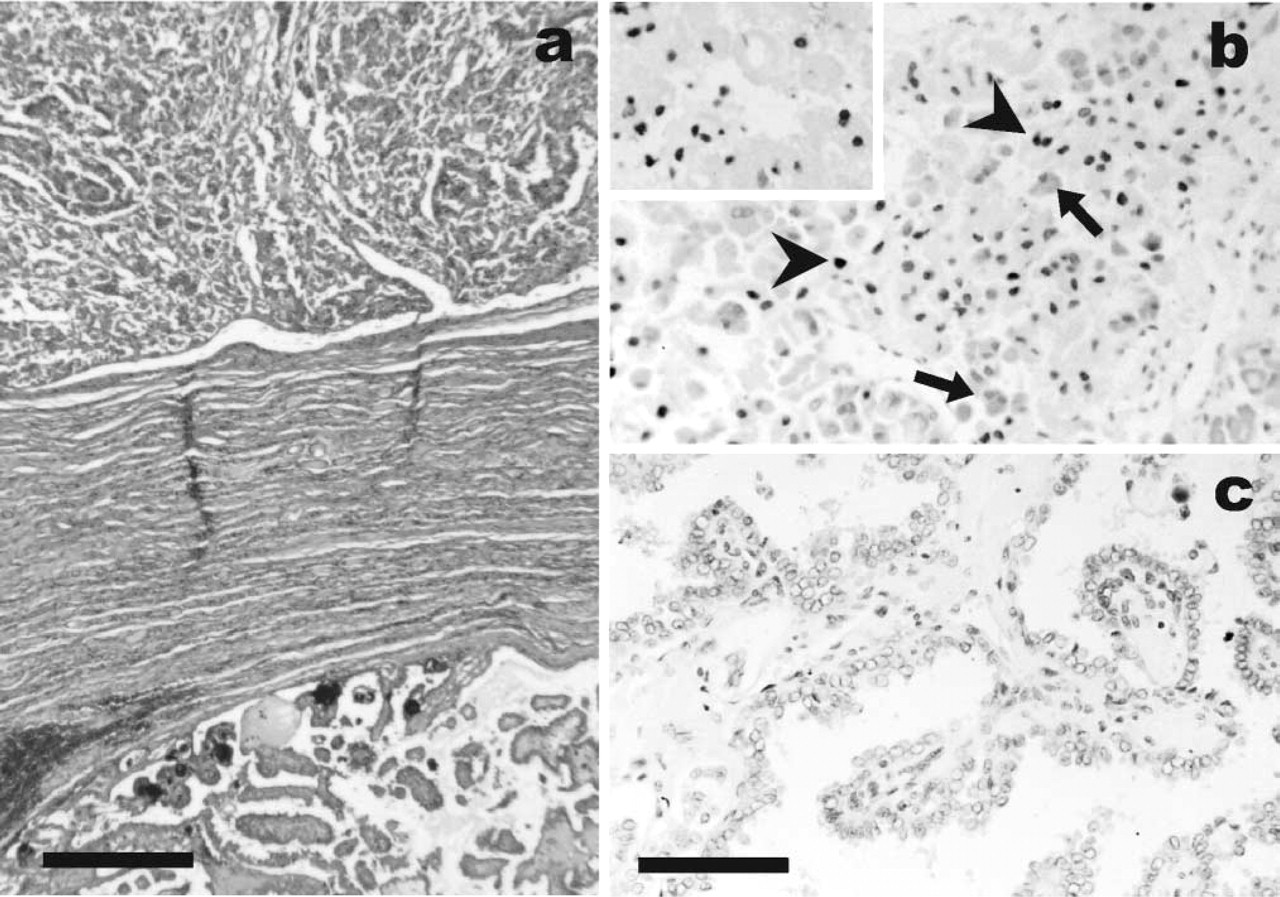
In thyroid lesions, surprisingly, strong nuclear reactivity was present in virtually all (18/19) OC tumors even in the absence of any morphological signs of apoptosis (Figures 2 and 3). This labeling was absent in OCs in the setting of nodular goiter or thyroiditis, and in the vast majority of non-oxyphilic benign and malignant lesions (4/26 cases were positive, generally with a focal pattern) (see Table 1). Peritumoral thyroid was negative. Negative control experiments (omission of labeled nucleotides, TdT enzyme, or anti-fluorescein antibody) confirmed the specificity of the staining. By comparing different pretreatment methods, despite a variable intensity of the staining probably due to fixation conditions, a positive reaction was observed even in the absence of protease digestion. A low proteinase K concentration provided best results. Microwave oven pretreatment as an alternative to protease digestion gave comparable results (although a generally weaker reaction was found). No differences in the intensity or the percentage of positive cells were observed among different oxyphilic histotypes. Therefore, no correlation between the presence of DNA fragmentation and proliferative activity was detected. Apoptosis-unrelated nuclear labeling appeared to be non-organ-specific. In fact, nuclear DNA fragmentation patterns were strongly detected in renal oncocytomas (2/3 cases) (Figure 4), in pituitary (2/2) and parathyroid (1/1) oxyphilic adenomas, and in the oncocytoma of the parotid gland, whereas Warthin tumors were all unreactive (Table 2).
Oxyphilic carcinoma (
Cytoplasmic ISEL Staining
In the thyroid gland, the ISEL staining procedure produced peculiar cytoplasmic staining in OCs but not in non-oxyphilic cells. This finding was observed in almost all OCs of both hyperplastic and neoplastic lesions. Only one case of oxyphilic carcinoma was totally unreactive (Table 1). A finely granular pattern was usually observed, although in few cases perinu-clear dots were also present (Figure 5). The positivity was generally diffuse and homogeneously distributed within individual lesions; a few cases presented a patchy distribution. Moreover a variable degree of intensity was present, both among different OC lesions and within individual samples. Control sections (omission of TdT enzyme or fluoresceinated d-UTP) were completely negative. As with the nuclear staining, microwave oven or absence of proteinase K pretreatment gave comparable signals, even if weaker than proteinase digestion. No reactivity was present in non-oxyphilic cells, even with higher protease concentrations. In non-thyroidal oxyphilic tumors, the described cytoplasmic pattern was focally found to be restricted to kidney oncocytomas (2/3), salivary gland Warthin's tumor (1/3), and pituitary adenoma (1/2) (Table 2). Generally, in lesions having also diffuse nuclear reactivity, we observed a co-expression of nuclear and cytoplasm positivity in the same cells.
Total DNA Fragmentation
Total DNA extracted from oxyphilic tumors from both thyroidal and non-thyroidal lesions, compared to adjacent normal tissue, presented a smear of fragments ranging from very high molecular weight to less than 100 bp (Figures 2c and 4c). Non-oxyphlic tumors and the non-neoplastic counterpart presented in most cases a low degree of fragmentation, related to formalin-fixation and paraffin-embedding procedures.
Discussion
We report on the unexpected finding of a very high occurrence of nuclear DNA fragmentation in OC tumors and of a high percentage of positive nuclei within individual tumors (up to 100% in some cases). The parallel absence in the same tumors of immunoreactivity for activated caspase led us to exclude the involvement of a true apoptotic pathway, while suggesting the possibility of a peculiar genomic DNA susceptibility to damage in OC tumors, probably due to a particular oxidative status. DNA fragmentation in the same tumors was also confirmed by selective extraction and electrophoresis.
Nuclear and cytoplasmic staining in oxyphilic and non-oxyphilic lesions of the thyroid a
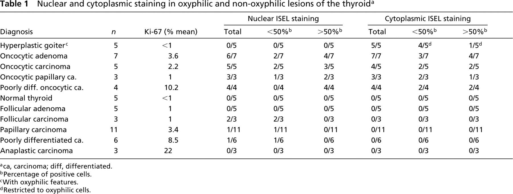
ca, carcinoma; diff, differentiated.
Percentage of positive cells.
With oxyphilic features.
Restricted to oxyphilic cells.
Great attention has been paid in determining the best pretreatment to prevent unspecific reaction (Strater et al. 1995; Negoescu et al. 1996; Panchalingam et al. 1996; Labat–Moleur et al. 1998). Indeed, the diffuse nuclear staining of most OCs with the ISEL method, even in the absence of any pretreatment, and the absence of this phenomenon in peritumoral thyroid tissue excluded the possibility of protease digestion as a cause of unspecific detection (Gal et al. 2000). We suggest that this peculiar genomic DNA fragmentation pattern in OC tumors may be the consequence of or even the key to a rigid response of oxyphilic tumors to ischemic stimuli, leading to a necrotic rather than an apoptotic response to stress conditions. In fact, these tumors lack expression of anti-apoptotic proteins (e.g., bcl-2) and are characterized by mitochondrial abnormalities (e.g., cytochrome C oxidase deficiency) potentially capable of blocking apoptotic processes (Muller–Hocker 1999). This model fits well with the observation of the common ischemic necrosis in oxyphilic thyroid nodules either spontaneously or after fine-needle aspiration procedures (Maximo and Sobrinho–Simoes 2000). Interestingly, a similar pattern of ISEL reactivity related to DNA fragmentation was also detected in extra-thyroid OC tumors, such as renal oncocytomas.
The question remains of whether the predisposition of OCs to DNA damage is an acquired defect primarily due to mitochondrial abnormalities. The role of mitochondrial disfunction in triggering apoptosis has not been fully elucidated, but several experimental models demonstrated functional changes of mitochondrial membranes as early events in programmed cell death (Bouchon et al. 2000; Halestrap et al. 2000; Lemasters et al. 1999; Salvioli et al. 2000; Shen et al. 2000; Voehringer et al. 2000). In several such models, mitochondrial alterations do not appear to be specific for apoptosis but are rather related to drug and hypoxia-mediated death programming (Riva et al. 1998; Diaz et al. 1999; Gajate et al. 2000).
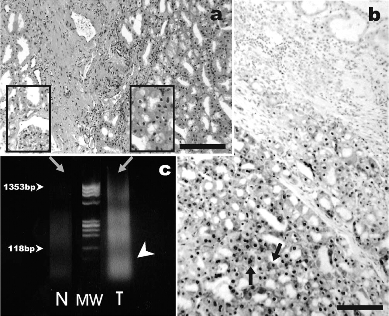
Renal oncocytoma. Tumor cells (
Nuclear and cytoplasmic ISEL staining in non-thyroidal oxyphilic lesions
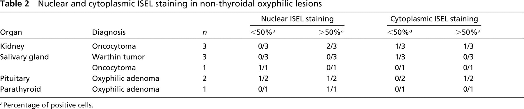
Percentage of positive cells.
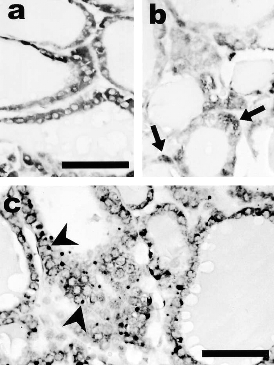
Benign nodular goiter with oxyphilic features. Intense cytoplasmic staining is observed by the ISEL method, both diffusely granular (
The cytoplasmic ISEL positivity in OCs deserves separate comment. Because no avidin-based procedure (possibly revealing endogenous biotin) is used in the ISEL method and no reaction was observed in appropriate negative control experiments, the cytoplasmic staining appears to be related to TdT function. Taking into consideration that the main difference between oxyphilic and non-oxyphilic cells is mitochondrial packaging and that mitochondria are the only source of DNA in eukaryotic cytoplasm, it appears likely that TdT recognizes peculiar mitochondrial DNA-damaged sites in OCs. Interestingly, our observation appears to fit with the recent report of mitochondrial DNA damage, detected with the ISEL method (in comparison to other molecular techniques) in cases of Alzheimer disease. In such lesions, cytoplasmic staining of neurons was observed on paraffin-embedded tissues, thus providing evidence for the ability of nick end-labeling to detect mitochondrial DNA fragmentation (de la Monte et al. 2000). The peculiarity of the cytoplasmic staining of OCs needs to be further investigated by ISEL–electron microscopic investigations (Migheli et al., 1995), but its specificity is stressed by the fact that mitochondrion-rich tissues (e.g., liver or kidney) do not normally show such a cytoplasmic reaction (data not shown). Even a possible relationship between mitochondrial and genomic DNA damage in OCs could not be excluded.
In conclusion, this study demonstrates for the first time an extensive DNA fragmentation in OC tumors of the thyroid and also of other organs, possibly related to interaction of ischemic factors and of mitochondrial cytoplasmic packaging rather than to the programmed cell death/apoptosis pathway. A surprising cytoplasmic staining, possibly related to mitochondrial DNA fragmentation, was also found in most OC lesions.
Footnotes
Acknowledgments
Supported by grants from Italian Ministry of University and Research (ex 60% to MP) and from the Associazione Italiana per la Ricerca sul Cancro (AIRC, Milan).