
Abstract
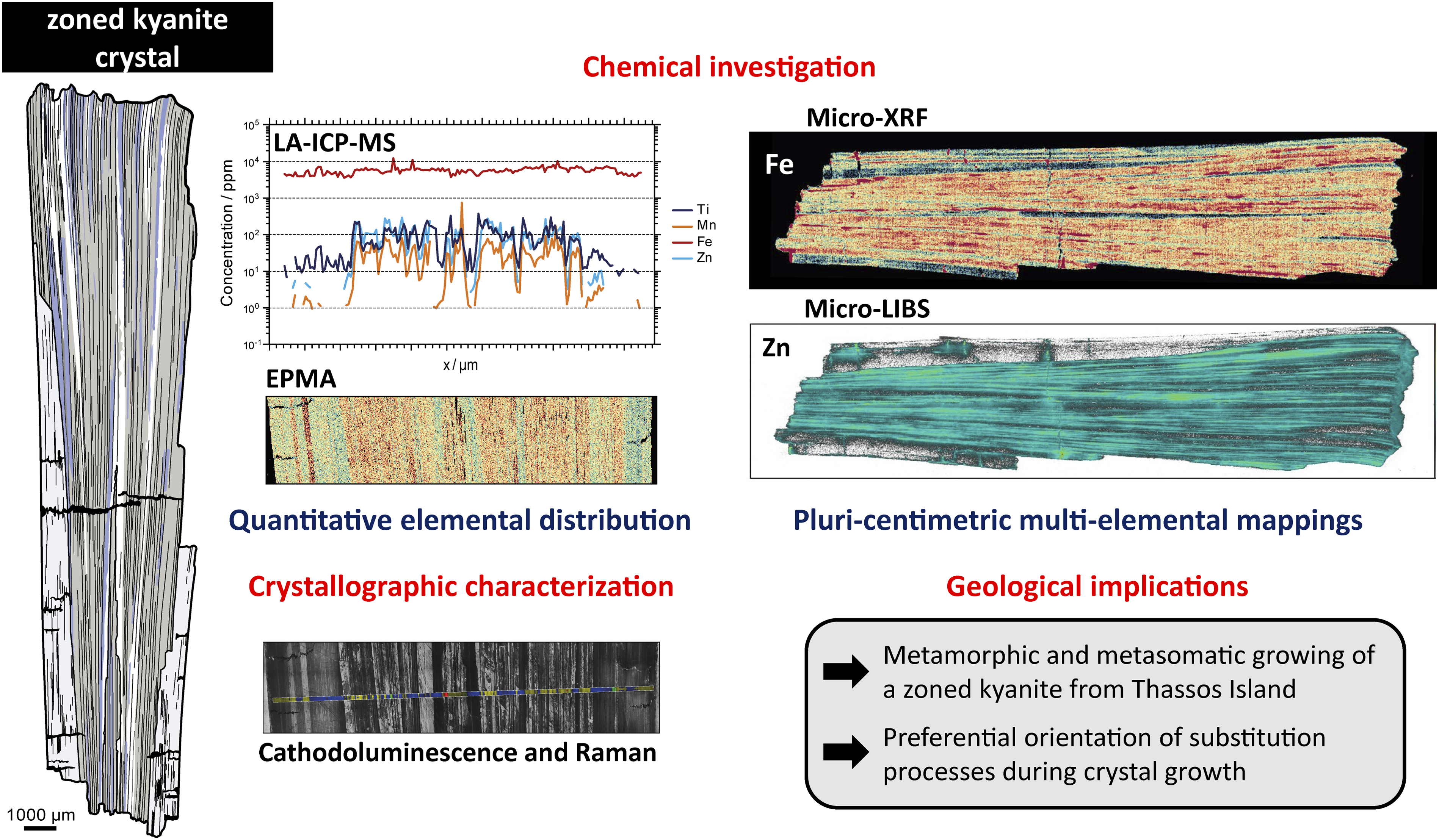
Comprehensive mineralogical and petrographic studies require analytical methods capable to report the distribution of major to trace elements within crystals in order to unravel their formation conditions and subsequent evolution. Additionally, the investigation of transition elements (e.g., Ti, V, Cr, Mn, Fe, and Zn) is essential for the comprehension of substitution processes within colored minerals. This study is conducted on a zoned kyanite crystal from a deformed quartz vein found within garnet–kyanite–biotite–hematite–plagioclase±staurolite±sillimanite paragneiss of Thassos Island, Greece. Herein, we show the efficiency of combining conventional, for example, cathodoluminescence, electron probe microanalysis (EPMA), laser ablation inductively coupled plasma mass spectrometry (LA-ICP-MS), and new methods, for example, micro-laser-induced breakdown spectroscopy (µLIBS), micro-X-ray fluorescence (µXRF), and Raman spectroscopy, to determine the chemical and crystallographic features of minerals. The simple chemistry of this crystal offers an ideal case to compare and valuate the potential of combined spectroscopy techniques to analyze minerals. We demonstrate that µLIBS and µXRF are perfectly adapted to perform multi-element imaging of major to trace elements down to the ppm level within a pluricentimetric crystal (2.3 x 0.5 cm) prior to quantitative analyses. We also highlight the benefit of cathodoluminescence and Raman mapping in the investigation of crystallographic features within minerals. The multispectroscopic approach enabled us to correlate growth stages of kyanite with the polymetamorphic history of the sample. Our results also highlight the spatial dependence of Ti for the generation of blue zonation by Fe2+–Ti4+ substitutions with Al3+.
Keywords
Get full access to this article
View all access options for this article.
References
Supplementary Material
Please find the following supplemental material available below.
For Open Access articles published under a Creative Commons License, all supplemental material carries the same license as the article it is associated with.
For non-Open Access articles published, all supplemental material carries a non-exclusive license, and permission requests for re-use of supplemental material or any part of supplemental material shall be sent directly to the copyright owner as specified in the copyright notice associated with the article.