Restricted accessResearch articleFirst published online 2022-4
Infrared Spectroscopy-Determined Bone Compositional Changes Associated with Anti-Resorptive Treatment of the oim/oim Mouse Model of Osteogenesis Imperfecta
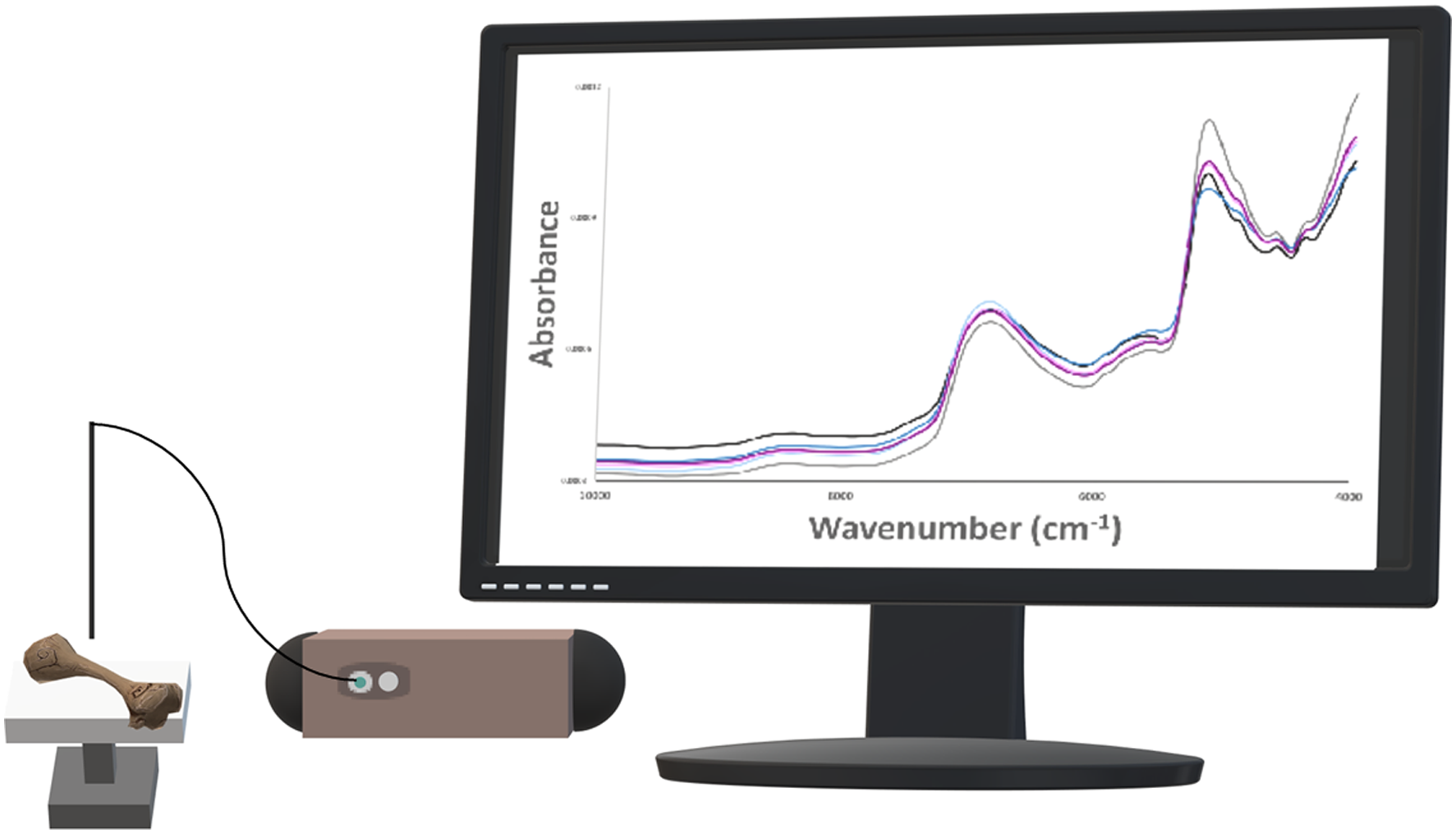
Applications of vibrational spectroscopy to assess bone disease and therapeutic interventions are continually advancing, with tissue mineral and protein composition frequently investigated. Here, we used two spectroscopic approaches for determining bone composition in a mouse model (oim) of the brittle bone disease osteogenesis imperfecta (OI) with and without antiresorptive agent treatment (alendronate, or ALN, and RANK-Fc). Near-infrared (NIR) spectral analysis using a fiber optic probe and attenuated total reflection Fourier transform infrared spectroscopy (ATR FTIR) mode were applied to investigate bone composition, including water, mineral, and protein content. Spectral parameters revealed differences among the control wildtype (WT) and OIM groups. NIR spectral analysis of protein and water showed that OIM mouse humerii had ∼50% lower protein and ∼50% higher overall water content compared to WT bone. Moreover, some OIM-treated groups showed a reduction in bone water compared to OIM controls, approximating values observed in WT bone. Differences in bone quality based on increased mineral content and reduced carbonate content were also found between some groups of treated OIM and WT bone, but crystallinity did not differ among all groups. The spectroscopically determined parameters were evaluated for correlations with gold-standard mechanical testing values to gain insight into how composition influenced bone strength. As expected, bone mechanical strength parameters were consistently up to threefold greater in WT mice compared to OIM groups, except for stiffness in the ALN-treated OIM groups. Furthermore, bone stiffness, maximum load, and post-yield displacement showed the strongest correlations with NIR-determined protein content (positive correlations) and bound-water content (negative correlations). These results demonstrate that in this study, NIR spectral parameters were more sensitive to bone composition differences than ATR parameters, highlighting the potential of this nondestructive approach for screening of bone diseases and therapeutic efficacy in pre-clinical models.
LimJ.GrafeI.AlexanderS.LeeB.“Genetic Causes and Mechanisms of Osteogenesis Imperfecta”. Bone2017; 102: 40–49.
3.
MariniJ.C.ForlinoA.BächingerH.P.BishopN.J., et al.“Osteogenesis Imperfecta”. Nat. Rev. Dis. Primers2017; 3: 17052.
4.
MorelloR.“Osteogenesis Imperfecta and Therapeutics”. Matrix Biol2018; 71–72: 294–312.
5.
MontiE.MottesM.FraschiniP.BrunelliP., et al.“Current and Emerging Treatments for the Management of Osteogenesis Imperfecta”. Ther. Clin. Risk Manage2010; 6: 367–381.
6.
RalstonS.H.GastonM.S.“Management of Osteogenesis Imperfecta”. Front. Endocrinol2020; 10: 924–924.
7.
ForlinoA.CabralW.A.BarnesA.M.MariniJ.C.“New Perspectives on Osteogenesis Imperfecta”. Nat. Rev. Endocrinol2011; 7(9): 540–557.
8.
TournisS.DedeA.D.“Osteogenesis Imperfecta: A Clinical Update”. Metabolism2018; 80: 27–37.
9.
RussellR.G.G.“Determinants of Structure–Function Relationships Among Bisphosphonates”. Bone2007; 40(5, Suppl. 2): S21–S25.
10.
ThompsonK.RogersM.J.CoxonF.P.CrockettJ.C.“Cytosolic Entry of Bisphosphonate Drugs Requires Acidification of Vesicles after Fluid-Phase Endocytosis”. Mol. Pharmacol2006; 69(5): 1624–1632.
11.
CoxonF.P.ThompsonK.RoelofsA.J.EbetinoF.H.RogersM.J.“Visualizing Mineral Binding and Uptake of Bisphosphonate by Osteoclasts and Non-Resorbing Cells”. Bone2008; 42(5): 848–860.
12.
BockO.FelsenbergD.“Bisphosphonates in the Management of Postmenopausal Osteoporosis: Optimizing Efficacy in Clinical Practice”. Clin. Interv. Aging2008; 3(2): 279–297.
13.
BoyceA.M.“Denosumab: An Emerging Therapy in Pediatric Bone Disorders”. Curr. Osteoporos. Rep2017; 15(4): 283–292.
14.
Hoyer-KuhnH.FranklinJ.AlloG.KronM., et al.“Safety and Efficacy of Denosumab in Children with Osteogenesis Imperfect: A First Prospective Trial”. J. Musculoskelet. Neuronal. Interact2016; 16(1): 24–32.
15.
BargmanR.PoshamR.BoskeyA.L.DiCarloE., et al.“Comparable Outcomes in Fracture Reduction and Bone Properties with RANKL Inhibition and Alendronate Treatment in a Mouse Model of Osteogenesis Imperfecta”. Osteoporos. Int2012; 23(3): 1141–1150.
16.
TauerJ.T.RobinsonM.-E.RauchF.“Osteogenesis Imperfecta: New Perspectives from Clinical and Translational Research”. JBMR Plus2019; 3(8): e10174.
17.
ChipmanS.D.SweetH.O.McBrideD.J.JrDavissonM.T., et al.“Defective Pro Alpha 2(I) Collagen Synthesis in a Recessive Mutation in Mice: A Model of Human Osteogenesis Imperfecta”. Proc. Natl. Acad. Sci. U.S.A1993; 90(5): 1701–1705.
18.
BoskeyA.L.MarinoJ.SpevakL.PleshkoN., et al.“Are Changes in Composition in Response to Treatment of a Mouse Model of Osteogenesis Imperfecta Sex-Dependent?” Clin. Orthop. Relat. Res2015; 473(8): 2587–2598.
19.
CamachoN.P.LandisW.J.BoskeyA.L.“Mineral Changes in a Mouse Model of Osteogenesis Imperfecta Detected by Fourier Transform Infrared Microscopy”. Connect. Tissue Res1996; 35(1–4): 259–265.
20.
YaoX.CarletonS.M.KettleA.D.MelanderJ., et al.“Gender-Dependence of Bone Structure and Properties in Adult Osteogenesis Imperfecta Murine Model”. Ann. Biomed. Eng2013; 41(6): 1139–1149.
21.
SharirA.BarakM.M.ShaharR.“Whole Bone Mechanics and Mechanical Testing”. Vet. J2008; 177(1): 8–17.
22.
GaoH.JiB.JagerI.L.ArztE.FratzlP.“Materials Become Insensitive to Flaws at Nanoscale: Lessons from Nature”. Proc. Natl. Acad. Sci. U.S.A2003; 100(10): 5597–5600.
23.
NijhuisW.H.EastwoodD.M.AllgroveJ.HvidI., et al.“Current Concepts in Osteogenesis Imperfecta: Bone Structure, Biomechanics, and Medical Management”. J. Child. Orthop2019; 13(1): 1–11.
24.
CarrieroA.ZimmermannE.A.PalusznyA.TangS.Y., et al.“How Tough is Brittle Bone? Investigating Osteogenesis Imperfecta in Mouse Bone”. J. Bone Miner. Res2014; 29(6): 1392–1401.
25.
MisofB.M.RoschgerP.BaldiniT.RaggioC.L., et al.“Differential Effects of Alendronate Treatment on Bone from Growing Osteogenesis Imperfecta and Wild-Type Mouse”. Bone2005; 36(1): 150–158.
26.
A.L. Boskey, S.B. Doty. “Mineralized Tissue: Histology, Biology and Biochemistry". In: J.R. Shapiro, P.H. Byers, F.H. Glorieux, P.D. Sponseller, editors. Osteogenesis Imperfecta: A Translational Approach to Brittle Bone Disease. San Diego: Academic Press, 2014. Chap. 4, Pp. 31–43.
27.
MandairG.S.MorrisM.D.“Contributions of Raman Spectroscopy to the Understanding of Bone Strength”. BoneKEy Rep2015; 4: 620–620.
28.
DehringK.A.CraneN.J.SmuklerA.R.McHughJ.B., et al.“Identifying Chemical Changes in Subchondral Bone Taken from Murine Knee Joints Using Raman Spectroscopy”. Appl. Spectrosc2006; 60(10): 1134–1141.
29.
GoodyearS.R.GibsonI.R.SkakleJ.M.WellsR.P.AspdenR.M.“A Comparison of Cortical and Trabecular Bone from C57 Black 6 Mice Using Raman Spectroscopy”. Bone2009; 44(5): 899–907.
30.
MeganckJ.A.BegunD.L.McElderryJ.D.SwickA., et al.“Fracture Healing with Alendronate Treatment in the Brtl/+ Mouse Model of Osteogenesis Imperfecta”. Bone2013; 56(1): 204–212.
31.
CamachoN.P.CarrollP.RaggioC.L.“Fourier Transform Infrared Imaging Spectroscopy (FTIRIS) of Mineralization in Bisphosphonate-Treated oim/oim Mice”. Calcif. Tissue Int2003; 72(5): 604–609.
32.
MasciM.WangM.ImbertL.BarnesA.M., et al.“Bone Mineral Properties in Growing Col1a2(+/G610c) Mice: An Animal Model of Osteogenesis Imperfecta”. Bone2016; 87: 120–129.
33.
GrankeM.DoesM.D.NymanJ.S.“The Role of Water Compartments in the Material Properties of Cortical Bone”. Calcif. Tissue Int2015; 97(3): 292–307.
34.
AilavajhalaR.QueridoW.RajapakseC.S.PleshkoN.“Near Infrared Spectroscopic Assessment of Loosely and Tightly Bound Cortical Bone Water”. Analyst2020; 145(10): 3713–3724.
35.
KramerR.Z.VenugopalM.G.BellaJ.MayvilleP., et al.“Staggered Molecular Packing in Crystals of a Collagen-Like Peptide with a Single Charged Pair”. J. Mol. Biol2000; 301(5): 1191–1205.
36.
WangY.von EuwS.FernandesF.M.CassaignonS., et al.“Water-Mediated Structuring of Bone Apatite”. Nat. Mater2013; 12(12): 1144–1153.
37.
GrankeM.DoesM.D.NymanJ.S.“The Role of Water Compartments in the Material Properties of Cortical Bone”. Calcif. Tissue Int2015; 97(3): 292–307.
38.
NymanJ.S.RoyA.ShenX.AcunaR.L., et al.“The Influence of Water Removal on the Strength and Toughness of Cortical Bone”. J. Biomech2006; 39(5): 931–938.
39.
UnalM.YangS.AkkusO.“Molecular Spectroscopic Identification of the Water Compartments in Bone”. Bone2014; 67: 28–236.
40.
Büning-PfaueH.“Analysis of Water in Food by Near Infrared Spectroscopy”. Food Chem2003; 82(1): 107–115.
41.
AilavajhalaR.OswaldJ.RajapakseC.S.PleshkoN.“Environmentally Controlled Near Infrared Spectroscopic Imaging of Bone Water”. Sci. Rep2019; 9(1): 10199.
42.
BozkurtA.OnaralB.“Safety Assessment of Near Infrared Light Emitting Diodes for Diffuse Optical Measurements”. Biomed. Eng. Online2004; 3(1): 9.
43.
TanakaY.“Impact of Near-Infrared Radiation in Dermatology”. World J. Dermatol2012; 1(3): 30–37.
44.
ShanasN.QueridoW.DumontA.YonkoE., et al.“Clinical Application of Near Infrared Fiber Optic Spectroscopy for Noninvasive Bone Assessment”. J. Biophotonics2020; 13(4): e201960172.
45.
PadalkarM.V.PleshkoN.“Wavelength-Dependent Penetration Depth of Near Infrared Radiation into Cartilage”. Analyst2015; 140(7): 2093–2100.
46.
FarisF.ThornileyM.WickramasingheY.HoustonR., et al.“Non-Invasive in Vivo Near-Infrared Optical Measurement of the Penetration Depth in the Neonatal Head”. Clin. Phys. Physiol. Meas1991; 12(4): 353–358.
47.
JepsenK.J.GoldsteinS.A.KuhnJ.L.SchafflerM.B.BonadioJ.“Type-I Collagen Mutation Compromises the Post-Yield Behavior of Mov13 Long Bone”. J. Orthop. Res1996; 14(3): 493–499.
48.
JepsenK.J.SilvaM.J.VashishthD.GuoX.E.van der MeulenM.C.“Establishing Biomechanical Mechanisms in Mouse Models: Practical Guidelines for Systematically Evaluating Phenotypic Changes in the Diaphyses of Long Bones”. J. Bone Miner. Res2015; 30(6): 951–966.
49.
QueridoW.AilavajhalaR.PadalkarM.PleshkoN.“Validated Approaches for Quantification of Bone Mineral Crystallinity Using Transmission Fourier Transform Infrared (FTIR), Attenuated Total Reflection (ATR) FTIR, and Raman Spectroscopy”. Appl. Spectrosc2018; 72(11): 1581–1593.
50.
WischmannJ.LenzeF.ThielA.BookbinderS., et al.“Matrix Mineralization Controls Gene Expression in Osteoblastic Cells”. Exp. Cell Res2018; 372(1): 25–34.
51.
RajapakseC.S.PadalkarM.V.YangH.J.IspiryanM.PleshkoN.“Non-Destructive NIR Spectral Imaging Assessment of Bone Water: Comparison to MRI Measurements”. Bone2017; 103: 116–124.
52.
HechtK.T.WoodD.L.SutherlandG.B.B.M.“The Near Infra-Red Spectrum of the Peptide Group”. Proc. R. Soc. A1956; 235(1201): 174–188.
53.
UnalM.CreecyA.NymanJ.S.“The Role of Matrix Composition in the Mechanical Behavior of Bone”. Curr. Osteoporos. Rep2018; 16(3): 205–215.
54.
LiC.SeifertA.C.RadH.S.BhagatY.A., et al.“Cortical Bone Water Concentration: Dependence of MR Imaging Measures on Age and Pore Volume Fraction”. Radiology2016; 280(2): 653.
55.
BargmanR.HuangA.BoskeyA.L.RaggioC.PleshkoN.“RANKL Inhibition Improves Bone Properties in a Mouse Model of Osteogenesis Imperfecta”. Connect. Tissue Res2010; 51(2): 123–131.
56.
AllenM.R.TerritoP.R.LinC.PersohnS., et al.“In Vivo UTE-MRI Reveals Positive Effects of Raloxifene on Skeletal-Bound Water in Skeletally Mature Beagle Dogs”. J. Bone Miner. Res2015; 30(8): 1441–1444.
57.
WolframU.SchwiedrzikJ.“Post-Yield and Failure Properties of Cortical Bone”. BoneKEy Rep2016; 5: 829.
58.
BermanA.G.WallaceJ.M.BartZ.R.AllenM.R.“Raloxifene Reduces Skeletal Fractures in an Animal Model of Osteogenesis Imperfecta”. Matrix Biol2016; 52–54: 19–28.
59.
JepsenK.J.SchafflerM.B.KuhnJ.L.GouletR.W., et al.“Type I Collagen Mutation Alters the Strength and Fatigue Behavior of Mov13 Cortical Tissue”. J. Biomech1997; 30(11–12): 1141–1147.
60.
MartinR.B.IshidaJ.“The Relative Effects of Collagen Fiber Orientation, Porosity, Density, and Mineralization on Bone Strength”. J. Biomech1989; 22(5): 419–426.
61.
AbueiddaD.W.SabetF.A.JasiukI.M.“Modeling of Stiffness and Strength of Bone at Nanoscale”. J. Biomech. Eng2017; 139(5): 051006.
62.
ErivanR.VillatteG.CueffR.BoisgardS.DescampsS.“Rehydration Improves the Ductility of Dry Bone Allografts”. Cell Tissue Bank2017; 18(3): 307–312.
63.
NymanJ.S.NiQ.NicolellaD.P.WangX.“Measurements of Mobile and Bound Water by Nuclear Magnetic Resonance Correlate with Mechanical Properties of Bone”. Bone2008; 42(1): 193–199.
64.
VashishthD.GibsonG.J.KhouryJ.I.SchafflerM.B., et al.“Influence of Nonenzymatic Glycation on Biomechanical Properties of Cortical Bone”. Bone2001; 28(2): 195–201.
65.
MilesC.A.AveryN.C.RodinV.V.BaileyA.J.“The Increase in Denaturation Temperature Following Cross-Linking of Collagen is Caused by Dehydration of the Fibres”. J. Mol. Biol2005; 346(2): 551–556.
66.
AndriotisO.G.ChangS.W.VanleeneM.HowarthP.H., et al.“Structure-Mechanics Relationships of Collagen Fibrils in the Osteogenesis Imperfecta Mouse Model”. J. R. Soc. Interface2015; 12(111): 20150701–20150701.
67.
HongA.L.IspiryanM.PadalkarM.V.JonesB.C., et al.“MRI-Derived Bone Porosity Index Correlates to Bone Composition and Mechanical Stiffness”. Bone Rep2019; 11: 100213.
68.
AfaraI.O.PrasadamI.CrawfordR.XiaoY.OloyedeA.“Near Infrared (NIR) Absorption Spectra Correlates with Subchondral Bone Micro-CT Parameters in Osteoarthritic Rat Models”. Bone2013; 53(2): 350–357.
Supplementary Material
Please find the following supplemental material available below.
For Open Access articles published under a Creative Commons License, all supplemental material carries the same license as the article it is associated with.
For non-Open Access articles published, all supplemental material carries a non-exclusive license, and permission requests for re-use of supplemental material or any part of supplemental material shall be sent directly to the copyright owner as specified in the copyright notice associated with the article.