
Abstract
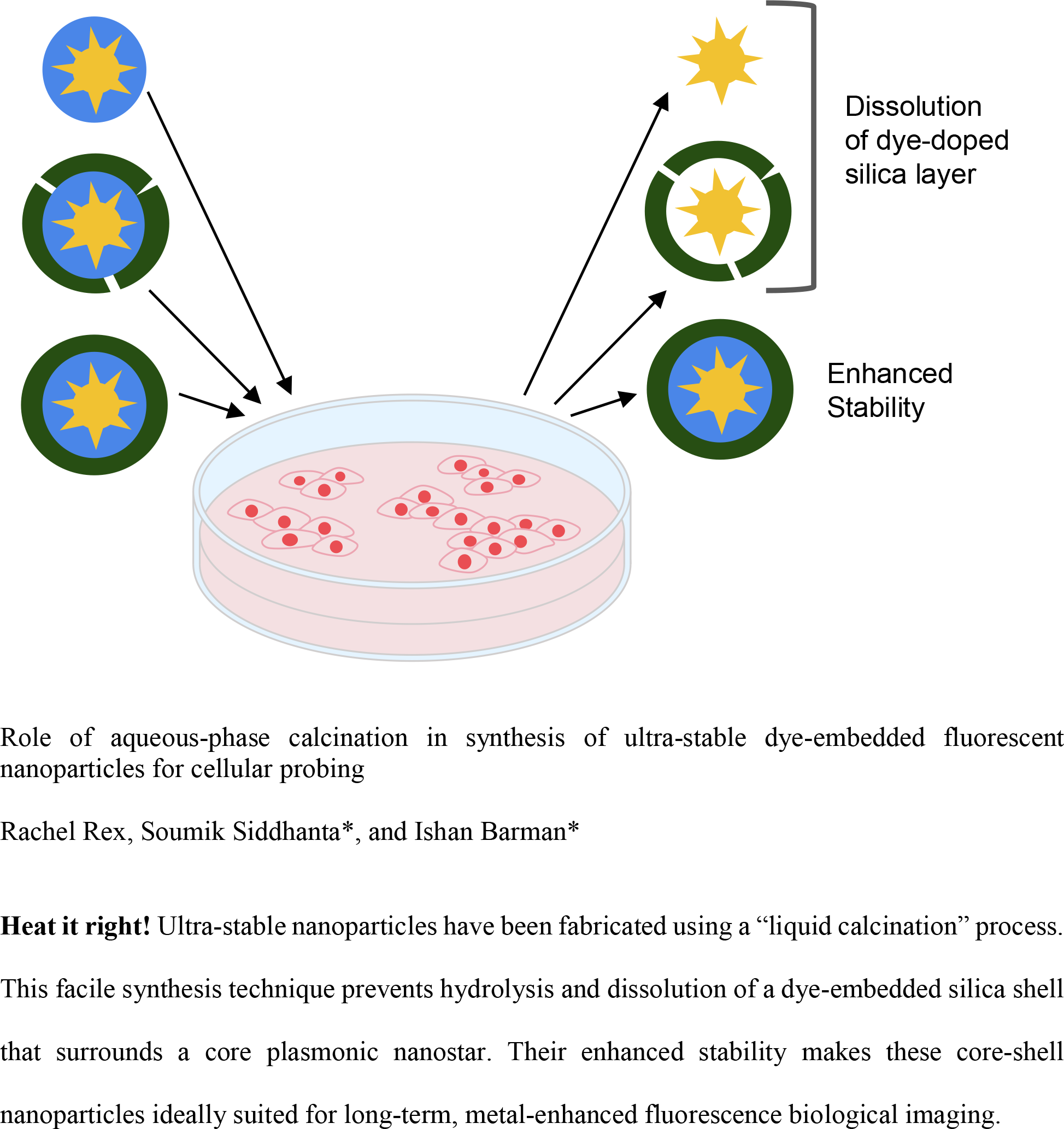
Fluorescence imaging is a major driver of discovery in biology, and an invaluable asset in clinical diagnostics. To overcome quenching limitations of conventional fluorescent dyes and further improve intensity, nanoparticle-based constructs have been the subject of intense investigation, and within this realm, dye-doped silica-coated nanoparticles have garnered significant attention. Despite their growing popularity in research, fluorescent silica nanoparticles suffer from a significant flaw. The degradation of these nanoparticles in biological media by hydrolytic dissolution is underreported, leading to serious misinterpretations, and limiting their applicability for live cell and in vivo imaging. Here, the development of an ultra-stable, dye-embedded, silica-coated metal nanoparticle is reported, and its superior performance in long-term live cell imaging is demonstrated. While conventional dye-doped silica nanoparticles begin to degrade within an hour in aqueous media, by leveraging a modified liquid calcination process, this new construct is shown to be stable for at least 24 h. The stability of this metal-enhanced fluorescent probe in biologically relevant temperatures and media, and its demonstrated utility for cell imaging, paves the way for its future adoption in biomedical research.
Keywords
Get full access to this article
View all access options for this article.
References
Supplementary Material
Please find the following supplemental material available below.
For Open Access articles published under a Creative Commons License, all supplemental material carries the same license as the article it is associated with.
For non-Open Access articles published, all supplemental material carries a non-exclusive license, and permission requests for re-use of supplemental material or any part of supplemental material shall be sent directly to the copyright owner as specified in the copyright notice associated with the article.