
Abstract
Background
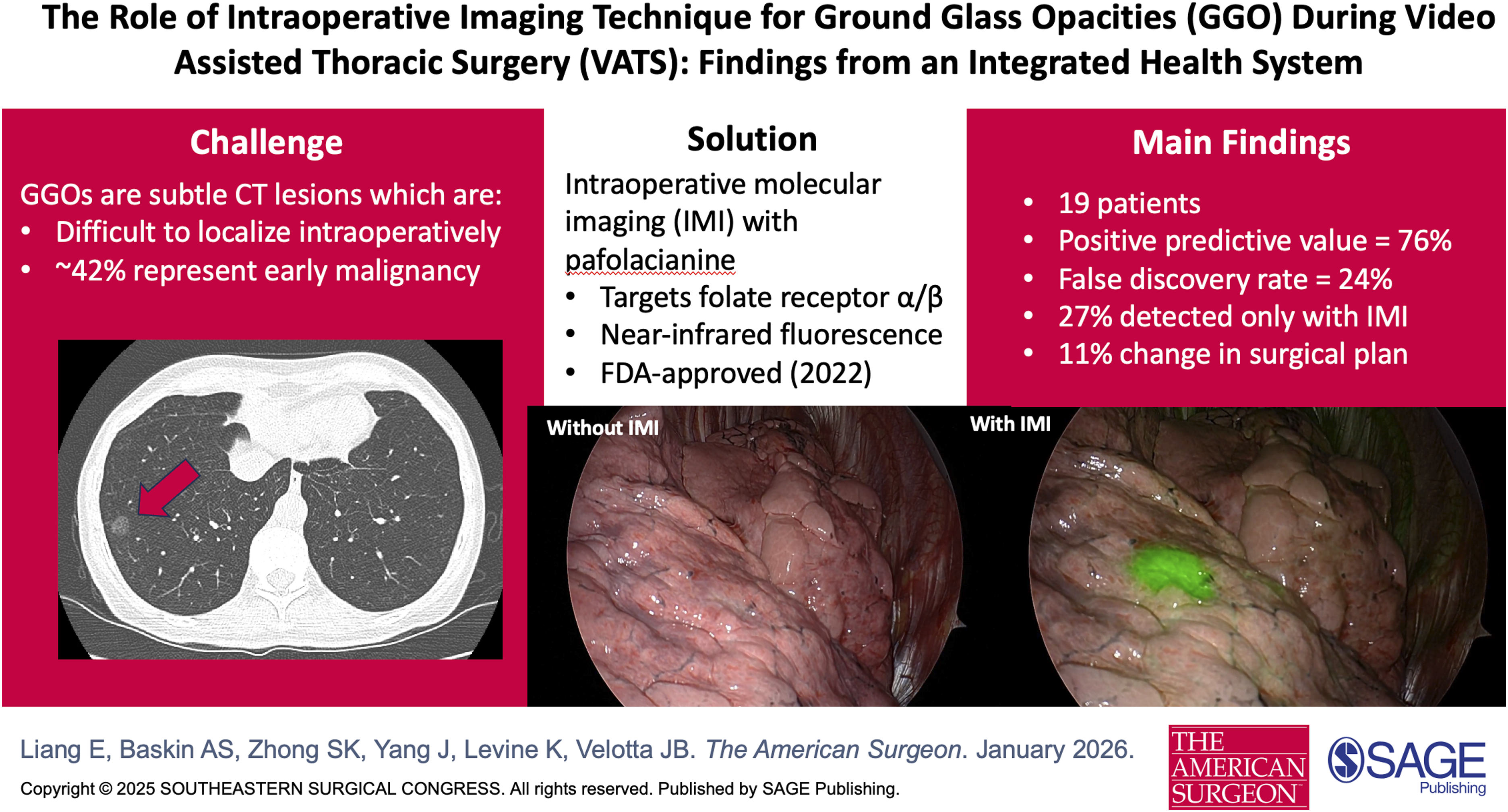
Intraoperative visualization of ground glass opacities (GGOs) remains challenging, with past techniques limited by safety concerns and poor margin assessment. We evaluate the utility of pafolacianine, an intraoperative molecular imaging (IMI) agent for GGO visualization.
Methods
We conducted a prospective study from an integrated health system of patients undergoing video-assisted thoracoscopic surgery (VATS) lung resections for GGOs from 6/1/2025 to 8/31/2025. Pafolacianine was administered intravenously one hour preoperatively. Lung parenchyma was inspected for fluorescence using Stryker 1788 imaging before resection. Real-time intraoperative and postoperative surveys were completed, and chart review was performed to collect sociodemographic, pathologic, and surgical outcome data.
Results
Twenty-one patients (median age 70 years), predominantly female (62%), Asian (48%), and never-smokers (71%), underwent VATS with a median hospital stay of 1 day. Intraoperative molecular imaging prompted escalation to completion lobectomy in 10% of patients. Among 19 non-metastatic patients, 26 lesions (median diameter 14 mm) were resected, the majority of which were pure GGOs (69%). Intraoperative molecular imaging detected all malignant lesions, with 27% identified exclusively by IMI. Adenocarcinoma accounted for 62% of lesions, while 23% were benign. Intraoperative molecular imaging had a positive predictive value of 0.76 (95% CI, 0.54-0.90) and false discovery rate of 0.24 (95% CI, 0.10-0.46). All margins were negative, and no adverse events were reported.
Discussion
This exploratory real-world study suggests that pafolacianine facilitates optimal intraoperative visualization of challenging GGO lesions, supporting its potential to improve surgical decision-making, tumor detection, and margin assessment during VATS lung resections.
Get full access to this article
View all access options for this article.
References
Supplementary Material
Please find the following supplemental material available below.
For Open Access articles published under a Creative Commons License, all supplemental material carries the same license as the article it is associated with.
For non-Open Access articles published, all supplemental material carries a non-exclusive license, and permission requests for re-use of supplemental material or any part of supplemental material shall be sent directly to the copyright owner as specified in the copyright notice associated with the article.