



Abstract
Background
Patients with atypical ductal hyperplasia (ADH) diagnosed on core needle biopsy (CNB) are recommended for surgical excision. However, the rate of upstaging to malignancy on final pathology varies widely. This study sought to determine the rate of upstaging for ADH diagnosed on CNB and to evaluate risk factors associated with upstaging.
Methods
Patients diagnosed with ADH by CNB from 1/2014 to 9/2024 were identified from a prospectively maintained database. Pathology, imaging, demographics, family history of breast cancer, genetic testing, and risk of future malignancy were collected. Univariate and multivariable analysis were performed to evaluate predictors of upstaging.
Results
456 patients with ADH on CNB met inclusion criteria. Median age was 53 years (range 19-89 years). Overall, 110 (24.1%) patients were upstaged on surgical excision. The highest risk of upstaging was observed in patients with multifocal ADH (71.4%). Upstaged patients were older (P < .001), more likely to have a mammographic mass or architectural distortion (P = .03), abnormal MRI (P = .001), ADH bordering on DCIS or multifocal ADH on biopsy (P < .001). Multivariable analysis identified abnormal MRI as an independent predictor of higher risk (P = .046) and focal ADH as a predictor of lower upstage risk (P = .03). At a median follow-up of 52.5 months, 21 (4.6%) patients developed subsequent DCIS, and 20 (4.4%) patients developed subsequent invasive cancer.
Conclusion
ADH on CNB was upstaged on surgical excision in 24.1% of patients. Multifocal ADH and abnormal MRI findings strongly predict upstaging to malignancy. The 9.6% rate of subsequent malignancy necessitates long-term surveillance for all patients with ADH.
Keywords
Introduction
Atypical ductal hyperplasia (ADH) is a high-risk, non-malignant breast lesion that confers a four-to-five-fold increase in lifetime risk of developing invasive breast carcinoma. Pathologically, ADH is characterized by epithelial proliferation in breast ducts and shares cellular and molecular characteristics with low-grade ductal carcinoma in situ (DCIS). The distinction between ADH and DCIS is primarily quantitative: ADH is limited to lesions measuring <2 mm or involving no more than two ductal spaces, whereas low-grade DCIS exceeds both criteria. 1 Importantly, distinguishing these entities on core needle biopsy (CNB) can be difficult due to the intrinsic limitations of tissue sampling. This limitation is compounded by the tendency for ADH to occur incidentally within coexisting breast tumors, such that its presence on CNB may indicate an undersampled component of more extensive disease. Accordingly, the resulting diagnostic ambiguity has historically led to the recommendation for surgical excision of ADH to mitigate the risk of missing an underlying carcinoma. Rates of upstaging on surgical excision from ADH to DCIS or invasive breast carcinoma vary widely in the contemporary literature, ranging from 4.9% to 59.1%, underscoring the significant heterogeneity and ongoing uncertainty in management.2–13 Prior studies have attempted to define predictors of ADH upgrade on surgical excision but have been constrained by small sample sizes and the inclusion of patients with mixed or overlapping diagnoses, such as atypical lobular hyperplasia (ALH) and ADH with coexisting lobular carcinoma in situ (LCIS). We performed a comprehensive analysis of lesions with ADH on CNB and evaluated preoperative, radiologic, and pathologic risk factors that are predictive of upstaging to DCIS or invasive breast carcinoma upon surgical excision.
Methods
This study was performed with the approval of our Institutional Review Board. Patients with a diagnosis of ADH on CNB from January 2014 to September 2024 were identified from a prospectively maintained institutional database using the Deep6 AI software. Patients with ADH and concomitant cancer on CNB were excluded from analysis. Demographics, histopathological features on CNB, including focality and the presence of concomitant diagnoses, mammographic, ultrasound, and MRI findings, family history of breast cancer, genetic testing, and subsequent pathological findings on surgical excision were reviewed. Student’s t-test and Pearson’s chi-square tests were used to compare continuous and categorical variables for statistical significance, respectively. Multivariable logistic regression was used to identify independent predictors of upstage. A P-value <0.05 was considered significant. Statistical analyses were performed using STATA software.
Results
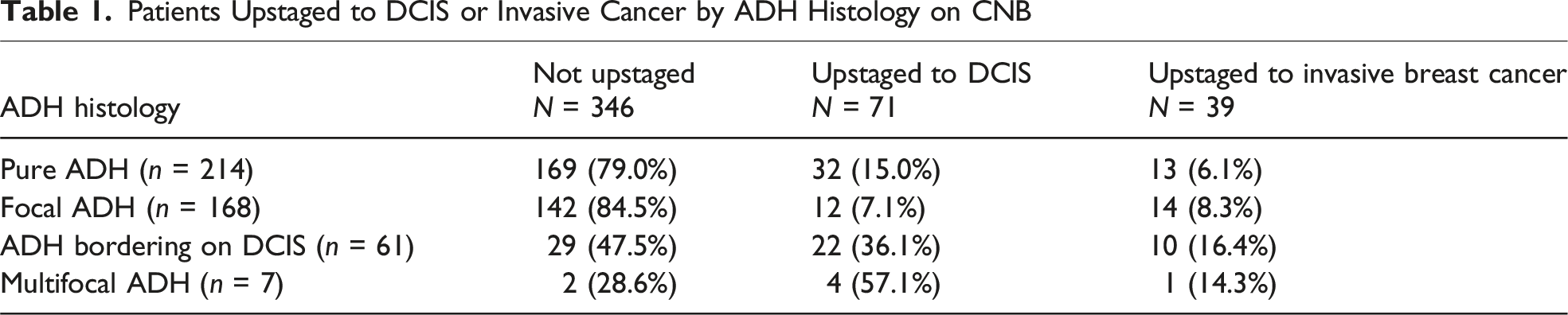
Patients Upstaged to DCIS or Invasive Cancer by ADH Histology on CNB
Characteristics of Patients With ADH by Upstage Status

Outcomes of Patients With ADH by Upstage Status

Multivariable Logistic Regression of Risk of Upstaging Among Patients With ADH on Core Needle Biopsy

Discussion
The prevalence of DCIS or invasive carcinoma in ADH specimens varies widely in the literature, complicating efforts to identify which patients are most likely to benefit from surgical excision. Prior studies have reported widely variable upstaging rates ranging from 4.9% to 59.1%; however, these analyses are largely limited by small sample sizes and the inclusion of heterogeneous CNB diagnoses, often combining ADH with additional high-risk or atypical lesions.2–13 Our study provides a comprehensive analysis of ADH diagnosed on CNB and demonstrates an overall upstage rate of 24.1%, with substantially higher rates observed among patients with multifocal ADH or ADH bordering on DCIS.
Histologically, ADH and DCIS are essentially indistinguishable, with the differentiating factor being the size of the lesion. The amount of sampling on the CNB can therefore be the only distinction between ADH and DCIS. This breadth of range underscores the difficulty in defining a universally accepted low-risk group. Among our patient cohort, approximately one in four patients diagnosed with ADH on CNB had a malignancy requiring excision, mirroring a national average of 29% and validating the representativeness of our study population for further risk analysis. 13 This upstage rate, coupled with the fact that an ADH diagnosis increases a patient’s risk for breast cancer by 4 to 5 times, is the rationale for the current recommendation of precautionary surgical excision.12,14 While this data can support surgery, it simultaneously indicates that >70% of patients undergo unnecessary surgery. The challenge lies in safely identifying the large subset of patients who are not upstaged and who could benefit from less invasive active surveillance.
The most recent established guidelines from organizations such as the National Cancer Comprehensive Network and the American Society of Breast Surgeons have identified key pathological, radiological, and medical history factors that categorize patients as low-risk for upstaging to DCIS or invasive cancer upon surgical excision. These include the absence of other high-risk lesions, the presence of solely mammographic calcifications rather than a mass or architectural distortion, smaller lesion size on imaging (<1 cm) with fewer foci, more biopsy cores obtained, and smaller volume of ADH on biopsy specimens.15–17 Nevertheless, the recommendations remain cautious, generally advocating for surgical excision while placing considerable emphasis on interdisciplinary consultation and patient-led shared decision-making. This article seeks to shed light on additional, previously underappreciated characteristics that should be considered when planning treatment, thereby refining the selection of patients for observation vs surgical management.
In our study, focal ADH was an independent predictor of a low upstage rate, with an overall rate of 15.5%, which was lower than that observed in pure ADH (21.0%) and dramatically lower than the 71.4% seen in multifocal ADH. The low odds ratio of 0.23 suggests that focal ADH represents a distinct pathological entity less likely to harbor underlying malignancy and, when considered alongside other patient-specific risk factors, should be incorporated into low-risk stratification frameworks to guide individualized management. Conversely, ADH bordering on DCIS represents a high-risk pathological diagnosis, demonstrating an upstage rate of 52.5% in our cohort, and therefore should continue to be regarded as a strong indication for surgical excision. Multifocal ADH identified on biopsy emerged as the highest-risk subtype, with an upstage rate of 71.4% on univariate analysis. This finding was further supported by multivariable analysis, in which multifocal ADH demonstrated a large odds ratio of 2.04, indicating an increased likelihood of upstaging compared with pure ADH. In addition, patients who underwent more than one biopsy in the ipsilateral breast due to the presence of multiple lesions had the highest likelihood of upstaging, with an odds ratio of 8.09. However, neither of these results reached statistical significance, likely due to the small sample size. Nonetheless, a diagnosis of multifocal ADH on CNB or the presence of multiple lesions in the ipsilateral breast warranting biopsy should be considered a high preoperative predictor of upstaging, necessitating surgical excision.
Abnormal breast MRI findings were identified as a significant independent predictor of upstaging in our cohort, with an odds ratio of 3.21 (P = .046), indicating that patients with abnormal MRI findings were more than three times as likely to be upstaged compared with those with unremarkable MRIs. In our study, patients with abnormal MRI findings demonstrated an upstage rate of 43.1%, consistent with prior reports suggesting that contrast-enhanced imaging improves the prediction of ADH upstaging and underscoring the important role of advanced imaging in contemporary risk stratification.18,19
Our study also offers insight into long-term risk, with 9.6% of patients developing subsequent DCIS or invasive breast cancer during follow-up, without a predilection for ipsilateral vs contralateral disease. This finding reinforces that a diagnosis of ADH confers a persistent lifetime risk of breast cancer that warrants ongoing surveillance, irrespective of whether surgical excision was deferred or malignancy was identified at the time of surgical excision.
This study has several important limitations that warrant consideration. Foremost, the relatively small overall sample size limits statistical power and may have constrained our ability to detect significant associations on multivariable analysis, particularly for higher-risk subgroups such as multifocal ADH and ADH bordering on DCIS. Although both entities demonstrated markedly elevated upstage rates and large effect sizes, these findings did not reach statistical significance on multivariable analysis, likely reflecting limited event numbers rather than a true absence of association. In addition, the retrospective design introduces the potential for selection bias, variability in imaging interpretation, and heterogeneity in pathologic assessment across cases. These limitations underscore the need for larger, prospective studies to validate risk stratification frameworks and refine criteria for safely omitting surgical excision in patients with ADH.
Footnotes

Funding
The authors received no financial support for the research, authorship, and/or publication of this article.
Declaration of Conflicting Interests
The authors declared no potential conflicts of interest with respect to the research, authorship, and/or publication of this article.