
Abstract
Herein this study, pure and manganese- (Mn-) doped ZnO (2 wt. %) nanoparticles have been synthesized using the chemical precipitation method and characterized for the photodegradation of methyl green (MG) pollutant dye under natural sunlight. The structural analysis via XRD patterns has revealed that both intrinsic and Mn-doped ZnO (2 wt. %) samples have hexagonal wurtzite structures with appropriate phase purity, clearly indicating the absence of any external impurity. The incorporation of Mn in the host ZnO lattice has decreased the crystallite size (21.10 → 18.76 nm), and nanoparticle-type surface features with sizes in the 50–100 nm range have been observed through FESEM-based surface morphological studies. Both aforementioned observations have merit in providing more active area and a high surface area to volume ratio for photocatalytic reaction. The investigation of photophysical properties indicates that in Mn-doped ZnO nanoparticles, the absorption peak is blue-shifted by 5 nm (365 → 360 nm), due to the widening of the bandgap. The degradation kinetics of MG dye follow the pseudo-second-order kinetics, and the degradation efficiency has been observed to be 62.78% mediated by pure ZnO and 66.44% by Mn-doped ZnO (2 wt. %) photocatalyst under 60 minutes of sunlight irradiation. Specifically, the rate of photocatalytic reaction (K) ~0.01792 min-1 and
1. Introduction
Water pollution as a consequence of rapid industrial development, urbanization, and the bulging human population has become the most decisive and challenging environmental problem across the globe [1, 2]. In particular, the extensive discharge of untreated organic dyes from the leather, textile, and apparel industries is rapidly contaminating the already dwindling water resources [3, 4]. Distressingly, these synthetic dyes are intrinsically chemically and physically stable compounds; hence, based on their stability and solubility in water, synthetic dyes (if not treated) pile up in industrial effluents and wastewater [5]. Rationally, there exists a dire need for the development of highly efficient, economical, easily accessible, eco benign, and sustainable green solutions (based on a renewable energy approach) to mitigate the water pollution crisis.
In the recent past, various research strategies/technologies have been adopted for the remediation of industrial wastewater and dye-contaminated water bodies, including plasma-based advanced oxidation process [6], ozonation [7], membrane filtration [8], bioelectrochemical system [9], heterogeneous Fenton catalysts [10], ion exchange removal [11], adsorption, and electrocoagulation [12]. Among these, the photocatalysis process mediated by metal oxide semiconductors is an environmentally safe process that simulates the natural photosynthesis process to speed up the chemical reactions requiring light [13]. In particular, the countries where ample amount of sunlight is available, photocatalysis involving sunlight may prove to be the most economical and desirable process. Further, photocatalytic degradation offers benefits over traditional wastewater treatment methods in terms of high effectivity, cost-effectiveness, and energy efficiency [14]. In literature, various synthetic routes have been proposed, such as hydrothermal [15] and metal-organic chemical vapor deposition method (MOCVD) [16] for the synthesis of metal oxide photocatalysts.
Among the numerous metal oxide semiconductors, TiO2 and ZnO are known to be exceptional photocatalysts due to their notably high photosensitivity, nontoxic nature, and wide bandgap (WBG) [17]. However, ZnO is anticipated to be an appropriate substitute for TiO2 due to its similar bandgap energy, lower cost, and effective capability to degrade organic pollutants in aqueous solutions [18]. ZnO nanoparticles absorb more light photons than TiO2 nanoparticles under the same ambient conditions [19]. ZnO is a versatile inorganic compound with distinctive physicochemical and optoelectronic properties such as high electron mobility, large exciton binding energy, greater chemical and thermal stability, and strong oxidation capability [20, 21]. Recently nanosized ZnO particles have received considerable attention in photocatalytic applications due to high specific surface area and low-cost production possibility [22]. However, ZnO exhibits high recombination of photogenerated electron-hole pairs. One effective route to reduce the recombination rate is by doping the ZnO with transition-metal cations, which creates traps that immobilize the charge carriers and thus reduce the recombination rate [23]. Manganese is considered one of the best dopant materials because of its high solubility, abundant electron states, and large magnetic moment [24]. Successful doping of ZnO with manganese (Mn) has been previously reported to cause a hyperchromic shift in the optical absorption of ZnO, which may be attributed to the shrinkage of the native optical bandgap of ZnO [25]. Ruh Ullah and Dutta have previously experimentally demonstrated that coupling of ZnO with Mn (ZnO : Mn2+) leads to improved photodegradation towards methylene blue dye owing to a substantial increase in defect sites caused by Mn2+ doping [26].
In our previous study, we described an effective route for the degradation of methyl green (MG) dye under visible light illumination by pristine- and strontium- (Sr-) doped zinc oxide (ZnO) photocatalysts [27]. So far, to reduce the recombination rate, most of the R&D efforts have been directed at narrowing the bandgap of photocatalysts by introducing dopant(s) into the structure of the host semiconductor and resultantly harvesting visible instead of UV light [28]. Improving the photocatalytic activity of WBG photocatalysts, without sacrificing the high bandgap energy, may also improve the disinfection process by UV light and further reduce operating costs by process intensification [29].
2. Experimental Procedures
2.1. Chemical Reagents
For the synthesis of pure and Mn-doped ZnO nanoparticles, the chemical precipitation synthesis route has been adopted. The chemicals used in the present study were all analytical-grade reagents and were used without further refinement. For instance, the zinc and manganese precursors, i.e., zinc nitrate hexahydrate (molar
2.2. Synthesis of Pure and Mn-Doped ZnO
In this chemical synthesis procedure, one molar (1 M) Zn(NO3)2.6H2O solution has been prepared in deionized water and subjected to vigorous stirring (for one hour) by placing it on a hot plate (T ~100°C). Later, the base solution, i.e., unimolar NaOH aqueous solution, has been prepared, under gentle magnetic stirring for 30 mins at room temperature, to obtain a homogenous alkaline solution. The precipitating agent, i.e., NaOH solution, has been gradually (dropwise) introduced into zinc nitrate solution, accompanied by constant magnetic stirring, until the alkalinity of the solution increases significantly (specifically, pH reaches 12). At this stage, the white milky suspension is achieved, wherein the precipitates are allowed to settle down, and the supernatant solution is decanted, carefully. The white precipitates, so obtained, have been washed (five times) with deionized water to remove impurities, if any. Later, the precipitates have been filtered and dried in an oven (overnight, at T ~300°C) to remove the moisture completely. Lastly, the as-prepared ZnO powder (pure) has been finely grounded in a mortar with the help of a pestle to obtain ZnO nanostructures. The synthesis procedure (adopted in the present study) for pure and metal-doped ZnO nanoparticles is well-reported in the literature [30, 31].
For the preparation of Mn-doped ZnO nanostructures, the same aforementioned synthesis procedure has been followed, except for the further addition of manganese precursor, i.e., 2 wt. % manganese nitrate in zinc nitrate solution. Figure 1 summarizes (in pictorial form) the procedure adopted for the synthesis of Mn-doped ZnO photocatalyst.

The synthesis route of Mn-doped ZnO nanoparticles via the chemical precipitation method.
2.3. Photocatalytic Activity
Herein this study, we have investigated the photocatalytic activity (PCA) of pure and Mn-doped ZnO nanoparticles for the effective degradation application of MG dye under natural sunlight irradiation. The MG dye has been specifically selected as a model dye for our investigation, on account of its extensive discharge as an industrial effluent coupled with its adverse effects on human health. The MG dicationic dye exhibits peak absorbance in the visible range, specifically at
The vials containing MG dye and the synthesized photocatalysts were later sequentially irradiated with sunlight under constant stirring, per 10 minute time intervals for about an hour. The reaction mixture has been irradiated by natural sunlight, specifically between the hours of 11 a.m. and 12 p.m. on a bright sunny day (27th September 2021, Lahore, Pakistan), where variation in sunlight intensity was monitored to be nearly minimum. As it is well understood that society has become increasingly conscious of the adverse impacts of rapid industrial development on the global ecosystem and resultantly, sustainable development has become a popular catchphrase, recently. In general, the UV light used in photocatalysis requires an input of energy for its generation, which increases both the cost and environmental footprint of the processes as compared to nonpolluting and renewable natural sunlight [32]. Contrariwise, in tropical countries, ample sunlight is readily available throughout the year; therefore, using natural sunlight as a light source is the logically more promising and sustainable approach which may add substantially to the economic and practicability of the photocatalytic process to meet the actual needs of water decontamination [33].
The progress of photocatalytic decolorization of MG dye has been experimentally monitored by extracting 3 mL of analytical samples, each after 10 min intervals. Chiefly, after photodegradation and decolorization, the characteristic dark green color of the MG dye aqueous medium was observed to fade away and, in the long run, turned pale green as the irradiation period progressively increased. Specifically, in our study, the quantification of photocatalytic degradation of MG dye has been achieved via UV-vis spectroscopy in the near UV-visible wavelength range of 300-800 nm, wherein the concentration of dye is estimated by registering the absorption spectrum of its aqueous solution. Figure 2 displays a pictorial rendition of the photocatalytic activity of (a) pure ZnO and (b) Mn-doped ZnO towards MG dye. The degradation efficiency has been determined by the following mathematical expression [34]:

Photocatalytic activity of pure and Mn-doped ZnO towards methyl green (MG) dye, under natural sunlight irradiation.
The absorption spectrum of the pure and Mn-doped nanostructures has been examined by AE-S60-2U UV-vis spectrophotometer (A & E Lab Instruments, Guangzhou, China). The UV-vis measurement has been accomplished after the rigorous dispersion of metal oxide nanoparticles in deionized water. The surface morphology of the pure and Mn-doped ZnO (2 wt. %) nanoparticles has been studied by a high-resolution Nova Nano-450 field emission scanning electron microscope (FESEM). The XRD patterns of the synthesized photocatalysts have been recorded using D-8 Discover (Bruker, Germany). The structural characterization of pure and Mn-doped ZnO has been performed by XRD over the range between 20° and 80° with the diffractometer functioning of Cu Kα1 radiations (
3. Results and Discussion
To study the structural properties, phase purity, and crystallite size estimation, the X-ray diffraction patterns of pure and Mn-doped (2 wt. %) ZnO nanoparticles have been examined in the range of

(a) The XRD patterns of pure and Mn-doped (2 wt. %) ZnO nanoparticles before and (inset) after photocatalysis and (b) the relative peak shift towards higher angles with Mn-doping in the ZnO host matrix.
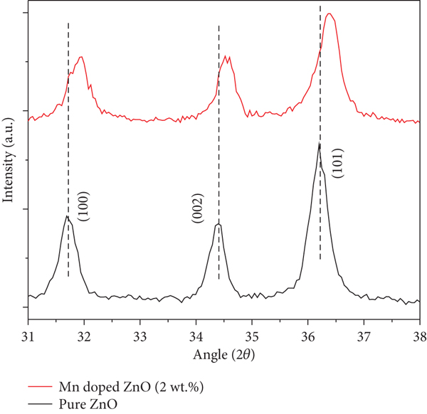
The synthesis of semiconductor materials with controlled doping is a tedious task since either the doping ions are found segregated at nanocrystal surfaces or they even form secondary phases, rather than being incorporated in the core [37]. Albeit, the XRD pattern of the Mn-doped ZnO seems nearly identical to that of the pure ZnO. However, from Figure 3(a), it may be primarily inferred that the diffraction peak intensity has decreased with the doping of Mn2+ content in the ZnO matrix. This observation indicates that the dopant ions have substituted the inner lattice of Zn2+ ions, as it has caused the crystallinity to degenerate (i.e., lattice disorder) and increase in the concentration of defects in the sample [38–40]. Secondly, interestingly, we have also observed a shift of XRD peaks in the case of Mn-doped ZnO towards higher 2θ values compared to those of pure ZnO. Specifically, Figure 3(b) demonstrates a slight shift in the center of the three most intense (100), (002), and (101) diffraction peaks observed for the Mn-doped ZnO nanostructures. This interesting observation indicates that Mn has been successfully incorporated into the host Zn2+ lattice sites. The ionic radius of the substitute Mn2+ (~0.80 Å) is slightly higher than that of Zn2+ (~0.74 Å) [41], so a slender shift towards lower angles was expected indicating the increase of the lattice parameters of the host lattice, as observed in numerous prior studies [39, 42, 43]. In contrast, in various other studies [44–46] and likewise in our present study, it has been experimentally observed that the diffraction peaks have shifted towards higher angles, instead. Shatnawi et al. also observed a decrease in the average crystallite size, and based on the X-ray photoelectron spectroscopy (XPS), results attributed it to the introduction of pronounced lattice defects (oxygen vacancies) with an increase in the Mn-doping level in ZnO [47]. Othman et al. have also observed (a) a shift in diffraction angle towards the higher angles, (b) a decrease in Zn-O bond length, and (c) a decrease in crystallite size with increasing Mn content in ZnO [48]. This interesting experimental observation may be sourced by the existence of multiple ionization valence states of Mn, such as Mn3+ and Mn4+ with relatively smaller ionic radii, ca. 0.58 Å, and 0.53 Å, respectively [49].
The X-ray diffractometer (XRD) remains a dominant quantitative analysis tool for the estimation of crystallite size. In general, the Scherrer formula is ubiquitously used to estimate nanostructural parameters, but it only considers the effect of crystallite size on the XRD peak broadening and ignores the important intrinsic strain contribution [50, 51]. Crystallite size and lattice strain measure the size of coherently diffracting domains and the distribution of lattice constants from lattice dislocations, respectively [52]. The origin of the lattice strain is mainly attributed to the point defect, grain boundary, and stacking faults as a result of doping which may ultimately cause lattice expansion or lattice contraction in the nanocrystals [53]. In the present study, the Williamson-Hall (W-H) method has been adopted for the estimation of the crystalline domain size of the synthesized samples, i.e., pure and Mn-doped ZnO. The simplified W-H diagnostic tool assumes that the broadening in Bragg’s peaks is the sum of peak broadening due to finite crystallite size and induced strain [54, 55]. The W-H equation is given as [56]
The crystallite size of pure and Mn-doped ZnO has been estimated to be ~21.10 nm and 18.76 nm, respectively, as shown in Table 1. It has been observed that the crystallite size of the Mn-doped ZnO sample has decreased as compared to pure ZnO.
The estimation of crystallite size and microstrain in pure and Mn-doped ZnO samples using the W-H method.
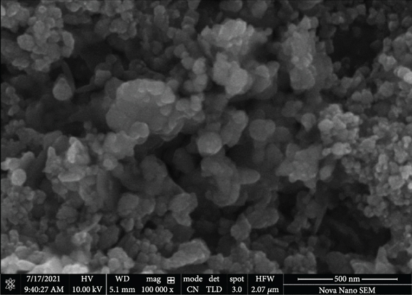
Field emission scanning electron microscopy (FESEM) analysis has been conducted to investigate the morphological properties of pure and Mn-doped ZnO nanoparticles. FESEM images have been noted at different magnifications to study the effect of Mn-doping on the shape, size, and distribution of particles. Specifically, FESEM images of both samples at 25,000x and 100,000x magnifications are displayed in Figures 4(a) and 4(b) for pure ZnO and Figures 4(c) and 4(d) for Mn-doped ZnO, respectively. In general, the FESEM micrographs depict nanoparticle-based surface morphology with inhomogeneity in particle size and shapes. However, it may be observed that particles with sizes in the 50–100 nm range are the most frequent in both photocatalysts. The nanoparticle-based surface morphology has merit in providing more surface-to-volume ratio for photocatalytic reaction. Admittedly, the FESEM of the synthesized photocatalysts (in powder form) show aggregation; however, it must be undermined that in the aqueous suspension form during the PCA study, the continuous stirring effectively prevents aggregation and thus maintain a large active surface area of the synthesized photocatalysts.

FESEM topographical analysis (surface view) of (a, b) pure ZnO and (c, d) Mn-doped ZnO at 25kx and 100kx magnification scales, respectively.


The light absorption property of the pure and Mn-doped ZnO nanoparticles has also been investigated in the near UV-visible (300–800 nm) wavelength range, as shown in Figure 5. It may be easily observed that both synthesized particles have considerably higher absorption in the UV-A region (300–400 nm) and relatively feeble absorption in the visible range (400–700 nm). Specifically, the pure ZnO nanoparticles exhibit a strong excitonic absorption band at 365 nm, which is envisaged to be the characteristic absorption peak of ZnO nanostructures [57, 58], whereas a slight hypsochromic shift in the absorption peak of the Mn-doped ZnO (~360 nm) is evident as compared to that of pure ZnO nanoparticles. Resultantly, the energy bandgap (Eg) of Mn-doped ZnO nanoparticles (3.4 eV) has increased as compared to that of pure ZnO (3.3 eV), as evidently observed by the Tauc plot depicted in Figure 5 (inset).
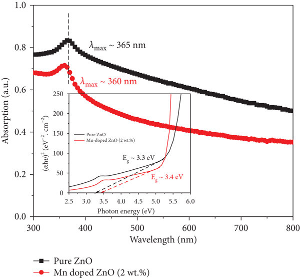
The UV-vis absorption spectra of pure and Mn-doped ZnO nanoparticles and (inset) the bandgap estimation of synthesized samples via Tauc plot.
Wu et al. have observed similar results, i.e., a blue shift in the absorption edge of ZnO and a broadening of Eg, which indicates that Mn-doping not only produces elemental energy levels but also affects the intrinsic defect energy levels of ZnO [59]. Husain et al. suggest that the blue shift in optical absorption or increase in the Eg, by successive doping of Mn in ZnO, may be attributed to the orbitals hybridized between the Mn atom and host band [60]. Viswanatha et al. also find it reasonable to expect the bandgap (Eg) to increase with increasing concentration of Mn since the bandgap of MnO (Eg~4.2 eV) is greater than that of ZnO (Eg~3.3 eV) [61]. It must be undermined that the crystallite size of as-synthesized ZnO (calculated from XRD) is significantly higher than the Bohr exciton radius of ZnO (i.e., 2.34 nm) [62]; therefore, the increase in bandgap or blue shift in absorption may not be attributed to quantum confinement effect. Instead, similar bandgap widening has been attributed to Burstein-Moss effect which is widely reported (for ZnO) in literature [25, 63, 64]. Nevertheless, based on the efficient absorption of pure and Mn-doped ZnO in the near UV wavelength range, we have investigated the capability of both photocatalysts in the presence of solar light irradiation instead of visible light, alone.
The Brunauer–Emmett–Teller (BET) method is ubiquitously utilized in nanotechnology and is envisaged to be an ideal experimental tool to estimate the specific surface area of the nanomaterials [65]. In the present study, a linear BET multipoint plot of

Linear plot of BET equation to determine the surface area of (a) pure ZnO and (b) Mn-doped ZnO nanoparticles.

The specific surface area of both synthesized photocatalysts may thus be mathematically determined by the following equation:
Figure 7 depicts the UV-vis absorption spectrum of MG dye (solution state in DI water) which implies the strong distinctive absorption peak positioned at λ
max~620 nm, accompanied by two prominent shoulder peaks at 315 and 425 nm, respectively [16, 72, 73]. The shoulder peaks correspond to benzoic rings in the dye structure [74]. Specifically, the weaker absorption peak in the near UV region (i.e., at 315 nm) may be ascribed to n-π

The solution state (DI water) absorption spectrum of methyl green pollutant dye.
During the PCA study, primarily, the light absorption characteristics of the pure MG aqueous solution (devoid of any photocatalyst) have been examined, under natural sunlight irradiation (illumination

The photodegradation of MG dye mediated by pure and Mn-doped ZnO nanoparticles (a) normalized


It may be easily observed from Figure 8(a) that both photocatalysts exhibit significant photocatalytic properties toward the degradation of MG pollutant dye. However, the photocatalytic performance of ZnO has been observed to improve substantially with Mn-doping. For detailed analysis, the photocatalytic degradation kinetics of MG dye has been investigated in time “t,” to estimate the photocatalytic reaction rate constant (
The linear expression for the pseudo-first-order is mathematically expressed as [79]
The value of “K1” has been calculated (from Figure 8(b)) by estimating the slope of the plot between irradiation time and
Contrariwise, the pseudo-second-order relation may be expressed as
The slope of graphs (presented in Figure 8(c)) helps to find out the values of the second-order rate constant (
Figure 8(d) compares the photocatalytic performance of both catalysts. It may be observed that after 60 min irradiation of natural sunlight, ~62.78% and ~66.44% photocatalytic degradation of MG aqueous solution has been achieved mediated by pure and Mn-doped (2 wt. %) ZnO photocatalyst, respectively. It may be established from Figure 8 that Mn-doped ZnO nanoparticles exhibit distinctly higher PCA efficiency of MG dye as compared to pure ZnO. Earlier, Li. et al. have also obtained similar results in a study that investigates the potential of pure and Mn-doped ZnO photocatalysts towards methyl orange dye (MO) degradation. In their study, the superior photocatalytic activity of Mn-doped ZnO has been ascribed to the dopant ions, which not only provide impurity energy levels but also serve as electron trapping sites to promote the separation and restrain the recombination of photogenerated charge carriers [80].
It is one of the major drawbacks of ZnO photocatalyst that the rate of electron-hole recombination is significantly higher [81]. Doping ZnO lattice with transition metals typically introduces intermediate energy states, wherein electrons might get trapped, thereby impeding the probabilities of electron-hole pair recombination [82]. For instance, Ibanescu et al. have previously confirmed a relatively blue shift in absorption with the manganese doping in ZnO and a reduction in the possibility of electron-hole pair recombination [83]. Similarly, Gupta et al. have also confirmed that the synergistic interactions with multiple charge transfer pathways may considerably enhance the PCA efficiency of the Mn-doped ZnO photocatalyst [84].
The photodegradation mechanism of methyl green dye mediated by synthesized photocatalysts (pure and Mn-doped ZnO) under sunlight may be described by the following successive reactions:
In the first step, when a photon having energy equaling or exceeding the bandgap energy (Eg) of photocatalysts (pure and Mn-doped ZnO) is absorbed, an electron (e−) is excited from the valence band (VB) and gets transferred to the empty conduction band (CB), creating a hole vacancy (h+) in the VB (as in Equation (10)). In the second step, these photoinduced charge carriers may migrate to the active sites of the surface of the photocatalyst, where they act as reducing/oxidizing agents to facilitate redox reactions on the surface. Specifically, the electrons react with oxygen to produce superoxide anion, whereas the holes react with water and hydroxide ions to produce hydroxyl radicals (as expressed in Equations (13) and (14), respectively) [85]. However, numerous studies have previously indicated that pure ZnO (as a photocatalyst) exhibits low-charge separation efficiency [85, 86]. Hence, photoinduced e−/h+ pairs in ZnO may inevitably recombine producing heat as wasted energy. Moreover, since the recombination process is much faster than the charge transportation to the active sites, this ultimately results in reduced photocatalytic efficiency of ZnO [87]. The recombination process inhibits the photocatalytic performance of ZnO, since only a limited number of photoinduced charge carriers produce the active radicals (•OH and
Contrariwise, when ZnO is doped with manganese atoms, the dopant ions replace Zn ions in the host lattice and tend to act like electron scavengers. The difference in the bandgap of pure and Mn-doped ZnO is a direct indication of the variation in the electronic structure of hexagonal ZnO with Mn2+ substitution [89]. The widening of the bandgap may be attributed to the Burstein-Moss effect, which causes a shifting in the position of the Fermi level into the conduction band (as shown in Figure 9). Specifically, the shift of Fermi level is the consequence of the Pauli exclusion principle, since the enhanced charge carrier concentration (sourced by Mn-doping) results in the occupancy of the low energy levels closer to the conduction band [64]. Resultantly, the low energy transitions are blocked, since the Pauli exclusion principle forbids excitation into the preoccupied states, and resultantly, the apparent bandgap is increased (where apparent

Schematic representation of the Burstein-Moss effect in Mn-doped ZnO nanoparticles.
Table 2 compares the photocatalytic activity of the synthesized photocatalysts (pure and Mn-doped ZnO) with the literature.
Comparison of photocatalytic performance of synthesized photocatalysts with literature.
4. Conclusion
In this study, we have utilized the chemical precipitation method to synthesize pure and Mn-doped ZnO (2 wt. %) nanoparticles for photodegradation (of methyl green pollutant dye). The XRD structural analysis has revealed the hexagonal wurtzite structure of both as-synthesized intrinsic and extrinsic (Mn-doped) ZnO photocatalysts. The investigation of photophysical properties by UV-vis indicates that the absorption peak in Mn-doped ZnO nanoparticles is blue-shifted by 5 nm (365 → 360 nm), due to the widening of the bandgap, which has been attributed to the Burstein-Moss effect instead of quantum confinement. Furthermore, the BET analysis has revealed that the specific surface area (
Footnotes
Data Availability
All data has already been provided in the form of ORIGIN plots. Further requested data can be made available upon the reader’s request.
Ethical Approval
Neither there are potential risks and ethical issues involved in the current research, nor the authors have engaged in any form of malicious harm to another person or animal.
Conflicts of Interest
The authors declare no conflict of interest.
Acknowledgments
The authors would like to thank the Deanship of Scientific Research Qassim University for funding the publication of this project.