
Abstract
In this study, TiO2 nanomaterials were prepared using a solvothermal method and codoped with Co2+ and Fe3+ ions for the photocatalytic degradation of organic pollutants under visible light. The physicochemical properties of the obtained materials were studied by powder X-ray diffraction, field emission electron scanning microscopy, energy-dispersive X-ray spectroscopy, and nitrogen adsorption isotherms. Optical absorption was characterized by UV-vis absorption spectroscopy. The photocatalytic activities of the prepared materials were evaluated through methylene blue (MB) degradation under visible light irradiation. Results showed the excellent performance of MB degradation investigated on TiO2 samples codoped with Co2+ and Fe3+ in comparison with undoped and Co2+-doped TiO2 samples. The codoped TiO2 samples degraded 85%–90% of MB after 120 min, whereas all the prepared TiO2 samples were composed of pure anatase phase and had a spherical-like shape and mean crystalline size ranging from 6.2 nm to 7.8 determined by Scherrer’s equation. The optical absorption of the TiO2 codoped with Co2+ and Fe3+ was significantly enhanced toward the visible light region. The pseudo-second-order kinetic model fits well for the degradation of MB on as-prepared TiO2 codoped with Co2+ and Fe3+.
1. Introduction
The development of metal oxide nanoparticles has been extensively attracted as adsorbents and photocatalysts for the treatment of dye-containing wastewater [1–3]. Titanium dioxide (TiO2) nanoparticles have considerable attention because of their long-term stability, low cost, and nontoxicity [4]. However, the major drawback of TiO2 is a large bandgap energy (~3.2 eV and 3.0 eV for anatase and rutile phases, respectively), and it requires ultraviolet (UV) irradiation (wavelength,
In this study, we aimed to synthesize and characterize TiO2 nanoparticles codoped with Co2+ and Fe3+ ions using the solvothermal method. The effect of the doping concentration of Co2+ was investigated to determine the optimal condition for preparing a photocatalyst with high activity in the visible light region. The obtained nanomaterials were investigated by various physicochemical methods, including XRD, FE-SEM, EDXS, nitrogen adsorption-desorption isotherms, and UV-vis absorption. Photocatalytic activities of prepared nanomaterials were evaluated for degradation of methylene blue (MB). Our results indicated that the prepared TiO2 materials codoped with Co2+ and Fe3+ showed a highly photocatalytic efficiency compared to single-doped TiO2. This nanomaterial could be an effective alternative for the treatment of wastewater in the textile industry.
2. Materials and Methods
2.1. Chemicals and Preparation
All chemicals were of analytical grade and used as received without further purification. Tetraisopropyl orthotitanate (
Single doping of TiO2 with Co2+ and codoping with Co2+ and Fe3+ were conducted by the solvothermal synthesis. The obtained samples were referred to as
2.2. Material Characterization
X-ray diffraction (XRD) patterns of the prepared TiO2 samples were recorded with a D8 ADVANCE Bruker Diffractometer within
The photocatalytic activity of the prepared TiO2 samples was evaluated by degradation of MB under visible light irradiation. A 250 W Osram mercury lamp equipped with a UV cut-off filter was used as a visible light source. In a typical experiment, 40 mg of the prepared TiO2 sample was added into a 100 mL quartz photoreactor containing 50 mL of 14 mg·L-1 MB solution. The reaction mixture was then stirred in the dark at a constant speed of 150 rpm for 30 min to reach the adsorption-desorption equilibrium. The resulting mixture was irradiated under the visible light source for up to 120 min. After predefined intervals (30, 60, 90, and 120 min), the samples were removed from the photoreactor and centrifuged. The residual concentration in the supernatant was measured with a UV-vis spectrometer (Agilent 8453, USA) at a wavelength of 665 nm. MB concentration was determined using a linear regression equation obtained by plotting a calibration curve of MB within a range of known concentrations. The photocatalytic ability of the TiO2 samples was evaluated through the percentage of MB degradation as follows:
3. Results and Discussion
3.1. Characterization of the Synthesized Materials
The XRD patterns of the synthesized samples (undoped, single-doped, and codoped TiO2) are shown in Figure 1. The results demonstrated the formation of monocrystalline TiO2 in the anatase phase in all samples. The XRD pattern of undoped TiO2 samples (Figure 1(a)) shows typically crystalline peaks at

XRD patterns of (a) undoped TiO2, (b) TiO2-1%Co, (c) TiO2-2.5%Co, (d) TiO2-5%Co, and co-doped TiO2: (e) TiO2-1%Co-2.5%Fe and (f) TiO2-2.5%Co-2.5%Fe.
Mean crystalline size and phase of the prepared TiO2 samples.
The morphology of the synthesized TiO2 samples was further observed through FE-SEM imaging. Figure 2 presents the FE-SEM images of the representative samples: undoped TiO2 (Figures 2(a) and 2(b)), TiO2-1%Co (Figures 2(c) and 2(d)), TiO2-5%Co (Figures 2(e) and 2(f)), and TiO2-1%Co-2.5%Fe (Figures 2(g) and 2(h)) at magnifications of 100 k and 200 k. The results indicated that the synthesized TiO2 samples consisted of numerous crystalline particles that agglomerated to form tiny clusters. The FE-SEM images demonstrated that the particles were spherical and had uniform size distribution. The calculations from the FE-SEM images confirmed that the average sizes were 26.5, 23.8, 27, and 20.8 nm for undoped TiO2, TiO2-1%Co, TiO2-5%Co, and TiO2-1%Co-2.5%Fe samples, respectively. These results showed deviations from the data obtained from XRD due to the agglomeration of the nanoparticles in the FE-SEM images. However, the results were in agreement with the XRD results, confirming that Co and Fe doping can suppress TiO2 particle growth and the TiO2-1%Co-2.5%Fe sample had the smallest particle size with a narrow distribution of the particle size (inset in Figure 2).

FE-SEM images of undoped TiO2 (a, b), TiO2-1%Co (c, d), TiO2-5%Co (e, f), and TiO2-1%Co-2.5%Fe (g, h) samples with magnifications of 100 k and 200 k. Inset shows a particle size distribution of TiO2-1%Co-2.5%Fe samples.
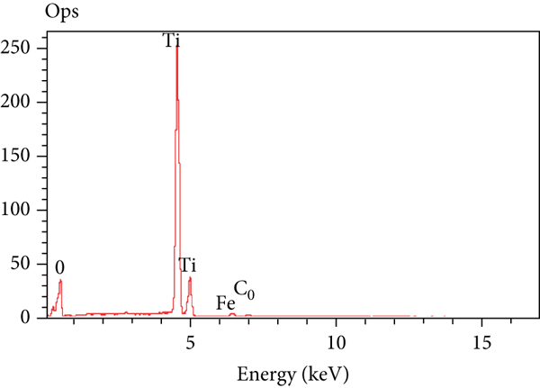
The presence of doping metals on the TiO2 samples was determined through EDXS analyses. The EDXS spectra of the representative TiO2 samples (undoped TiO2, TiO2-1%Co, TiO2-5%Co, and TiO2-1%Co-2.5%Fe) are presented in Figure 3. As shown in Figures 3(a), O and Ti were found to be the major components of undoped TiO2 with 65.35 at% and 33.65 at%, respectively. Co was detected as the doping component of TiO2-1%Co and TiO2-5%Co samples because tiny peaks were observed in the EDXS spectra (Figures 3(b) and 3(c)), which were attributed to the presence of Co in the samples (0.21 at% and 0.89 at%, respectively). The contents of Co and Fe in TiO2-1%Co-2.5%Fe samples were 0.28 at% and 0.74 at%, respectively (Figure 3(d)). The results confirmed that Co and Fe were successfully doped in the TiO2 samples.

EDXS spectra of (a) undoped TiO2, (b) TiO2-1%Co, (c) TiO2-5%Co, and (d) TiO2-1%Co-2.5%Fe samples.


Nitrogen adsorption-desorption isotherms were determined for the representative samples to analyze and quantify the pore structure and surface area of doped and codoped TiO2 samples. Figure 4 presents the nitrogen adsorption-desorption isotherms of three selected samples: TiO2-1%Co (Figure 4(a)), TiO2-5%Co (Figure 4(b)), and TiO2-1%Co-2.5%Fe (Figure 4(c)). Results in Figure 4 indicate that the three samples had a similar hysteresis loop, which could be associated with capillary condensation in mesopores. The hysteresis loops of these samples, which were observed in the

Nitrogen adsorption-desorption isotherms of representative samples: (a) TiO2-1%Co, (b) TiO2-5%Co, and (c) TiO2-1%Co-2.5%Fe samples.


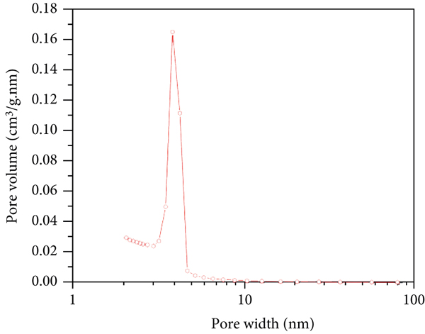
Pore size distributions of representative samples: (a) TiO2-1%Co, (b) TiO2-5%Co, and (c) TiO2-1%Co-2.5%Fe samples.


Surface characteristics of the representative TiO2 samples.
aBET surface area. bTotal pore volume determined using desorption curves of the isotherms. cPeak pore sizes from the pore size distributions.
Table 3 compares the BET surface areas of doped and codoped TiO2 materials of the present work with those of other doped and codoped TiO2 materials reported previously. The BET surface areas of doped and codoped TiO2 materials of our work were within 164–174 m2/g, which were higher than those of Fe-doped TiO2 materials [18, 19], Co-doped TiO2 materials [20], Cr- and Fe-doped TiO2 materials [18], and Fe- and La-doped TiO2 materials [19] (Table 3). The larger BET surface areas of the doped and codoped TiO2 materials of the present work were possibly due to their smaller particle sizes ranging from 6.2 nm to 7.5 nm (Table 3), depending on the doping amount. The doped TiO2 materials of our work were prepared under more favorable conditions (solvothermal treatment at 180°C without calcination). The other TiO2 materials were prepared by hydrothermal treatment or sol-gel method, followed by calcination, which resulted in the formation of larger particle sizes and significant loss of the BET surface area (Table 3).
Comparison of the BET surface areas of the TiO2 samples prepared in this work with those of previous works.
The UV-vis absorption spectra of undoped TiO2, doped TiO2, and codoped TiO2 samples were recorded to study their optical properties (Figure 6). As shown in Figure 6 (curve a), the undoped TiO2 sample was characterized by a narrow absorption spectrum, ranging from 250 nm to 370 nm in the UV region. No visible light absorption was observed for the undoped TiO2 sample. Co-doped TiO2 (Figure 6, curves b–d) and Co- and Fe-doped TiO2 samples (Figure 6, curves e and f) had broader absorption spectra, which shifted toward longer wavelengths in the visible light region. The absorption spectra of Co-doped TiO2 samples had maximum peaks at about 330–340 nm, but the shoulder of the peaks shifted to the visible range (400-700 nm). TiO2 samples codoped with Co and Fe were characterized by a broader maximum peak at 400–500nm and stronger intensity of the absorption shoulder in the visible region compared with those of single-doped TiO2 samples. Figure 6 shows that the absorption edges of TiO2 codoped with Co and Fe moved remarkably, with a redshift to the visible region in comparison with undoped TiO2. These optical absorption characteristics of TiO2 codoped with Co and Fe were possibly due to absorption induced by dopants. The absorption edges can be extrapolated by making a tangent line of the UV-vis absorption spectra [21]. Therefore, the bandgap energies of the prepared TiO2 samples were estimated from the UV-vis spectra via the following equation:
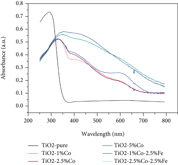
UV-vis spectra of undoped TiO2, TiO2-1%Co, TiO2-2.5%Co, TiO2-5%Co, TiO2-1%Co-2.5%Fe, and TiO2-2.5%Co-2.5%Fe samples.
The bandgap energy levels of Co-doped TiO2 samples were estimated to be 2.03 eV, 2.00 eV, and 1.99 eV (for TiO2-1%Co, TiO2-2.5%Co, and TiO2-5%Co, respectively), which were higher than those of Co- and Fe-codoped TiO2 samples (their bandgap energies are 1.65 eV and 1.59 eV for TiO2-1%Co-2.5%Fe and TiO2-2.5%Co-2.5%Fe, respectively). The calculated bandgap energies of single-doped and codoped TiO2 samples were lower than that of undoped TiO2 samples (~3.25 eV). The results revealed that the dopant elements were successfully incorporated into the TiO2 crystal lattice and extended the optical absorption toward the visible light region. TiO2 samples codoped with Co and Fe had stronger optical absorption of the visible light than single-doped and undoped TiO2 samples. This means that the TiO2 codoped with Co and Fe can absorb visible light in a much wider range of wavelengths than undoped TiO2 and single-doped TiO2 samples, which is beneficial for increasing the photocatalytic efficiency under visible light.
3.2. Photocatalytic Activities of the Prepared TiO2 Materials for Degradation of MB
Before the photocatalytic reaction, the adsorption for MB removal on the synthesized TiO2 materials was conducted under dark conditions. Results obtained showed that the percentages of MB removal were only 5%-6% after 60 min for all the synthesized materials, but desorption of MB was observed by prolonging adsorption time for those materials. Therefore, the photocatalytic activities of the prepared TiO2 samples were evaluated by degrading MB solution under visible light irradiation. The reaction mixture was first stirred in the dark for 30 min to reach the adsorption-desorption equilibrium and to ensure that the degradation of MB obtained is due to the photocatalytic reaction with the presence of the synthesized TiO2 materials, but not due to the adsorption. Figure 7 shows the percentage of MB degradation over all of the TiO2 samples (undoped TiO2, TiO2-1%Co, TiO2-2.5%Co, TiO2-5%Co, TiO2-1%Co-2.5%Fe, and TiO2-2.5%Co-2.5%Fe samples) as a function of irradiation time. For comparison, control experiments were performed under the same conditions but without the presence of a photocatalyst (Figure 7, curve MB). As shown in Figure 7 (curve MB), almost no degradation of MB was observed without the photocatalyst (only 7.95% of MB was degraded after 120 min of exposure to visible light irradiation), indicating that MB was unable to self-degrade. Undoped TiO2 and Co-doped TiO2 with low doping concentration (TiO2-1%Co and TiO2-2.5%Co) showed pure photocatalytic activity. The degradation efficiencies of MB on undoped TiO2 samples were comparable with those of Co-doped TiO2 samples with low Co doping concentration for all the tested time points (Figure 7, curves undoped TiO2, TiO2-1%Co, and TiO2-2.5%Co). The degradation percentages of MB were 10.95%, 17.85%, and 17.5% after 120 min of irradiation for undoped TiO2, TiO2-1%Co, and TiO2-2.5%Co, respectively. However, the degradation of MB is enhanced with higher Co doping concentration. The degradation percentages of MB increased three times on TiO2-5%Co samples and were higher than those on TiO2-1%Co and TiO2-2.5%Co samples for all the tested time points. About 39.8% of MB was degraded within 30 min on TiO2-5%Co and then gradually increased to ~50% by increasing irradiation time up to 120 min. Moreover, the significantly enhanced degradation of MB was observed on codoped TiO2 samples (Figure 7, curves TiO2-1%Co-2.5%Fe and TiO2-2.5%Co-2.5%Fe). The highest degradation of MB was obtained by TiO2 codoped with 1%Co and 2.5%Fe for all the time points tested. The degradation of MB was almost complete on codoped TiO2 samples for the tested time; 90% and 85% of MB were degraded on TiO2-1%Co-2.5%Fe and TiO2-2.5%Co-2.5%Fe after 120 min, respectively.

The degradation of MB using different catalysts: undoped TiO2, TiO2-1%Co, TiO2-2.5%Co, TiO2-5%Co,TiO2-1%Co-2.5%Fe, and TiO2-2.5%Co-2.5%Fe samples under visible light irradiation.
The results showed that TiO2 samples codoped with Co and Fe exhibited higher photocatalytic degradation of MB under visible light than the undoped and single-doped TiO2 samples, which could be attributed to their lower bandgap values compared to those of the undoped and single-doped TiO2 samples. These observations indicated that codopant ions had a favorable effect on the photocatalytic performance of the prepared TiO2 materials. These ions can provide additional energy levels within the bandgap of TiO2. The bandgap of TiO2 consists of a contribution from the 2p orbitals of O for the valence band and the 3d orbitals of Ti toward the conduction band, which have a large difference in energy, leading to the activation of TiO2 in the UV light region with extremely high energy. The band structures of codoped TiO2 materials are mainly affected by the 3d energy states of the transitional metal ions (Co2+ and Fe3+). In fact, the UV-vis absorption studies (Figure 6) revealed that the respective absorption bands of codoped TiO2 samples effectively shifted toward wavelengths longer than 400 nm. Thus, the partially filled Co/Fe 3d bands were located below the conduction band of TiO2. Hence, when photons with wavelengths longer than 400 nm are used for illumination, the electrons in the Co 3d and Fe 3d bands, instead of electrons in the valence band of TiO2, are excited to the conduction band, while Co2+ and Fe3+ loses one electron and becomes Co3+ and Fe4+ (see Scheme 1). This phenomenon can induce more photogenerated electrons and holes to participate in the photocatalytic reaction.

Band structure of TiO2 and TiO2 codoped with Co2+ and Fe3+. VB: valence band; CB: conduction band.
As calculated above, the bandgap energy values of codoped TiO2 samples (1.59 eV-1.65 eV) were lower than those of single-doped TiO2 samples (1.99-2.03 eV) and undoped TiO2 (3.25 eV), indicating the lower energy of photons necessary to generate electron transition and holes (
The plausible reaction mechanism of the photodegradation is given below:
It is suggested that the photodegradation of MB can be divided into three main steps: (1) the initial step is the formation of electrons (
Thus, this work proposed a study to understand the role of Co and Fe in codoped TiO2 nanoparticles in yielding better performance as a visible light-driven photocatalyst.
The kinetics of photodegradation of MB on as-prepared TiO2 materials was further investigated using the pseudo-first-order kinetic model [22] as follows:

Plots of (a) first- and (b) second-order reaction rates for the degradation of MB over as-prepared TiO2 materials.

where
The observations revealed that the degradation of MB on the undoped TiO2 and Co single-doped TiO2 samples with 1% Co and 2.5% Co doping can be described by the pseudo-first-order equation. Meanwhile, the pseudo-second order fits better compared to the pseudo-first order for describing the degradation of MB on the TiO2 doped with 5%Co and codoped with Co and Fe, considering that
Table 4 compares the degradation of MB on the TiO2 materials as prepared in this work with those published in previous works [24–28]. It was found that the TiO2 codoped with 1%Co and 2.5%Fe in this work showed the highest photocatalytic efficiency with 90% of MB degraded after visible light irradiation of 2 h. The metal oxide (Bi2O3) [24] and mixed metal oxides (e.g., CeO2/V2O5 [24], CeO2/CuO [25], and ZnO/CdO [26]) exhibited much lower degradation efficiency of MB compared with the TiO2 materials of our work, which degraded about 64.2%-75% of MB for a longer irradiation time of 4 h. The complex metal oxide CaBi6O10 [24] and nanocomposite g-C3N4-CdS [27] demonstrated relatively high degradation percentages of MB (~97% and 90.45% for CaBi6O10 and g-C3N4-CdS, respectively), which could be comparable with that of the TiO2 codoped with 1%Co and 2.5%Fe of our work, but these materials require a quite long irradiation time of 4 h (for CaBi6O10) and 3 h (g-C3N4-CdS) to obtain the complete degradation of MB under visible light. Moreover, a similar study reported that TiO2 doped with 10% F [28] also revealed a high degradation percentage of MB (~90%), but after a much longer irradiation time of 4 h, compared to that of the TiO2 codoped with Co and Fe of our work. The comparison revealed that the photocatalytic efficiency of the materials depends not only on their band structure but also on their surface characteristics. The excellent photocatalytic performance of the TiO2 codoped with Co and Fe of our work could be attributed to their larger BET surface area compared to those of the other materials. As a result, the TiO2 codoped with Co and Fe has more active sites and simultaneously can absorb much more visible light and generate more electron-hole pairs, which in turn enhanced the photocatalytic activity and reduced the reaction time as compared to the other materials.
Comparison of MB degradation of the TiO2-codoped samples of this work with other materials.
4. Conclusions
In this work, we presented the characterization of TiO2 nanoparticles codoped with Co2+ and Fe3+ synthesized through solvothermal treatment at 180°C without further calcination at high temperatures. Undoped TiO2 and Co single-doped TiO2 materials were also prepared using the same procedure to compare with codoped TiO2 in terms of optical absorption and photocatalytic efficiency for MB degradation. The doped and codoped TiO2 materials of the present work exhibited better adsorption characteristics (e.g., their BET surface areas of 164-174 m2/g) than doped and codoped TiO2 materials of previous works; as-prepared codoped TiO2 materials had significantly enhanced optical adsorption toward the visible light region. Among the TiO2 samples studied, the codoped TiO2 materials showed the highest photocatalytic activity for the degradation of MB; they degraded about 90% of MB within 120 min under visible light irradiation. The TiO2 samples codoped with Co and Fe in this work showed higher photocatalytic efficiency for the degradation of MB than those of metal oxides, mixed metal oxides, and other materials in the previous works. The pseudo-second-order kinetic model fits well for describing the degradation of MB on the TiO2 codoped with Co and Fe. Hence, this study provides a simple route to synthesize an effective photocatalyst for the degradation of dye compounds under visible light for wastewater treatment.
Footnotes
Data Availability
All the data used to support the findings of this study are included within the article.
Conflicts of Interest
There are no conflicts of interest to declare.
Acknowledgments
This study was funded by the Vietnam National Foundation for Science and Technology Development (NAFOSTED) under grant number 104.03-2019.313.