
Abstract
Silver liquid thin film, formed by silver nanoparticles stacking and spreading on the surface of the liquid, is one of the important parts of magnetic fluid deformable mirror. First, silver nanoparticles were prepared by liquid phase chemical reduction method using sodium citrate as reducing agent and stabilizer and silver nitrate as precursor. Characterization of silver nanoparticles was studied using X-ray diffractometer, UV-vis spectrophotometer, and transmission electron microscope (TEM). The results showed that silver nanoparticles are spherical and have a good monodispersity. Additionally, the effect of the reaction conditions on the particle size of silver is obvious. And then silver liquid thin films were prepared by oil-water two-phase interface technology using as-synthesized silver nanoparticles. Properties of the film were investigated using different technology. The results showed that the film has good reflectivity and the particle size has a great influence on the reflectivity of the films. SEM photos showed that the liquid film is composed of multilayer silver nanoparticles. In addition, stability of the film was studied. The results showed that after being stored for 8 days under natural conditions, the gloss and reflectivity of the film start to decrease.
1. Introduction
Magnetic fluid deformable mirror is an important element for wave front correction in adaptive optics. It is widely used in the field of high-resolution imaging technology, laser plastic surgery, and laser communications [1, 2]. The reflective film plays an important role in the deformable mirror system and silver liquid thin film is a common material in this field [3, 4]. On the one hand, the film exhibits the metallic luster and high reflectivity. On the other hand, the film is liable to flow and not easy to be fractured [5]. In the presented work, preparation and characterization of silver liquid thin films formed by silver nanoparticles were reported, and the film can stack and spread on the surface of the magnetic liquid. There are two steps in the preparation techniques of silver liquid thin film, preparation of silver nanoparticles, and formation of silver liquid thin film. A number of approaches are available for synthesis of silver nanoparticles, for example, chemical reduction [6, 7], photochemical [8], electrochemical [9], sonochemical [10], sol-gel [11], and so forth. Silver nanoparticles were prepared by liquid phase chemical reduction method using sodium citrate as reducing agent and stabilizer and silver nitrate as precursor in this paper. And then silver liquid thin film was assembled by some of reaction process using as-synthesized silver nanoparticles as raw material. There are many methods for preparation of silver liquid thin film [12–15] too, such as LB film method, self-assembled monolayer method, vapor deposition method, and oil-water two-phase interface deposition method. The oil-water two-phase interface deposition method is commonly used currently. Many studies of the film preparation were reported using this method. Yogev and Efrima [16–18] got the first silver liquid thin film. The film can reach 3–4 microns thick wherein 40% of the silver content can be achieved. The essence of this process is that silver ions are reduced to silver. Gordon et al. [19] invented an easier way to prepare silver liquid thin films. It is not necessary to add any surface active agent in this method; the silver sol solution just needs to be mixed and dissolved with certain transition metal complexes of the organic phase. And after a period of vigorous shaking, the dark film forms. Then Gingras and coworkers [20] transformed the above-described method and proposed a new method. The mixture of the silver sol and organic ligand dissolved in organic solvent was violently shaken, and a liquid metal film can be obtained. In our laboratory, silver liquid thin films were prepared by the oil-water two-phase interface deposition method. As we know, the properties, such as size, shape, composition, and spatial distribution of nanoparticles of the silver film, depend on its microstructure [21, 22]. So reaction conditions for preparing silver nanoparticles, including reaction time and initial dropping amount of sodium citrate, would be investigated. In addition, the basic properties of silver liquid thin films, such as the microstructure, chemical composition, forming principle, and reflectivity, were studied.
2. Experimental Details
2.1. Preparation of Silver Liquid Thin Films
2.1.1. Preparation of Silver Nanoparticles
Silver nanoparticles were firstly prepared by liquid phase chemical reduction method. In this progress, silver nitrate was the precursor and sodium citrate was the reducing agent and stabilizer. And then the silver nanoparticles with preferable monodispersity and high stability can be obtained.
Silver nitrate was weighed and added into 100 mL deionized water and stirred for 30 min and heated to boiling in an oil bath. The sodium citrate was weighed and added into 20 mL deionized water, stirred for 30 min, and heated to boiling. First, the set amount of sodium citrate solution was slowly added dropwise into the boiling silver nitrate solution within one minute while vigorously stirring, and the solution was kept boiling for 5 min. And then, continue to drop the sodium citrate solution into the silver nitrate solution, and the total amount of the sodium citrate solution was 2 mL. The reaction solution was kept boiling until the end of the reaction. After completion of the reaction, the solution was transferred to a magnetic stirrer and was stirred until cooling down. In the reaction, the initial volume of the sodium citrate was set: 0.2 mL, 0.3 mL, 0.4 mL, 0.5 mL, and 0.6 mL. And the total reaction time was set: 10 min, 20 min, 30 min, 40 min, and 50 min. The mixture of silver nanoparticles was washed through centrifugation. And the impurities, such as unreacted silver nitrate and sodium citrate, were removed. Finally, the pure silver sol was obtained.
2.1.2. Preparation of Silver Liquid Thin Films
The 50 mL solution with 2,2-bipyridine and 1,2-dichloroethane was prepared, and the concentration was 5 × 10⁻² mol/L. The 50 mL nanosilver sol solution was measured. The two solutions were poured into a sealed polypropylene plastic cup; the mixture solution was shaken for 10 min. After the solution was kept stable for at least 30 min, a layer of thin film with a metallic luster coating the entire aqueous phase occurred. The excess organic phase and water are discharged. And the silver liquid thin film remained on the surface of the deionized water and was stored in a polypropylene Petri dish. The clean quartz substrate was ramped into the solution with a certain gradient to carry the silver thin film and taken out from the solution keeping the same gradient. Then the silver thin film was transferred onto the surface of the substrate, dried in an oven at 80°C for 30 min, and stored at room temperature in an airtight environment.
2.2. Characterisation Methods
Phase analysis measurements were performed in a Rigaku Ultima X-ray diffractometer (Japan RIGSKU Company). The quartz substrate coated with silver thin film was tested directly. The scan angle was from 30° to 120°, and the scanning speed was 10°/min.
UV-visible absorption characteristic analysis was carried out with a UV-2600 UV-Vis spectrophotometer. The silver sol was diluted, placed in a quartz cuvette, and tested directly. The optical slit width was set to 2 cm.
Microscopic morphology of silver nanoparticles was observed with a transmission electron microscope JEM-2100F at an accelerating voltage of 120 kV.
On the condition of natural environment, stability of silver liquid thin film was tested. The film gloss and light reflection capability were observed by way of taking pictures of every four days.
On the condition of 5.0 kV operating voltage, the surface morphology of silver liquid thin film on the surface of quartz substrate was observed with a scanning electron microscopy (SEM) JSM-6700F (Japan JEOL Company).
Reflectivity curves were obtained with U-4100 near-infrared spectrophotometer (Japan RIGSKU Company). The silver liquid thin film coated on the surface of quartz substrate was observed directly.
3. Results and Discussion
3.1. Characterization of Silver Nanoparticles
3.1.1. Phase Analysis of Silver Nanoparticles
Phase of silver nanoparticles was investigated by X-ray diffraction method and corresponding XRD pattern was shown in Figure 1. It indicates the crystalline structure of silver nanoparticles. Clear peaks of face centered cubic (fcc) phase at 38.15°(111), 44.28°(200), and 62.62°(220) were shown in the XRD pattern. The values of plane spacing d corresponding to these peaks are 2.3527 Å, 2.0367 Å, and 1.4823 Å, respectively. It is found that all the diffraction peaks in the pattern correspond to those of silver (JCPDS No. 04-0783). Therefore, the silver nanoparticles have high purity, and neither silver compound nor impurity has been intermixed in the silver nanoparticles.

XRD patterns of silver nanoparticles synthesized by chemical reduction method.
3.1.2. Morphology and Monodispersity Analysis of Silver Nanoparticles
UV-vis absorption spectroscopy is one of the widely used simple and sensitive techniques for the observation of nanoparticles synthesis and characterization. Morphology, particle size, and its distribution of silver nanoparticles are related to some parameters of UV-vis absorption spectrum, such as absorption peak number, position, FWHM, and peak value.
Figure 2 is UV-vis absorption spectroscopy of silver nanoparticles under different reaction time. One peak of 400–420 nm is shown in every curve, and half-widths of all absorption peaks are relatively narrow, and it indicates that the obtained silver nanoparticle size is relatively uniform and has good monodispersity. According to Mie scattering theory [23], size of the nanoparticles is related to the peak position of UV-vis absorption spectroscopy and the size change is related to the displacement of the peak. Generally, if the particles size is small, the peak position is near ultraviolet region. On the contrary, the peak position is close to infrared region. As is shown in Figure 2, a red shift of the plasmon resonance absorption peak occurs with the increase of reaction time, from 10 to 30 min. The maximum absorption peak moves from 400 to 420 nm. It indicates that the size of the silver nanoparticles increases with the increase of reaction time within 30 min. However, when the reaction time increases from 30 to 50 min, the maximum absorption peak position of silver nanoparticles has no obvious red shift, but the peak value gradually increases. When the reaction time exceeds 40 min, the peak value increasing trend slows. It shows that when the reaction time extends from 30 to 50 min, the particle size has no obvious growth, while the concentration of silver nanoparticles increases and gradually stabilizes when the reaction proceeds to 40 min. This phenomenon can be explained from the reaction mechanism of silver nanoparticles. When the reaction is continued to 30 min, the nucleation of silver nanoparticles forms and grows continually. When the reaction time is longer than 30 min, the surface of silver nanoparticles is coated by sodium citrate, which plays the role of a stabilizer. So the size of particles does not grow any more. So, during this period, the main phenomena in the reaction system are the increasing of the concentration of silver nanoparticles. When the reaction time reaches 40 min, the silver ions in the solution have been reduced to the elemental silver. The increasing rate of the concentration of silver nanoparticles is gradually slowing down.

UV-vis absorption spectra of silver nanoparticles synthesized under different reaction times. (a) 10 min, (b) 20 min, (c) 30 min, (d) 40 min, and (e) 50 min.
Figure 3 is UV-vis absorption spectroscopy of the silver nanoparticles with different initial dropping amount of sodium citrate. When the initial dropping amount of sodium citrate decreases from 0.6 to 0.2 mL, plasmon resonance absorption peak of the silver sol increases from 410 nm to 450 nm. It indicates that the size of silver nanoparticles increases gradually with the decrease of initial dropping amount of sodium citrate. The reason for this phenomenon is related to formation of nucleation in the process of reaction. A small amount of silver ions is reduced to silver atom with additive of initial dropping amount of sodium citrate. The silver atoms reunite and become silver nucleation. Then the silver ions are reduced by the excess sodium citrate continually. The silver atoms reduced in the later period do not form new nucleation but deposit on the original nucleus directly, so that the nucleus grows. It can be seen that the number of nucleation determines size of silver nanoparticles directly. If the total amount of silver nitrate is constant, the silver atoms increase with the increase of the initial dropping amount of sodium citrate in the process of reaction. And then more nucleation occurred, which leads to the smaller size of silver nanoparticles. The curve in Figure 3(e) has significant changes. It is attributed to the extension of storage time of the silver nanoparticles. During this period, the agglomeration of the particles occurs, size of the particles increases, and distribution of particle size becomes wider.

UV-vis absorption spectra of silver nanoparticles synthesized with different initial dropping amounts of sodium citrate. (a) 0.2 mL, (b) 0.3 mL, (c) 0.4 mL, (d) 0.5 mL, and (e) 0.6 mL.
As-synthesized silver nanoparticles are spherical and relatively uniform in size by UV-vis absorption spectroscopy. It has been demonstrated by the results from TEM images of silver nanoparticles, as is shown in Figures 4(a) and 4(b). Through the TEM images we can see that most of the nanoparticles are spherical and the size is relatively uniform, and the average particle size is about 70 nm.

High-magnification (a) and low-magnification (b) TEM images of silver nanoparticles under conditions of 40 min of reaction time and 0.2 mL of initial dropping amount of sodium citrate.
3.2. Preparation of Silver Liquid Thin Films
3.2.1. Microstructure of Silver Liquid Thin Film
Figure 5 is microstructure of the silver liquid thin film on the quartz substrate. It can be seen that the liquid film is composed of multilayer silver nanoparticles. The silver nanoparticles are mainly spherical and are evenly distributed within the field of view. Size of the nanoparticles is relatively uniform and about 70 nm. It indicates that the silver particle film had excellent monodispersity.

SEM surface images of silver liquid thin films. (a) ×20000, (b) ×200000.
3.2.2. Reflectivity of Silver Liquid Thin Film
Silver liquid thin films were prepared by silver nanoparticles with different sizes under the conditions of different initial dropping amounts of sodium citrate. Sizes of silver nanoparticles increase gradually with the decrease of initial dropping amount of sodium citrate by analyzing the UV-vis absorption spectroscopy. Based on this conclusion, the relationship between particle size and reflectivity was studied. Figure 6 is the reflectivity spectra of silver liquid thin films formed by silver nanoparticles with different diameter. As can be seen from Figure 6, the reflectivity of silver liquid thin films is getting higher gradually, ranging from 65% to 75%, and the reflectivity changes with the wavelength increase of the incident light. When the wavelength of the incident light is near infrared region, the reflectivity is relatively higher. When the size of silver nanoparticles increases, the reflectivity at different wavelengths increases significantly. The reason for the increase of reflectivity is the film thickness. In general, the thicker the film is, the higher the reflectivity is. Therefore, when the particle size increases, the film thickness increases, and then the reflectivity increases.

Reflectivity spectra of silver liquid thin films formed by silver nanoparticles with different diameter observed under conditions of different initial dropping amounts of sodium citrate. (a) 0.2 mL, (b) 0.3 mL, (c) 0.4 mL, and (d) 0.5 mL.
3.2.3. Stability of Silver Liquid Thin Film
Considering the durability and availability of the magnetic fluid deformable mirror, the stability of the silver liquid thin films needs to be surveyed. The film is placed on the natural environment for 1 day, 4 days, 8 days, 12 days, 16 days, and 20 days. The changes of appearance of the film were observed by naked eyes, as shown in Figure 7. It can be seen from Figure 7 that the silver liquid thin film has good gloss and reflectivity at the fourth day. During 4 to 7 days, the gloss and reflectivity are almost the same. When the film is stored for eight days, the surface of the film is still complete, but the light dims, and the gloss and reflection property decrease. At the twelfth day, the surface of film begins to damage and the reflection property declines further. The surface of the film has a complete burst at the twentieth day. Several large fragments form and the reflection property is lost completely. The reason of the rupture of the film surface may be due to the oxidization of the silver particles on the film surface during the period of storage, then the adhesive force between the nanoparticles decrease. Therefore, the surface properties of the film change, and after the long time storage the film is completely broken.
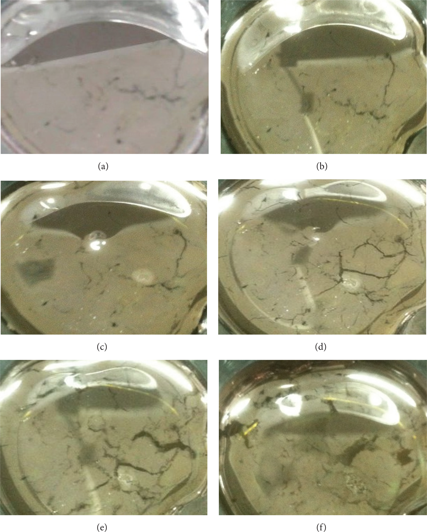
Photographs of silver liquid thin films stored for different days. (a) 1 day, (b) 4 days, (c) 8 days, (d) 12 days, (e) 16 days, and (f) 20 days.
4. Conclusions
The highly reflective silver liquid thin films were prepared using silver nanoparticles stacking and spreading on the surface of the liquid. Firstly, silver nanoparticles were prepared by liquid phase chemical reduction method. The results showed that the nanoparticle is spherical and has a good monodispersity. Reaction conditions, including reaction time and initial dropping amount of sodium citrate, have significant impact on size of silver nanoparticles. Size of silver nanoparticles increases with the increase of reaction time within 40 min. However, size of silver nanoparticles increases gradually with the decrease of initial dropping amount of sodium citrate. And then silver liquid thin films are prepared using as-synthesized silver nanoparticles by oil-water two-phase interface technology. The results showed that the film has good reflectivity, which is related to the size of silver nanoparticles. Microstructure analysis showed that the liquid film is composed of multilayer silver nanoparticles and the particles forming the film had a good monodispersity. And stability of the silver liquid thin films is good. The good gloss and reflectivity are keeping for about 8 days under natural conditions. The innovative work is that the reflective silver liquid thin film for the magnetic fluid deformable mirror is prepared using liquid phase chemical reduction method and oil-water two-phase interface method, and the future work is to analyze the compatibility of silver film and the magnetic fluid and the control properties of the magnetic fluid deformable mirror.
Conflict of Interests
The author declares that there is no conflict of interests regarding the publication of this paper.
Footnotes
Acknowledgments
This work was financially supported by National Natural Science Foundation of China (no. 51105371) and by Aviation Science Foundation of China (no. 20110188002).