
Abstract
The volume array coil in the magnetic resonance imaging (MRI) system is a typical application of the distributed sensor network in the biomedical area. Each coil provides a large coverage of the imaged object, and the signals are largely overlapped during the data acquisition. The intercoil image similarities can be explored for the distributed compressed sensing (CS) based image reconstruction. In this work, a singular value decomposition (SVD) based sparsity basis was developed for the CS-MRI with a volume array coil configuration. In this novel imaging method, the spatial correlation both of intracoil and intercoil exploited. The experimental results showed that is with eightfold undersampled k-space data acquisition, the target images could still be faithfully reconstructed using the proposed method, which offered a better imaging performance compared to conventional CS schemes.
1. Introduction
The application of compressed sensing (CS) to magnetic resonance imaging (MRI) has been actively studied in recent years. Essentially, CS requires that (a) the MR image is sparse in a mathematical transform basis and (b) the sensing matrix is incoherent with the sparsity basis, and then far fewer MR signals can be sampled than those required by the conventional (Nyquist-Shannon theorem based) approach for the image reconstruction with high quality [1].
To further reduce the acquisition time in MRI, the CS method has lately been developed to be combined with the partially parallel MR imaging (pMRI) method. Several novel techniques have been proposed for the CS-pMRI image reconstruction, such as the joint optimization methods [2–5] that formulate a large equation system by combining the CS and pMRI methods and the sequential methods [6–12] that add the CS method as a separate stage either before or after the pMRI stage. These existing CS-pMRI methods mainly exploit the intracoil data redundancies, without investigating the intercoil data similarities. For example, in the sequential methods [6–12], in the CS stage, individual coil images are reconstructed independently with the classical wavelet-based sparsity basis or/and the total variation (TV) regulations [1]. In the joint optimization methods [2–5], the sparsity constraints of all the coil images are incorporated into a large equation system. The intercoil data similarity is never studied although this property has the potential for further enhancing the CS reconstructions of MR images.
In the conventional radiofrequency array coil configuration for MRI, each coil has a limited coverage of the subject, and can only receive strong signals from the parts of the subject close to the coil. Owing to the significant signal variations in space, it is usually difficult to explore the intercoil data similarity information for better or fast imaging. However, for certain volume array coil systems [13, 14], the coils are positioned across the entire object, which provides a complete coverage of the imaged object. Then, each coil can observe clear, detailed structures of the object. This facilitates the exploitation of the intercoil similarities for the distributed CS based imaging reconstruction.
In this work, we proposed a singular value decomposition (SVD)-based sparsity basis for the volume array coil-based MR imaging. The new distributed CS method investigates the intercoil similarities of the images acquired by volume array coil systems; simulation results have showed that the proposed method offers better image quality than conventional CS approaches.
2. Theory
2.1. The SVD-Based Sparsity Basis for the Single-Coil-Based MR Image Reconstruction
In our previous work [15], a SVD-based sparsity basis was proposed for the single-coil-based MR image reconstruction. For clarity, a brief introduction of this method is given in this section.
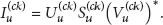
Suppose that F is a fully sampled k-space data and the image I can be reconstructed from F by a direct inverse Fourier transform. Using the SVD operation, the image I can be decomposed as

Here, U and V are two unitary matrices and S is a diagonal matrix, which is a sparse representation of the dense matrix I.
If the k-space is undersampled as

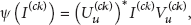
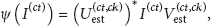
The unitary matrices


Figure 1 illustrates the SVD-based sparsity basis for the compressed sensing MRI.

(a) is the fully sampled MR image I and (b) is the MR image
2.2. Shared SVD-Based Sparsity Basis for Multiple-Coil-Based MR Image Reconstruction
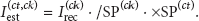
In the volume array coil system for MR imaging, suppose that I is the target image and
The matrices
For volume coil array-based MR imaging, the main difference between the images
Here, the
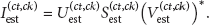
In this way, the coil t MR image
Compared with undersampled coil t image
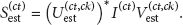
To summarize, the sparsifying transform and its inverse transform for multiple-coil-based MR imaging are defined as


where the sparsity basis is set as
2.3. Shared SVD-Based Sparsity Basis in the CS-SENSE Method
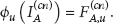
SENSE is an image-based reconstruction method for parallel imaging that employs the coil sensitivities to unfold the aliased pixels. In Cartesian SENSE [16], the acquisition process can be decoupled into two stages. First, it generates a set of aliased coil images with a reduced FOV, and we denote this operation as

where
Second, the Fourier transform ϕ is applied to each aliased coil image to get the k-space signal

The CS-SENSE method also uses the two-stage strategy [6, 7]. Compared with SENSE operation, in the CS-SENSE operation, the above Fourier transform step in (10) is replaced with the CS reconstruction method.
After a set of aliased coil images are generated in (9), there exists an opportunity that they can be further undersampled in k-space in (10). First, the full FOV image I is undersampled in the sensitivity modulation
Here, the operation
In the CS-SENSE method, the final reduction factor R is the product of the reduction factor
The reconstruction in (11) can be formulated as:
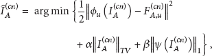
where α and β are weighting parameters.
In the distributed CS stage of the CS-SENSE method, to solve (12) for all the coil images, we first set
After the aliased coil images
% Shared SVD-based sparsity basis. % Reconstruct the aliased coil 1 MR image Obtain the aliased coil 1 image Use the SVD matrices of Reconstruct % Reconstruct other aliased coil images. For Get the estimation of Use the SVD matrices of Reconstruct End For Set Solve (9) using the SENSE method to get the estimated MR image
3. Experiments
3.1. Volume Array Coil System Models
Various models of volume array coil systems with nonlocalized sensitivity profiles have been proposed in recent years; two different models will be used in our experiments. The first model is a head coil system [13], as shown in Figure 2(a). This is a four-coil system. Each coil has a pair of main conductors parallel to the direction of the main magnetic field (B0 field) and located on opposite sides: a conductor is placed on the top to connect the two parallel conductors and a circular conductor base is added to form a closed circuit. The second coil model is a knee coil system [14], as shown in Figure 2(b). This system is designed on the three-dimensional orthogonality principle. It has three coils arranged 120° apart azimuthally and tilted to an angle of 54.7° to the

Two volume array coil systems. (a) Head coil system [13]. (b) Knee coil system [14]. For the head coil system, we set the length of the two main parallel conductors to 320 mm and the diameters of the two circular conductor bases to 280 mm and 300 mm. For the knee coil system, the three coils formed a cylinder with the height of the cylinder being 230 mm and the diameter of the circular bases being 150 mm.
3.2. Image Datasets
We used two MRI datasets to demonstrate the proposed method. For the head coil system, a brain image with size of 512 × 512 [15], which was acquired with a Bruker 2T MRI scanner, was simulated into four images (as shown in Figure 3). For the knee coil system, a knee image with size of 512 × 512 [18], which was acquired with a Siemens 1.5T MRI scanner, was simulated into three images (as shown in Figure 4).

Brain coil images obtained by simulating the sensitivity profiles of the head coil system in Figure 2(a).

Knee coil images obtained by simulating the sensitivity profiles of the head coil system in Figure 2(b).
3.3. Evaluation of Estimated Coil Images
Coil images were undersampled along the phased-encoding direction using reduction factors of 2, 3, and 4. Then to get the estimated coil images, the coil 1 images were reconstructed using the SVD matrices of the undersampled coil 1 image as sparsity bases; other coil images were estimated as
PSNR of the undersampled and the estimated images (dB).

Figure 5 provides another viewpoint of the undersampled and estimated coil images. It can be seen that the sparse representation computed by sparsifying the fully sampled coil image with the SVD matrices of the estimated coil image is closer to the singular values of the fully sampled coil images than that of the undersampled coil image.

(a) The difference between the singular value matrices of the fully sampled coil 2 knee image and its sparse representation that was sparsified with the SVD matrices of the undersampled coil 2 image with reduction factor 3. (b) The difference between the singular value matrices of the fully-sampled coil 2 knee image and its sparse representation that was sparsified with the SVD matrices of the estimated coil 2 image.
3.4. Reconstructions in the CS-SENSE Method
In the CS-SENSE method, k-space signals were first undersampled in the SENSE stage with a reduction factor 2 along the phase-encoding direction; they were then further undersampled with reduction factors of 2, 3, and 4 in the CS stage. Overall, the reduction factors were 2 × 2, 2 × 3, and 2 × 4. The SENSE method without L1 norm regulation was also tested as the baseline; the reduction factors were set as 4, 6, and 8 to correspond to the reduction factors in the CS-SENSE method.
Table 2 records the PSNR of the reconstructed coil images using the three different sparsity bases in the CS stage. The Daubechies-4 wavelet was used in the wavelet-based method. The coil images reconstructed by the SVD-based method had a much higher PSNR than the wavelet-based method. The shared SVD-based method used the reconstructed coil 1 images of the SVD-based method to obtain better sparsity bases for other coil images.
PSNR of the reconstructed coil images in the distributed CS stage (dB).
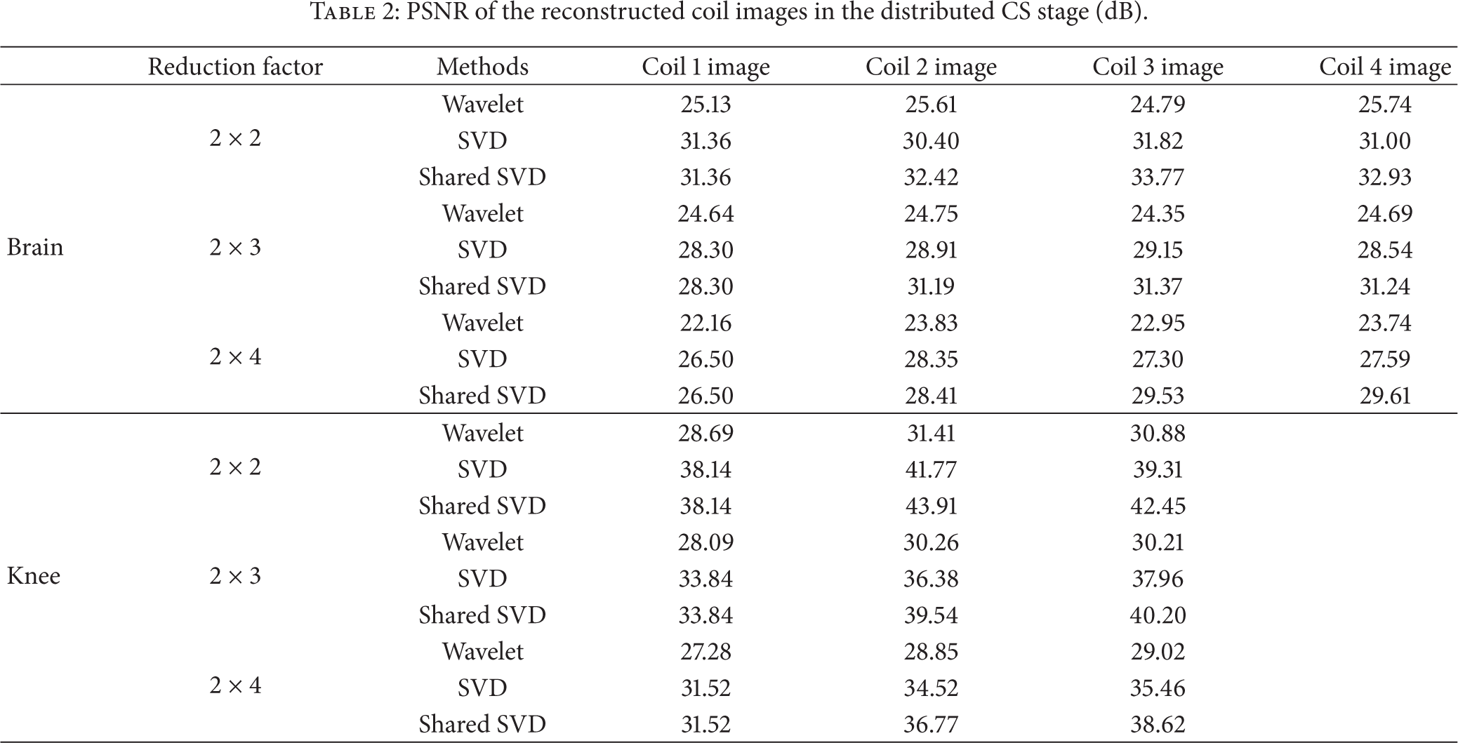
Table 3 records the PSNR of the reconstructed images in the later SENSE stage. The results of the SENSE method were also listed as baselines. The PSNR was further improved in the SENSE stage.
PSNR of the reconstructed images in the SENSE stage (dB).

Figures 6 and 7 show the reconstructed images under the reduction factor 2 × 4 using the CS-SENSE method with different sparsity bases. The error images are magnified five times for better illustration. The reconstruction errors using the CS-SENSE method with the wavelet sparsity basis are obvious at the edges. With the shared SVD method, the boundary artifacts are greatly suppressed, especially in the knee experiments.

Reconstructed brain images under the reduction factor 2 × 4 using the CS-SENSE method with different sparsity bases. (a) The wavelet-based method. (b) The SVD-based method. (c) The shared SVD-based method. The error maps are magnified five times for better illustration.

Reconstructed knee images under the reduction factor 2 × 4 using the CS-SENSE method with different sparsity bases. (a) The wavelet-based method. (b) The SVD-based method. (c) The shared SVD-based method. The error maps are magnified five times for better illustration.
The runtimes in the CS stage were recorded in Table 4. Reconstructions were performed on a laptop with Intel i7 CPU with 6 GB RAM. The software environment is Windows 7 and MATLAB 2008a. The iteration number in the FCSA method was set to 50 in all the three methods; thus, the prolonged runtimes of the wavelet-based method were caused mainly by the time-consuming sparsifying transforms. It takes an average of 0.629 seconds to decompose a matrix of 512 × 512 using the Daubechies-4 wavelet. In contrast, the SVD-based method sparsifies a matrix in two matrix multiplication operations as in (3a), which only takes 0.102 seconds on average.
Runtimes in the distributed CS stage (seconds).

4. Discussion
Figures 6 and 7 have shown that both the wavelet-based and SVD-based sparsity bases can produce clinically acceptable MR images with high reduction factors. Compared to the conventional phased array coil systems, the volume array coil systems considered in this work provide a complete coverage of the imaged object; thus, large intercoil similarities are available for further exploitation of the signal sparsity in both the wavelet and SVD domains.
As shown in Table 1 and in Figures 6 and 7, we can find that the shared SVD-based sparsity basis is capable of reconstructing MR images with a higher PSNR than the SVD-based sparsity and the wavelet sparsity basis. The wavelet sparsity has been widely used as a default sparsity basis for CS-MRI. The SVD-based sparsity basis, in our previous study, provided an alternative sparse representation for MR images. In this work, by making use of the intercoil similarities, the shared SVD-based sparsity basis led to much better reconstruction qualities compared to the classical wavelet sparsity basis. The strategy of the SVD-based sparsity basis is quite different from the wavelet-based sparsity basis. The wavelet sparsity basis transforms the MR image into a set of finer and coarser scales and then uses this set of scales as the sparse representation. The SVD-based sparsity basis decomposes the MR image that has been undersampled in k-space into one singular value matrix and two unitary matrices; the two unitary matrices are then used to set up sparsifying transform. The shared SVD-based sparsity basis improves the SVD-based sparsity basis, in the way that the coil images share the sparsity properties with each other.
Both the SVD-based methods can be further developed into the iterative procedures in the CS stage; that is, the SVD matrices of the reconstructed coil images can be used as the sparsity bases in the next reconstruction iterations. Because the shared SVD-based method offers better image qualities than the SVD-based method in the first iteration, it is expected that the shared SVD-based method can obtain optimal reconstruction results with less iterations than the SVD-based method requires.
Moreover, in the shared SVD-based method, because the coil image can “borrow” the sparsity bases from other coil images with its own sensitivity profiles, the inherent artifacts in the coil image can be reduced by the interventions of other coil images. In contrast, the SVD-based method always reconstruct one coil image from its own reconstruction results from the previous iteration results in this way, the inherent artifacts may be preserved or even amplified during iteration.
5. Conclusion
In this work, a shared SVD-based sparsity basis was proposed for the distributed compressed sensing MR image reconstruction of the volume array coil imaging. Owing to the intercoil data similarities, the SVD results of the multiple-coil MR images provided excellent sparsity bases for the reconstruction of multiple-coil MR images. The experimental results have shown that the target MR images can be reconstructed with high qualities under high reduction factors using the proposed method.
Footnotes
Acknowledgment
This work has been supported by the Fundamental Research Funds for the Central Universities, no. DUT13JR02.