
Abstract
Objectives:
To explore whether combined therapy with donepezil and acupuncture is better than treatment with donepezil or acupuncture individually in a rat model of Alzheimer’s disease.
Methods:
In this study, we randomly divided 40 7.5-month-old senescence-accelerated mouse prone 8 (SAMP8) male mice into four groups: SAMP8, SAMP8+D, SAMP8+MA and SAMP8+D+MA. An additional 10 7.5-month-old SAMR1 male mice were included as a healthy control group (SAMR1). Mice in the SAMP8+D group were given donepezil at a dose of 0.65 µg/g/day; mice in the SAMP8+MA group underwent manual acupuncture at GV20, GV26 and Yintang for 20 min per day; mice in the SAMP8+D+MA received both donepezil and manual acupuncture; and mice in the SAMR1 and SAMP8 groups underwent restraint only to control for the effects of handling. After the 15-day treatment, the Morris water maze test, micro-PET(positron-emission tomography), H&E (haematoxylin and eosin) staining, and immunohistochemistry were used to study the differences between donepezil (SAMP8+D), acupuncture (SAMP8+MA), and donepezil combined with acupuncture (SAMP8+D+MA) therapy for the treatment of Alzheimer’s disease.
Results:
We found that, compared with the untreated SAMP8 group, donepezil, manual acupuncture, and combined therapy with donepezil and manual acupuncture all improved spatial learning and memory ability, the level of glucose metabolism in the brain, and the content of Aβ amyloid in the cortex. Moreover, combined therapy outperformed treatment with donepezil or acupuncture individually in the SAMP8 mice.
Conclusion:
This study shows that the combination of manual acupuncture and donepezil in an Alzheimer’s disease animal model is superior to acupuncture and donezepil alone. However, randomised controlled trials should be undertaken to clarify the clinical efficacy of combination therapy.
Keywords
Introduction
Alzheimer’s disease is the most common neurodegenerative cause of dementia and is responsible for significant morbidity and mortality. It also places a significant economic burden on the global healthcare system. 1 Accumulation of Aβ amyloid and neurofibrillary tangles in the brain are the main pathological features of Alzheimer’s disease. 2 Although research on Alzheimer’s disease is growing, there is no effective therapy capable of curing the condition at present. 3
Nowadays, traditional Chinese therapy, especially acupuncture, is playing an increasingly important role in the treatment of Alzheimer’s disease.4,5 Preclinical research has shown that acupuncture could improve learning-memory function 6 and glucose metabolism in animal models of Alzheimer’s disease.7,8 Moreover, this therapy may be effective at treating patients with amnestic mild cognitive impairment, according to clinical research. 9 However, it remains unknown whether classical drug therapy for Alzheimer’s disease could be replaced by acupuncture, or whether acupuncture could be used in combination with drug therapy to treat the disease more effectively.
To explore these questions, we conducted research using an animal model of Alzheimer’s disease— the senescence-accelerated mouse prone 8 (SAMP8) mouse—which is characterised by learning and memory impairment and mood disorder similar to that present in Alzheimer’s disease. 10 The Morris water maze test, micro-PET (positron-emission tomography), H&E (haeomatoxylin-eosin) staining and immunohistochemistry were used to study differences in effectiveness of donepezil, acupuncture, and donepezil combined with acupuncture, in the treatment of Alzheimer’s disease. We anticipated that these findings would extend our previous studies on the use of acupuncture in neurological injury and evaluate the potential role of a combination of drug and acupuncture therapy as an alternative treatment for Alzheimer’s disease.
Methods
Experimental animals
SAMP8 and SAMR1 (control) breeding pairs were kindly provided by Professor Takeda at Kyoto University, Japan. 11 The animals were housed in a barrier facility at the Experimental Animal Centre of the First Teaching Hospital of Beijing University of Traditional Chinese Medicine and maintained at a controlled temperature (24±2°C) under a 12/12 hour dark/light cycle. The rats were provided with sterile drinking water and a standard pellet diet ad libitum. This study was carried out in strict accordance with the recommendations of the National Research Council’s ‘Guide for the Care and Use of Laboratory Animals’ (National Academies Press, Washington, DC, USA). All protocols involving operative and postoperative animal care were approved by the Beijing University of Chinese Medicine Institutional Animal Use and Care Committee and were carried out in accordance with the animal use and ethics committee approval statements for animal experiments provided by Beijing University of Chinese Medicine (Beijing, China). Forty 7.5-month-old SAMP8 mice (weighing 28–32 g) were randomly divided (according to a random number method) into four groups (n=10 each) that remained untreated (SAMP8 group) or received manual acupuncture (SAMP8+MA group), donepezil (SAMP8+D group), or both donepezil and manual acupuncture (SAMP8+D+MA group). An additional 10 SAMR1 mice were included as a healthy control group (SAMR1 group).
Acupuncture manipulation
In the SAMP8+MA group, acupuncture treatment was performed once a day for 15 days (with no treatment on the eighth day) at acupuncture points GV20, GV26 and Yintang, which were located as described previously. 12 The needles were rotated at the rate of two rotations per second for 30 s at each point. For the SAMP8+D group, the dosage of donepezil was 0.65 µg/g per day. 13 In the SAMP8+D+MA group, both treatments were combined. Rats in the other two groups (SAMP8 and SAMR1) were subject to restraint under the same conditions, but without needle insertion or drug administration, in order to control for any effect of animal handling.
Morris water maze behavioural test
The water maze consisted of a circular tank (90 cm diameter, 50 cm height) filled with water to a depth of 29 cm maintained at 24±1°C and rendered opaque with blue-black ink. A removable circular platform (9.5 cm diameter, 28 cm height) with its top surface 1 cm below the water was placed inside the pool. The area of the pool was conceptually divided into four quadrants (NE, NW, SW and SE) of equal size. Data were collected by a video camera (TOTA-450Ш, Japan), which was fixed to the ceiling of the room and connected to a video recorder and an automated tracking system (China Daheng Group, Beijing, China).
The mice were placed into the pool just below the surface of the water. They were considered to have escaped from the maze when they found the platform. Distal visual cues were placed around the room to help the mice learn the location of the hidden platform. All mice underwent a total of 6 days training, which included a 5-day hidden platform trial and a 1-day probe trial. For the first 5 days, the hidden (submerged) platform was maintained in the same position (in the middle of the NE quadrant) throughout the procedure. Once the mouse reached the platform, the time elapsed was recorded and it was allowed to remain there for 5 s. If it failed to find the platform in 60 s, then the time was recorded as 60 s as an upper limit. Each mouse was given two trials per day with an inter-trial interval of 2 hours. Swimming distance before reaching the platform was also recorded. On the sixth day, the platform was removed from the pool and the time spent swimming in the quadrant where the platform had formerly been was recorded over 60 s. The number of times the mouse crossed the platform, the swimming distance in the NE quadrant, and the total swimming distance were recorded.
Micro-PET imaging
Four animals were randomly selected from each group for micro-PET detection using 18F-fluorodeoxyglucose (18F-FDG) PET tracers provided by the Chinese Medicine Research Institute PET Room and a Siemens INVEON PET/CT imaging system. Before experiments began, the 20 mice underwent measurement of blood glucose, which was consistently within the normal range (~7.0–10.1 mmol/L). Following a 6 hour fast, the mice were placed into a suction chamber. Once completely anaesthetised using inhaled oxygen mixed with 1.5% isoflurane (1 L/min), approximately ~14.8–16.5 MBq 18F-FDG PET tracer was injected via the tail vein. After allowing 18F-FDG PET tracer uptake for 60 min, the mice were placed on the scan bed in a prone position, parallel to the long axis of the scanner, with their heads positioned within the scanner’s field of view. Micro-PET images were acquired at a frame rate of 30 s/frame and reconstructed using filtered back projection (FBP) and CT photon attenuation correction. Three-dimensional region of interest technology was applied to examine the whole brain (cortex, frontal lobe and hippocampus) in transverse, coronal and sagittal planes. The uptake rate per gram (%ID/g) was calculated within this region of interest.
Tissue sampling and processing
The remaining six mice per group were terminally anaesthetised by an intraperitoneal injection of 10% chloral hydrate (0.35 mL/100 g). Three minutes later, the chest was opened to expose the heart, and the left ventricle and ascending aorta were cannulated and rapidly injected with 100 mL saline. Then the right atrial appendage was cut and perfused with 4% paraformaldehyde until the liver turned white and backflow of clear liquid was observed. After perfusion was completed, the mice were decapitated. Whole brains were removed, stored on ice, fixed in 4% paraformaldehyde, and finally embedded in paraffin. For histopathological evaluation, 5 µm sections of brain tissue were cut using a Leica 1410 paraffin slicing machine (Leica, Germany), mounted and dried overnight at 40°C. Tissue sections were stained with haematoxylin and eosin (H&E) using routine procedures.
Immunohistochemistry
Additional sections of brain tissue were deparaffinised in xylene and alcohol, treated with citric acid antigen repair buffer, and then washed with phosphate-buffered saline (PBS) solution (pH 7.4) three times at 5-min intervals. Sections were incubated with 3% hydrogen peroxide solution for 20 min in the dark to block endogenous peroxidase and then washed again with PBS solution in the same manner. Then the sections were incubated with Aβ IgG antibody (1:50, ab10148, Abcam, Cambridge, UK) diluted with PBS overnight. After washing again with PBS solution and incubating with secondary antibody for 30 min at room temperature, sections were stained with DAB (3,3’-diaminobenzidine) and finally dehydrated in alcohol and xylene and then mounted. Microscopic images of brain tissue were captured at 10-40 times magnification, and the integral optical density (IOD) was calculated by analysing the corresponding brownish yellow areas in each microscopic image using Image-Pro Plus 6.0 (Media Cybernetics, USA) software.
Statistical analysis
The statistical analysis was performed by researchers who were not involved in the experimental operation. For the Morris water maze test, the swimming distance of the hidden platform was analysed using the Huynh-Feldt test, while one-way analysis of variance (ANOVA) was applied to the data obtained from the probe trial. The test of least significant difference (LSD) was used to compare group pairs. Mean values were considered to be significantly different when p<0.05. All statistical analyses were performed using the Statistical Package for the Social Sciences (SPSS) version 23.0 (SPSS Inc, Chicago, IL, USA).
Results
Effects of acupuncture and donepezil on spatial learning and memory ability
Figure 1 shows the results from the Morris water maze test. Figure 1A shows the distance swum by the mice before reaching the platform, in the hidden platform trial. At first, the task was relatively difficult for the mice. However, after 5 days of training, the distance swum before reaching the platform in all the groups was noted to have gradually shortened over time (p<0.01). As shown in figure 1A, mice of the SAMR1 group appeared to learn and remember the cues better, such that their swimming distances were shortened more quickly and were significantly less than the mice of the SAMP8 group from the third day (p<0.01). Interestingly, the distances swum before reaching the platform by the mice in the SAMP8+D+MA and SAMP8+MA groups were shorter than the SAMP8 mice (p=0.008 and p=0.03, respectively).
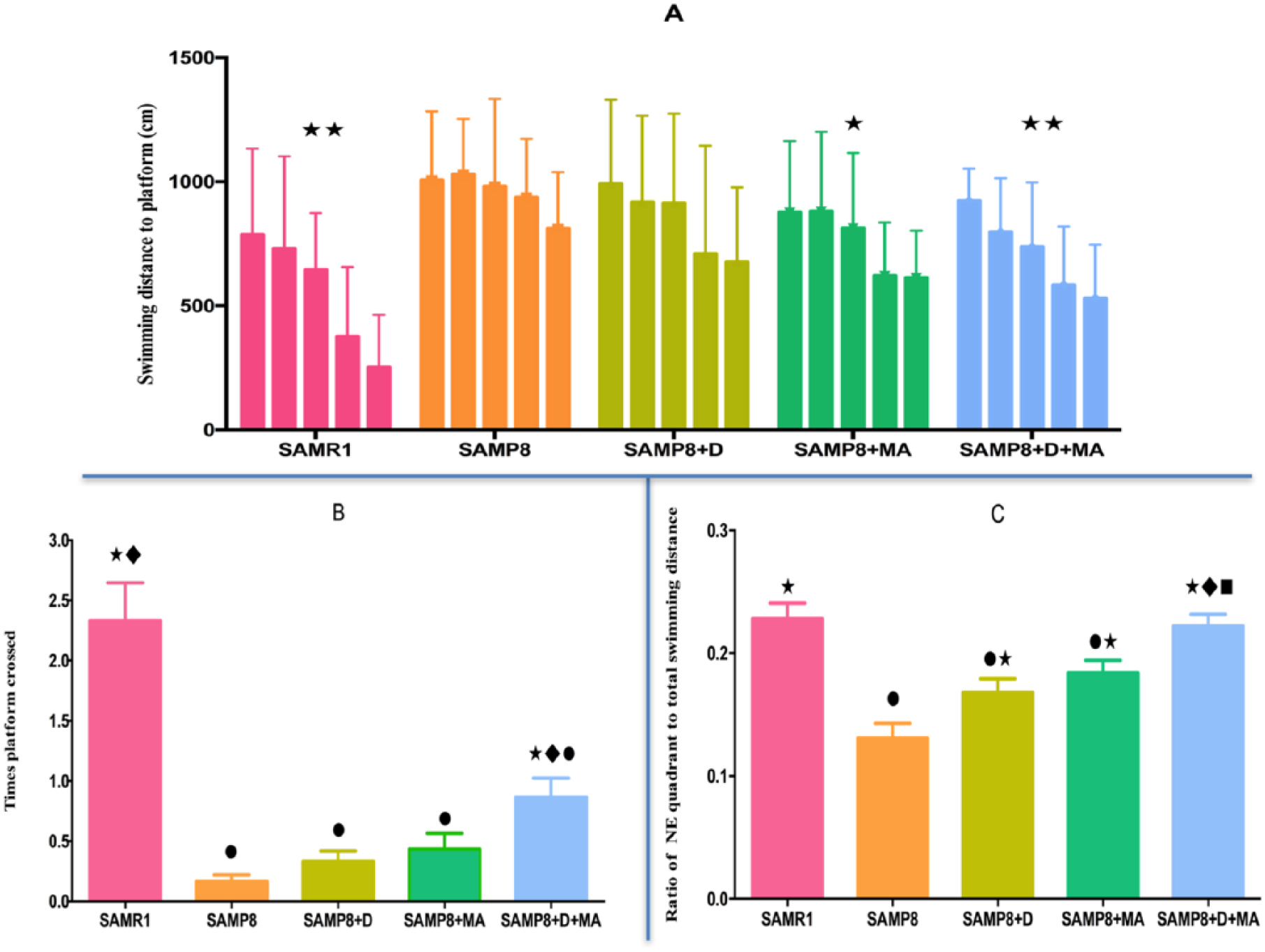
Results of the Morris water maze test for the hidden platform trial and probe trial. (A) The distance swum before reaching the platform in each group during the hidden platform trial. (B & C) The number of times the platform was crossed, and the ratio of NE quadrant to total swimming distance of each group. • p<0.05 compared with SAMR1 group; •• p<0.01 compared with SAMR1 group; ✶ p<0.05, ✶✶ p<0.01 compared with SAMP8 group; ♦ p<0.05, ♦ ♦ p<0.01 compared with SAMP8+D group.
Figure 1B shows the number of times the platform was crossed and figure 1C presents the ratio between the swimming distance in the NE quadrant and the total swimming distance, both markers of spatial memory ability, in the probe trial. Mice in all SAMP8 groups crossed the platform significantly fewer times than those in the SAMR1 group and there were no significant differences between SAMP8, SAMP8+D and SAMP8+MA groups, However, mice in the SAMP8+D+MA group crossed the platform significantly more frequently than those in the SAMP8 and SAMP8+D groups (p=0.007 and p=0.036, respectively). Furthermore, relative to the untreated SAMP8 group, the swimming distance ratio was increased in the SAMP8+D, SAMP8+MA and SAMP8+D+MA groups (p<0.05). Moreover, the ratio was greater in the SAMP8+D+MA group versus both the SAMP8+D and SAMP8+MA groups (p=0.001 and p=0.02, respectively).
Effects of acupuncture and donepezil on brain glucose metabolism
Figure 2 shows the effects of the different types of therapy on quantitative 18F-FDG uptake of the whole brain (cortex, frontal lobe and hippocampus), which was reduced in SAMP8, SAMP8+D and SAMP8+MA groups relative to healthy mice (SAMR1 group). 18F-FDG-uptake per gram was noted to be significantly higher in the two acupuncture-treated groups (SAMP8+MA and SAMP8+D+MA) compared with untreated SAMP8 mice (p=0.026 and p=0.015, respectively).
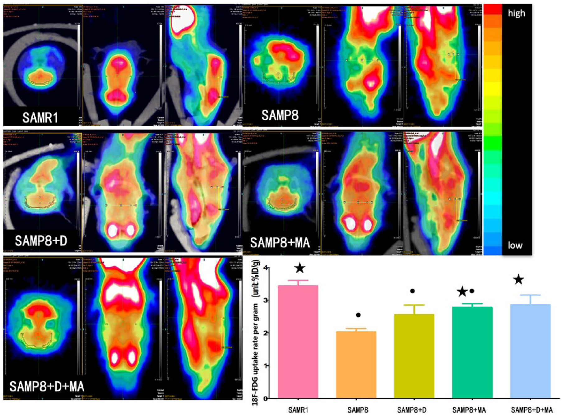
Results of micro-PET (positron-emission tomography) demonstrating the uptake rate per gram (%ID/g) of each group. The same colour standard and colour code (top = high, bottom = low) were used to display the metabolic rate of glucose. The left side of the image represents the right side of the animal. • p<0.05 compared with SAMR1 group; ★ p<0.05 compared with SAMP8 group.
Effects of acupuncture and donepezil on the neurons of the dentate gyrus
Representative H&E-stained images of brain tissue from each group are shown in figure 3. Mice in the SAMR1 group demonstrated clear-dyed neurons aligned in neat rows, with round nuclei and distinct kernels in the dentate gyrus. Conversely, neurons tended to be scattered and irregular, with indistinct kernels and nuclear pyknosis in the dentate gyrus of untreated SAMP8 mice. Subjectively, compared with the untreated SAMP8 mice, neurons were more neatly arranged in rows and clearer in structure with less nuclear condensation in the SAMP8+D, SAMP8+MA and SAMP8+D+MA groups. Tissues from SAMP8+D+MA and SAMR1 groups appeared to be most similar.
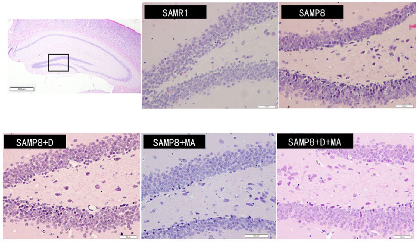
Representative H&E (haematoxylin-eosein) stained images of brain tissue from each group.
Effects of acupuncture and donepezil on cortical amyloid β deposition
Figure 4 shows the effects of the different therapeutic combinations on Aβ amyloid deposition. Compared with the SAMR1 group, the IOD of Aβ amyloid in the cortex was significantly higher in the SAMP8, SAMP8+D, SAMP8+MA and SAMP8+D+MA groups (p<0.001). However, compared with the SAMP8 and SAMP8+D groups, the IOD of cortical Aβ amyloid was significantly lower in the SAMP8+MA (p<0.001 and p=0.011, respectively) and SAMP8+D+MA (both p<0.001) groups. Moreover, Aβ amyloid deposition in the SAMP8+MA and SAMP8+D+MA groups did not differ significantly (p=0.174).
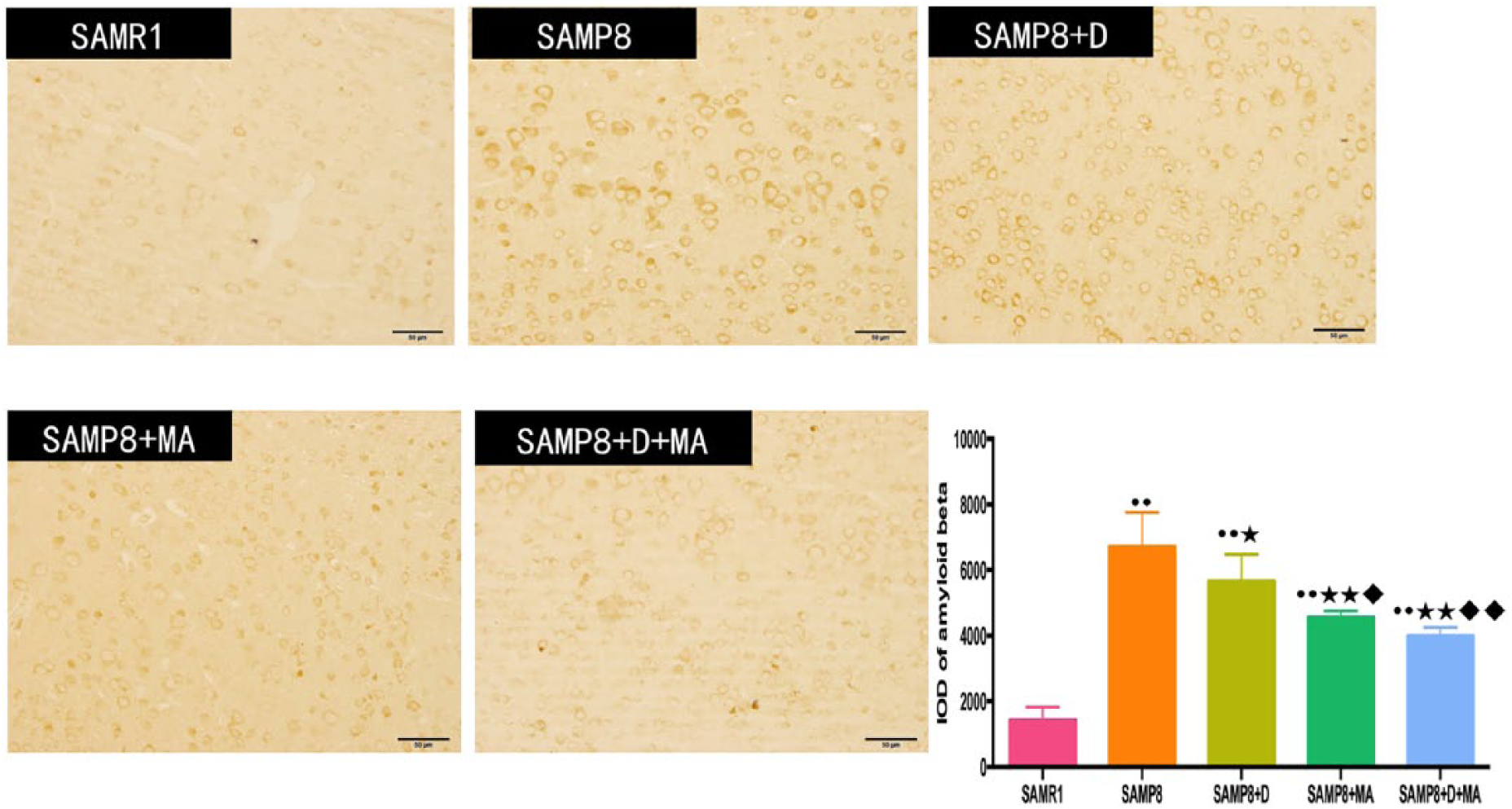
Representative images of β amyloid deposition in the cerebral cortex of each group. The integral optical density (IOD) of the cortex was calculated by analysing the corresponding brownish yellow areas in each microscopic image using Image-Pro Plus 6.0 software. •• p<0.01 compared with SAMR1 group; ✶p<0.05, ✶✶ p<0.01 compared with SAMP8 group; ♦p<0.05, ♦♦p<0.01 compared with SAMP8 +D group.
Discussion
In this study, we used the Morris water maze test, micro-PET, H&E staining and immunohistochemistry to examine changes in spatial learning and memory ability, whole-brain glucose metabolism, morphological changes in the dentate gyrus, and Aβ amyloid in the cortex of the senescence-accelerated mouse prone 8 (SAMP8) animal model of Alzheimer’s disease. For the first time, to our knowledge, we used these techniques to explore the effects of acupuncture combined with donepezil therapy.
Acupuncture treatment is a significant part of traditional Chinese medicine, and its history dates back to the origin of Chinese civilization. The therapeutic effects and safety profile of acupuncture are recognised by the Chinese and other populations around the world. 14 Nowadays acupuncture is used to treat many types of neurological disease, including various types of dementia (Alzheimer’s disease,15,16 vascular dementia17,18), Parkinson’s disease,19,20 depression,21,22 stroke23–25 and pain.26,27
Currently, drug treatment remains the mainstream therapy for Alzheimer’s disease. One aim of pharmacological therapy is to inhibit the breakdown of the chemical neurotransmitter, acetylcholine, by blocking the relevant enzyme. 28 Donepezil is a selective acetylcholinesterase inhibitor that is widely prescribed for Alzheimer’s disease. 29 It has been shown that donepezil is beneficial in the mild, moderate and severe stages of Alzheimer’s disease, in vascular dementia, and in dementia associated with Parkinson’s disease.30,31 Although donepezil has been used for more than 30 years in clinical practice, researchers and physicians continue to pursue further improvements in the treatment of Alzheimer’s disease.32,33 The results of the current study, using the Morris water maze test and micro-PET, showed that donepezil improved the learning and memory ability of the treated mice and increased the rate of glucose metabolism in the brain (both p<0.05 compared with SAMP8 group). There were no statistically significant differences in the number of times the platform was crossed, the rate of glucose metabolism or the IOD of cortical Aβ amyloid in SAMP8+D versus SAMP8+MA groups.
In clinical practice, once Alzheimer’s disease has been diagnosed, patients generally accept the use of drug therapy. 3 It is controversial to use only non-pharmacological approaches to treat the disease clinically. 34 Therefore, from a clinical practice perspective, our research is pragmatic because it addresses the use of classical drug therapy combined with non-drug therapy for the treatment of Alzheimer’s disease using an animal model. To our knowledge, this is the first study to evaluate the combination of donepezil—the classical drug treatment for Alzheimer’s disease—and the traditional Chinese therapy, acupuncture, to manage this particular condition.
In the Morris water maze test, the results of the hidden platform test reflects the animals’ learning ability and the probe test reflects their memory ability. 35 During the hidden platform test, the distance swum before reaching the platform was significantly reduced in the SAMP8+D+MA and SAMP8+MA groups from the third day, compared with the untreated SAMP8 group. During the probe test, the SAMP8+D+MA group performed significantly better than the SAMP8+D group in the time taken to cross the platform. Moreover, the SAMP8+D+MA group performed significantly better than SAMP8+D and SAMP8+MA groups in terms of the swimming distance in the NE quadrant and the ratio of the total swimming distance. These results suggest that donepezil combined with acupuncture may be superior to the individual components with respect to some outcome measures.
Clinically, imaging biomarkers are playing an increasingly important role in the diagnosis of Alzheimer’s disease and evaluation of the efficacy of treatment. 36 PET with 18F-FDG allows for the detection of neurodegenerative disorders in Alzheimer’s disease earlier than is otherwise possible. 37 Micro-PET is accessible for the non-invasive, quantitative and repetitive imaging of biological function in living animals. 38 Therefore, we used micro-PET to observe the changes in the level of glucose metabolism in the whole brain (cortex, frontal lobe and hippocampus). According to our results, the 18F-FDG uptake rate was increased in both acupuncture-treated groups relative to the SAMP8 group but did not differ significantly between SAMP8+D, SAMP8+MA and SAMP8+D+MA groups.
It is known that the accumulation of Aβ amyloid in the brain is one of the pathological characteristics of Alzheimer’s disease. 39 In addition, Aβ amyloid accumulation is considered to be a central disease-causing and disease-promoting event in Alzheimer’s disease. 40 Therefore, in the field of Alzheimer’s disease research, the content of Aβ amyloid in the brain is an important index for diagnosing and evaluating the condition.3,41 In this study, we used immunohistochemistry to qualitatively observe and quantitatively analyse the content of Aβ amyloid in the brain of SAMP8 mice. Levels of Aβ amyloid were reduced in all treated groups compared with the untreated SAMP8 group. Moreover, donepezil combined with acupuncture treatment significantly reduced the content of Aβ amyloid in the brain of SAMP8 mice. These results suggest that combination therapy may indeed inhibit or delay the progress of the pathogenesis of Alzheimer’s disease.
Conclusions
In summary, using the Morris water maze test, micro-PET, H&E staining and immunohistochemistry, this study has confirmed the hypothesis that donepezil combined with acupuncture improves spatial learning and memory ability, glucose metabolism and Aβ amyloid content in the brain of an Alzheimer’s disease animal model better than donepezil treatment or acupuncture individually. It is therefore proposed that donepezil combined with acupuncture may represent an alternative treatment for Alzheimer’s disease. However, the mechanism underlying the effects of acupuncture on this disease requires further exploration, and clinical trials should also be conducted to confirm the effects of combined therapy in humans.
Footnotes
Contributors
JJ designed the work and wrote the paper. GL and SS performed the animal experiments. YL performed the data analysis. ZL revised the paper and approved its publication. All authors reviewed the final version of the manuscript accepted for publication.
Funding
The authors disclosed receipt of the following financial support for the research, authorship, and/or publication of this article: National Natural Science Foundation of China (grant no. 81804178) and Beijing University of Chinese Medicine Research Program (grant no. 2017-JYB-JS-058).
Declaration of conflicting interests
The authors declared no potential conflicts of interest with respect to the research, authorship, and/or publication of this article.
Ethics approval
Approved by Beijing University of Chinese Medicine Institutional Animal Use and Care Committee.
Provenance and peer review
Not commissioned; externally peer reviewed.