
Abstract
Patients with primary headache often report pain that involves the front of the head, in the cutaneous distribution of the first (ophthalmic) division of the trigeminal nerve and at the same time or evolving, pain in the occipital and upper cervical region (1). Furthermore, other clinical features such as hypersensitivity of the skin of the face or scalp, neck muscle tenderness and hyperalgesia are often reported widely in the head (2, 3). The relatively poor localization of pain in primary headache disorders produces significant diagnostic problems and certainly impacts on the understanding of these disorders. In migraine alone, the extremely common premonitory symptom of neck discomfort or pain (4) leads to endless, and almost invariably useless, therapeutic attention to a structure that is as innocent of pathology as are the eyes innocent of pathology in patients with photophobia. An understanding of the anatomy and physiology of the trigeminal and upper cervical neurones that receive convergent input, the trigeminocervical complex, can render the clinical presentation clearer and the management strategies more effective, and must form an important basis for understanding head pain more broadly. Here we set out what is currently known of the anatomy and physiology of these pivotal structures. We have covered some aspects of these issues previously and here provide an updated view (5).
Clinical observations
Early neurosurgical studies in patients showed that stimulation of trigeminally innervated intracranial structures, such as the supratentorial dura mater and large cranial vessels, evoked painful sensations regardless of the stimuli applied and implied that the afferent input from dural structures is the neural substrate of head pain (6–9). Hence, afferent input, or at least perceived input, from dural structures is likely to be the neural substrate of pain in primary headache syndromes (10). It has been shown that spread and referral of pain can be induced by stimulation of structures in the neck that are innervated by the upper cervical roots. Posterior fossa tumours (11), stimulation of infratentorial dura mater (12), direct stimulation of cervical roots (11, 13), vertebral artery dissection (14, 15) and stimulation of subcutaneous tissue innervated by the greater occipital nerve (16, 17) may be perceived as frontal head pain. Similarly, direct stimulation of the supratentorial dura mater leads to pain mostly referred to the first (ophthalmic) division of the trigeminal nerve (12), but which may be also referred to dermatomes supplied by the upper cervical roots (18). Busch and colleagues (19) examined the R2 components of the nociceptive blink reflex responses (20) in 15 healthy subjects before and after unilateral nerve blockade of the greater occipital nerve with local anaesthetic. R2 response areas (AUC) decreased and the R2 latencies increased significantly after the nerve blockade only on the side of injection. AUC and latencies on the non-injection side remained stable. These data provide objective evidence for a functional influence of trigeminal nociceptive inputs from occipital inputs.
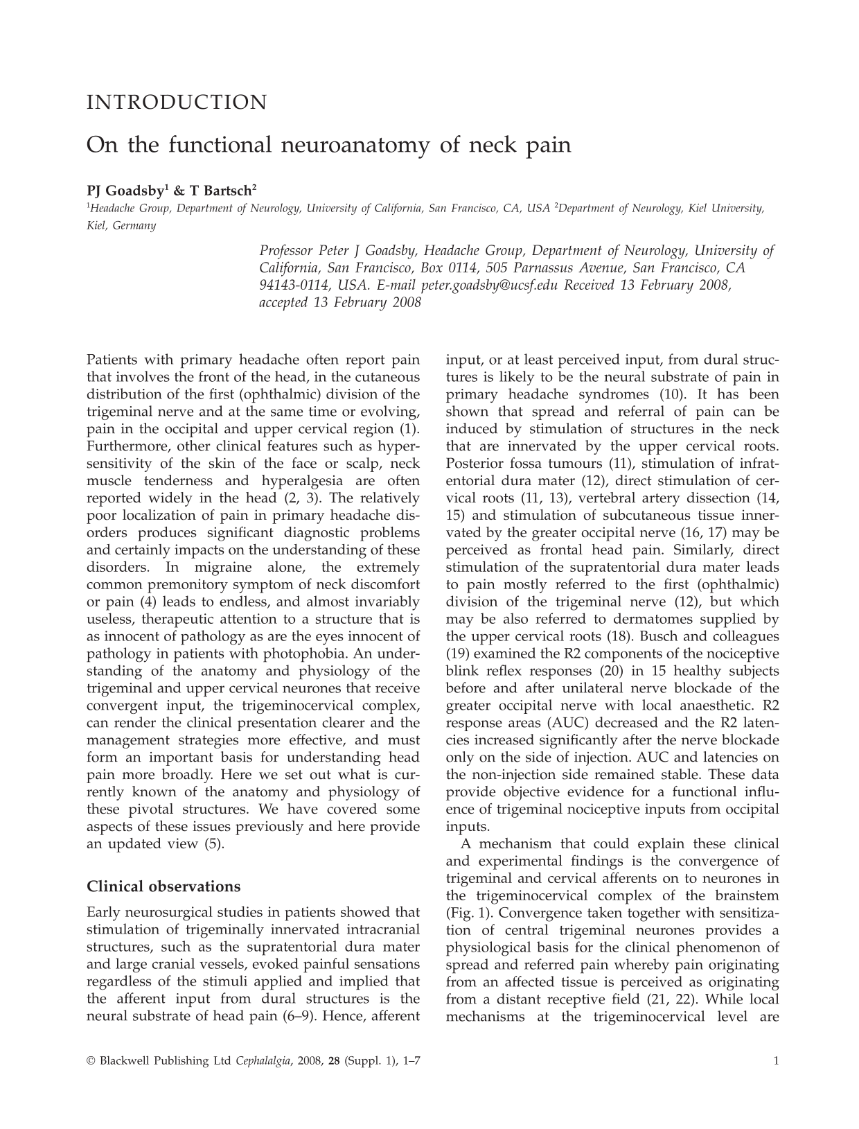
A mechanism that could explain these clinical and experimental findings is the convergence of trigeminal and cervical afferents on to neurones in the trigeminocervical complex of the brainstem (Fig. 1). Convergence taken together with sensitization of central trigeminal neurones provides a physiological basis for the clinical phenomenon of spread and referred pain whereby pain originating from an affected tissue is perceived as originating from a distant receptive field (21, 22). While local mechanisms at the trigeminocervical level are undoubtedly important, it is crucial to recall functional imaging evidence of altered thalamic processing in chronic migraine patients clinically well controlled by occipital nerve stimulation (23). It seems likely that the effects we describe in the trigeminocervical complex will have more, and more complex, correlates in other parts of the brain that will need to be explored to understand this physiology completely (Fig. 1).

Schematic drawing illustrating the convergence of dura mater and skin inputs from the trigeminal (ophthalmic, V1) distribution with cervical (muscle, joints, skin) afferents on to the same nociceptive second-order neurone in the trigeminocervical complex at the level of C2. These inputs ascend to brain stem modulatory sites, such as the periaqueductal grey matter and for further processing in the sensory thalamus, including ventroposteromedial thalamus. Descending influences can both inhibit (−) and facilitate (+) trigeminocervical nociceptive traffic.
Laboratory studies of the trigeminocervical complex
The nociceptive input from the dura mater to the first synapse in the brainstem is transmitted via small-diameter A- and C-fibre afferents in the ophthalmic division of the trigeminal nerve via the trigeminal ganglion to nociceptive second-order neurones in the superficial and deep layers of the medullary dorsal horn of the trigeminocervical complex (24–27). The trigeminocervical complex extends from the trigeminal nucleus caudalis to the segments of C2–C3 in the rat (28), cat (29) and monkey (30). Trigeminovascular nociceptive inputs project bilaterally in the brainstem from unilateral structures, such as peri-middle meningeal artery dura mater (31). These dural-sensitive trigeminal neurones show a high degree of convergent input from other afferent sources as they typically show also facial and corneal receptive fields (24, 25), although interestingly are less susceptible to wind-up than trigeminocervical neurones with peripheral inputs (32).
Cervical inputs
With regard to the innervation of the head, the upper cervical spinal roots also contribute to the sensory innervation of cranial and cervical structures. Occipital and suboccipital structures, such as vessels and the dura mater of the posterior fossa, deep paraspinal neck muscles (zygapophyseal) joints and ligaments, are innervated by the upper cervical roots and are recognized sources of neck and head pain (33–35). The nociceptive inflow from these suboccipital structures is also mediated by small-diameter afferent fibres in the upper cervical roots terminating in the dorsal horn of the cervical spine extending from the C2 segment up to the medullary dorsal horn (36–40). The major afferent contribution is mediated by the spinal root C2 that is peripherally represented by the greater occipital nerve (GON) (41–43). Similarly to the trigeminal sensory neurones, these cervical neurones show a high convergence of input from neck muscles and skin (26). Indeed, stimulation of the greater occipital nerve (40) or specifically of occipital muscle afferents will also activate neurones across the anatomical extent of the trigeminocervical complex (44).
Physiological evidence for convergence
Although an anatomical overlap of trigeminal and cervical afferents throughout the trigeminocervical complex from the level of the caudal trigeminal nucleus to at least the C2 segment was first suggested by Kerr (45), a direct coupling between meningeal afferents and cervical afferents in the spinal dorsal horn has not been described until recently (26). A population of neurones in the C2 dorsal horn was characterized that received convergent input from the supratentorial dura mater and the greater occipital nerve (GON). These neurones showed properties typical for dura-sensitive trigeminal neurones with a convergent input from the facial skin corresponding to the dermatome of the ophthalmic division of the trigeminal nerve including the cornea, but also showed a receptive field corresponding to the cervical skin of the C2/C3 dermatomes and to deep paraspinal muscles innervated by the GON. Interestingly, these trigeminocervical neurones showed convergent synaptic input not only from the supratentorial dura mater and from the ipsilateral GON, but also from the contralateral GON. Taken together with other studies that show similar contralateral projections of nociceptive afferents following labelling of the trigeminal and cervical dorsal root ganglia (46, 47), or after facial afferent (48) or GON stimulation (40), it seems that bi- or contralateral endings of nociceptive afferents of visceral and deep-somatic tissues are more common than previously acknowledged (49). This anatomical arrangement may find its functional correlate in the dull and poorly localized quality of head and neck pain (50).
The convergence of the GON and dural afferents in the trigeminocervical complex is noticeable as it is a convergence of a somatic spinal nerve (GON) and a visceral nerve (dura). Both systems show functional differences as visceral nerves show a greater portion of unmyelinated C-fibres (51) and show no wind-up to repetitive noxious stimulation (32, 52, 53), suggesting a different neuroplastic potential to noxious stimulation.
Central mechanisms of pain processing
Central sensitization
After strong noxious inputs, nociceptive second-order neurones in the spinal cord can be subject to a transient or long-lasting hyperexcitability to afferent stimulation. The current concept of this central sensitization considers an increased afferent barrage from primary nociceptive afferents, especially C-fibres, on to second-order neurones as crucial in the development of this hyperexcitability. In particular, stimulation of afferents from visceral and deep somatic tissues, such as muscle and joints, are more effective than cutaneous input in evoking such a central hyperexcitability (54, 55). The hypersensitivity of the afferent synaptic input in the spinal cord is thought to be due to the release of various neuropeptides, such as calcitonin gene-related peptide (CGRP), or to glutamate release and action at the NMDA receptor in response to afferent stimulation, but may also be due to decrease of local segmental spinal inhibition in response to the afferent stimulation (56, 57). The hyperexcitability is reflected in a reduction of the activation threshold, an increased responsiveness to afferent stimulation, an enlargement of receptive fields or the emergence of new receptive fields and the recruitment of ‘silent’ nociceptive afferents. The clinical correlates of this central hypersensitivity include the development of spontaneous pain, hyperalgesia and allodynia (56, 58).
Activity-dependent change in trigeminocervical neurones
Similar to the spinal cord, these activity-dependent changes in synaptic strength have been shown to take place also in dura-sensitive trigeminal neurones. Application of an ‘inflammatory soup’ onto the dura mater can induce a central sensitization of trigeminal second-order neurones in the caudal trigeminal nucleus with a subsequent increased responsiveness to dural and cutaneous facial stimulation (24). Some component of this response can be blocked by naproxen (59), as can trigeminocervical transmission be modulated by intravenous aspirin (60). We further analysed this functional interaction and found that convergent neurones responding to both dural and GON stimulation showed a central sensitization to GON stimulation with an increased excitability to dural input (26). Interestingly, the stimulation of neck muscle afferents in the GON by the C-fibre activator mustard oil was significantly more effective in increasing dural input than stimulation of cutaneous afferents in the GON. Furthermore, stimulation of the dura mater led also to a sensitization of these convergent neurones with a subsequent increased excitability to neck muscle and GON stimulation (61).
Some clinical implications of sensitization: It seems these neurones of the trigeminocervical complex are crucial for understanding the most common clinical patterns of pain referral between trigeminal and cervical dermatomes in migraine that does not necessarily involve a peripheral pathology in the cervical innervation territory (34). Interestingly, many headache forms benefit from a blockade of the greater occipital nerve, including migraine, cluster headache and hemicrania continua (62–64).
The clinical changes seen in primary headache syndromes, such as increased cutaneous sensitivity, hyperalgesia, allodynia, spread and referral of pain in the trigeminal and cervical dermatomes, are very suggestive of an altered trigeminocervical nociceptive system in terms of a facilitation or sensitization of central nociceptive neurones (65, 66). It could be shown that similar changes, such as extradural hypersensitivity and receptive field changes described in experimental models, can also be seen in headache patients, suggesting that a sensitization of central nociceptive neurones indeed takes place during migraine attacks (3, 67, 68).
Central pain modulation and headache
It is now well established that the nociceptive inflow to second-order neurones in the spinal cord and the trigeminocervical complex is subject to a modulation by descending inhibitory projections from brainstem structures such as the periaqueductal grey (PAG), nucleus raphe magnus (NRM) and the rostroventral medulla (RVM) (69–71), as stimulation of these regions produces profound antinociception (72). In particular, recent findings suggest that the ventrolateral division of the PAG (vlPAG) has a pivotal role in trigeminal nociception, as stimulation of the vlPAG modulates dural nociception and selectively receives input from trigeminovascular afferents (73–76).
It seems clear that pain-modulating circuits in the brainstem are not only involved in antinociception but under certain conditions also in the promotion of central sensitization and secondary hyperalgesia (77–79). These findings suggest the possibility that the level of excitability of dura-sensitive neurones in the trigeminocervical complex could be increased by, possibly dysfunctional, brainstem pain-modulatory structures.
Central modulation and primary headache
About 55% of patients suffering from familial hemiplegic migraine (FHM), an autosomal-dominant hereditary type of migraine, have a missense mutation in the CACNA1A gene (80). This mutation encodes for α1 subunit of the voltage-gated P-/Q-type calcium channel and the mutation produces important changes in its function in vivo (81). Human functional imaging data using positron emission tomography (PET) have shown for both episodic (82, 83) and chronic migraine (84) activation of brainstem structures not seen in experimental head pain (85).
In an experimental model of dural nociception pharmacological blockade of P/Q-type calcium channels in the ventrolateral periacqueductal grey matter (vlPAG) facilitated nociceptive input with increased responses to dural stimulation, as well as increased spontaneous activity, of neurones in the trigeminocervical complex (75, 86). This underlines the role of the vlPAG in dural antinociception and suggests that dysfunctional P/Q-type calcium channels in the PAG can increase the sensitivity and gain in dural-sensitive neurones in the trigeminocervical complex. Similarly increased spontaneous activity of dura-sensitive neurones in the trigeminocervical complex after blockage of P/Q-type calcium channels in the brainstem has been demonstrated (87). Given the generic importance of trigeminal cervical complex (TCC) neurones in primary headache, the interplay between brainstem modulatory systems and the TCC is likely to play an important role in the expression of many headache phenotypes.
Conflicts of interest
The authors declare no conflicts of interest.