
Abstract

Introduction
Ophthalmoplegic migraine is an extremely rare condition that is characterized by repeated attacks of headache associated with paresis of one or more ocular cranial nerves in the absence of a demonstrable intracranial lesion (1). Patients suffering from ophthalmoplegic migraine frequently have a history of migraine attacks. However the headache in ophthalmoplegic migraine often does not fulfil formal migraine criteria (2, 3). Whether ophthalmoplegic migraine has anything to do with migraine remains uncertain.
We describe a patient with a recurrent painful ophthalmoplegia that suggested an inflammatory, rather than a migrainous condition.
Case history
While on holiday at high altitude in Nepal, a 31-year old man suffered from moderate right-temporal localized headache associated with nausea and vomiting, but without photo- or phono-phobia. The headache did not force the patient to restrict his activities. In the course of several hours he developed diplopia and a right-sided ptosis. The headache and nausea disappeared within a day. The other symptoms were still present when he visited our hospital four days later.
His medical history included two episodes of severe headache, both associated with right-sided paresis of the oculomotor nerve (both objectively documented by a neurologist) at age 19 and 24 years. During these episodes the headache lasted for 1–2 days and was associated with nausea and vomiting. The headache forced him to stay in bed. On both occasions the paresis resolved spontaneously within several weeks, but a discrete pupil anisocoria (right pupil wider than left) persisted. Furthermore he suffered from monthly headache attacks associated with nausea and vomiting that lasted for several days at age 12 to 14 years. During these episodes he was forced to stay in bed. His mother, maternal grandmother and her sister and two brothers had a history of migraine.
Neurological examination revealed a complete paralysis of cranial nerves III and IV on the right side. Only abduction was possible with the right eye. When the patient was asked to look to the left and downward no torsion of the eye was seen, indicating a trochlear palsy as well as an oculomotor palsy on the right side (4). This was confirmed by an orthoptic examination. The patient's wife, who happened to be working as an orthoptist, had also found a paralysis of the cranial nerves III and IV when the symptoms started. MRI of the brain (performed 8 days after onset of symptoms, while symptoms were still present) showed enhancement and thickening of the distal cisternal portion of the right-sided fourth cranial nerve (Figs 1–3). There was no enhancement of the third cranial nerve. Unfortunately, we are unable to provide an axial image showing the origin of the right third cranial nerve. Cerebrospinal fluid (obtained 16 days after onset of symptoms) contained 1/3/UL lymphocytes, glucose of 4.3 mmol/l and protein of 0.26 g/l (all normal). IgG-index was increased 0.87 (normal 0.20–0.75), but the cerebrospinal fluid contained no oligoclonal bands.
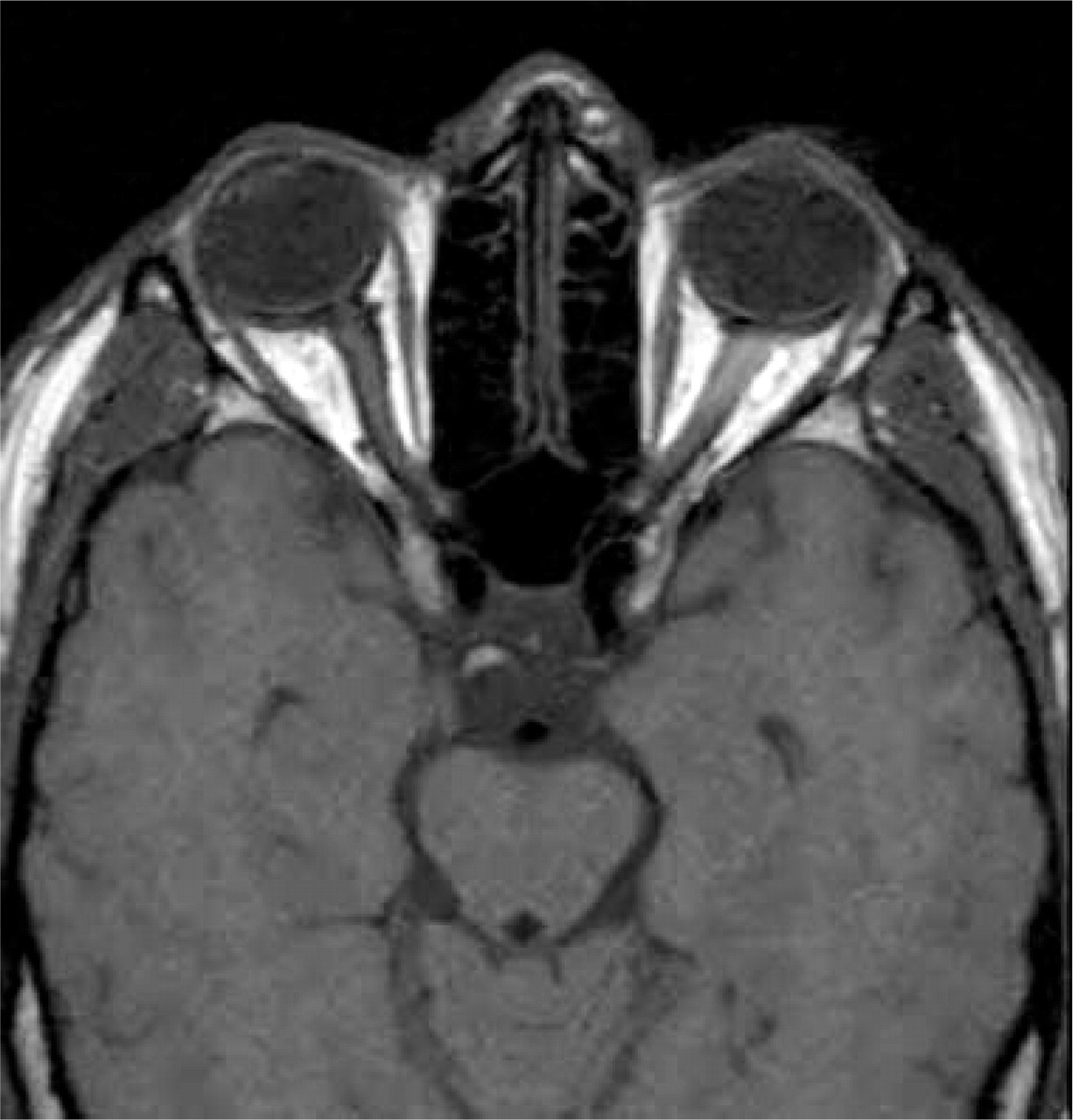
T1 weighted MRI sequence at the level of the pituitary stalk shows the thickened right fourth cranial nerve in the suprasellar cistern (TR/TE, 600/17). The thickening allows the visualization of this segment on this nonenhanced MRI.

Gd-DTPA T1 weighted MRI sequence shows the enhancing and thickened distal cisternal portion of the fourth cranial nerve (TR/TE, 600/17).

Gd-DTPA T1 weighted MRI sequence shows the enhancing fourth cranial nerve in the suprasellar cistern in this coronal image (TR/TE, 600/17).
The present episode of ophthalmoplegia resolved spontaneously in about three months. On re-examination four months after the start of the symptoms only the pre-existent pupil anisocoria remained. A repeated MRI (performed 111 days after onset of symptoms) no longer showed enhancement of the fourth cranial nerve (Fig. 4) and the IgG production in the cerebrospinal fluid (obtained 115 days after onset of symptoms) had normalized.

Gd-DTPA T1 weighted MRI sequence shows compared with Fig. 2 less enhancement of the fourth cranial nerve (TR/TE, 600/17).
Discussion
The three episodes experienced by the patient presented above fulfil the IHS criteria for ophthalmoplegic migraine (1). His most recent attack was associated with paresis of both the third and fourth cranial nerve. This unusual combination has never been described to date. In most cases of ophthalmoplegic migraine only the third cranial nerve is affected. Paresis of the abducens nerve occurs in 10% of cases. Trochlear nerve palsy is even more rare (4).
During the attack MRI showed enhancement of the affected fourth cranial nerve. Enhancement of the fourth cranial nerve on MRI has not been reported before. Wong and Sharpe (5) describe a patient with recurrent trochlear nerve palsy associated with migraine. MRI findings were normal. However it is not clear whether contrast-enhanced MRIs were made. Enhancement of the third cranial nerve on MRI may occur in patients with ophthalmoplegic migraine associated with oculomotor palsy (6–11). There is one report of reversible enhancement of the abducens nerve in a patient with ophthalmoplegic migraine associated with abducens nerve palsy (12). In all but one of these cases the enhancement resolved along with the signs and symptoms. In one patient persistence of recurrence of ophthalmoplegic migraine attacks was associated with long-lasting presence of the MRI abnormalities. Only after four years MRI findings normalized (10).
In our patient the third cranial nerve did not enhance after gadolinium although it was clinically involved. We do not have a good explanation for this. There is to our knowledge at least one other report of a patient suffering from ophthalmoplegic migraine involving the third cranial nerve without enhancement on MRI. This patient had an unusual history not only because of the normal MRI findings, but also because she had a permanent neurological deficit after an initial ophthalmoplegic migraine attack (13).
Stommel et al. (9) suggest an ischaemic mechanism of ophthalmoplegic migraine given the MRI findings of enhancement of the third cranial nerve at the point of exit from the brainstem. However, the third cranial nerve did not enhance on MRI imaging in patients with diabetic oculomotor palsy, even though this condition is thought to be due to ischaemia (14). Others proposed an infectious or inflammatory pathogenesis for ophthalmoplegic migraine (6, 8, 10). This suggestion is supported by the CSF results in our patient, which showed a slightly increased IgG-index during the episode. To our knowledge there are no other reports of CSF examinations in ophthalmoplegic migraine. Lance and Zagami (11) consider the possibility that ophthalmoplegic migraine is a recurrent neuritis or demyelinating neuropathy. An inflammatory process could trigger migraine headaches in people who are already migraineurs.
Several authors describe the outcome of uncontrolled trials of anti-inflammatory therapy in individual patients with ophthalmoplegic migraine. One patient failed to respond to prednisone therapy, but this patient did not fulfil formal criteria for ophthalmoplegic migraine since no headache was reported (15). In other cases a dramatic (within days) improvement of symptoms was reported following prednisone therapy (3, 16). However, the value of steroid treatment in ophthalmoplegic migraine remains questionable since symptoms tend to resolve spontaneously in weeks or months.
In conclusion the aetiology of ophthalmoplegic migraine remains obscure. In the case described above the patient's history of previous migraine attacks and his family history suggest a migrainous condition. However MRI results in our and other patients and the response to steroids in individual patients with ophthalmoplegic migraine, are more compatible with an inflammatory than a migrainous condition.
Footnotes
Acknowledgements
The authors would like to thank C.B. Majoie, neuroradiologist in the Academisch Medisch Centrum, Amsterdam for critically reviewing the MRIs.