
Abstract

C
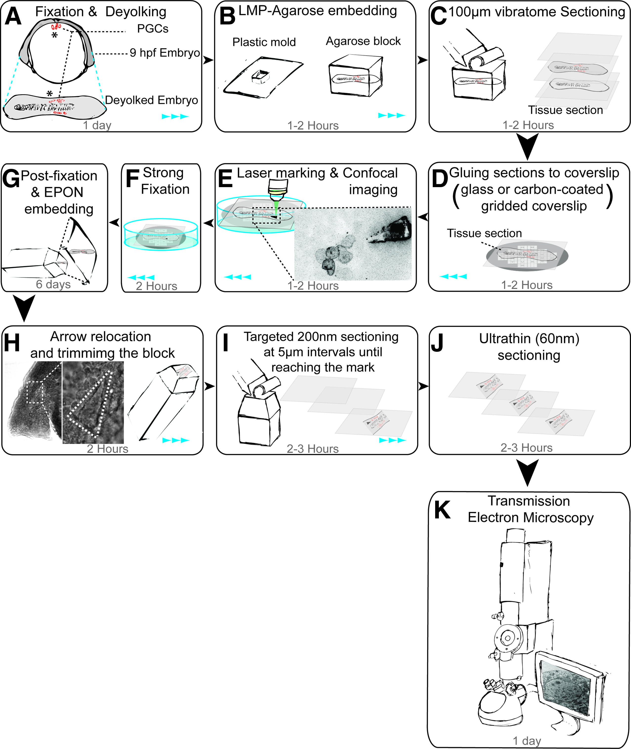
Schematic workflow of the correlative light and electron microscopy using laser marking.
Concise Workflow
Collect and fix embryos (8–12 hpf in this study) whose cells or subcellular structures of interest are fluorescently labeled (with GFP, YFP, or mCherry), dechorionate the embryos, 9 and fix with 2% PFA + 0.2% Glutaraldehyde in a 0.1 M PHEM buffer (60 mM of PIPES, 25 mM of HEPES, 2 mM MgCl2, 10 mM of EGTA), pH 6.9 at room temperature in a glass dish.
• After 10 min of fixation, deyolk the embryos under a fume hood as shown in Supplementary Movie S1 (Supplementary Data are available online at www.liebertpub.com/zeb).
• Continue fixation for 2 h at room temperature.
• Transfer to 1%PFA in the 0.1 M PHEM buffer using a glass pipette and select embryos for embedding based on the fluorescence signal (Fig. 1A).
Embedding of embryos in 4% low melting point (LMP) agarose in small plastic molds (Fig. 1B and Supplementary Data) and section to 100-μm-thick slices using a vibratome.
Collection of slices into flat-bottom plates (e.g., Corning® Costar® cell culture plates) that contain the 0.1 M PHEM buffer and screening for the slices containing fluorescently labeled cells [e.g., using a low magnification on an epifluorescence microscope (Fig. 1C)].
Gluing the selected slices to glass or carbon-coated gridded coverslips (the latter that can be produced or purchased, help to localize the region of interest when trimming the block) using a drop of LMP agarose (Fig. 1D). Transfer the samples to 0.1 M PHEM buffer.
Generation of a laser mark using an 850-nm pulsed laser on a two-photon setup (We used 100% power and 1000–2500 iterations. Power and iterations can be adjusted based on depth of cells and laser power output) (Fig. 1E). To prevent sample photo bleaching, we used the “test bleach” mode in ZEN software, equivalents can be used in other setups.
Performing confocal imaging of the cells for three-dimensional reconstruction (e.g., using a 63× objective with NA: 1.0, 0.8 μm z-slice and pinhole 1 AU) (Fig. 1E and Supplementary Movie S2).
For an overview of the samples in lower magnification, capture a photo using 5× and 20× objectives (the 20× objective is used for measurement of X/Y/Z coordinates of the cells). Depth of the cells (Z) from surface of the tissue was measured by subtracting the microscope stage position while focusing on the cells from that of the stage position when focusing on the tissue surface.
Strong fixation with 2% PFA + 2% glutaraldehyde in 0.1 M PHEM at room temperature for 2 h under the fume hood. Continue by exchanging solution with 1% PFA in the 0.1 M PHEM buffer at 4°C (Fig. 1F). Continue to the next step as soon as possible, as prolonged storage can degrade the quality of the sample.
Postfixing in 1% osmium tetroxide, containing 1.5% potassium cyanoferrate, and embedding in EPON after dehydration 10 (Fig. 1G).
Relocating the laser mark in EPON by illuminating the block under the microscope in brightfield and trimming the block to the area containing the laser mark (keeping the sample in the transmission electron microscopy (TEM) sample holder helps to avoid altering the position or the orientation of the sample) (Fig. 1H and Supplementary Movie S3).
Targeted sectioning by generating 200-nm-thick slices at 5 μm intervals and examining them for the presence of the laser mark (Fig. 1I).
Ultrathin sectioning (60 nm) at position of the laser mark (Fig. 1J) and TEM imaging (Fig. 1K).
Compare the light microscope and TEM images to identify the cells of interest (Supplementary Fig. S1).
Footnotes
Acknowledgments
We acknowledge the support of the University of Münster, the Cells in Motion (CIM) excellence cluster, the German research foundation (DFG), and the European Research Council (ERC). We thank Michal Reichman-Fried for comments on the article.
Disclosure Statement
No competing financial interests exist.
References
Supplementary Material
Please find the following supplemental material available below.
For Open Access articles published under a Creative Commons License, all supplemental material carries the same license as the article it is associated with.
For non-Open Access articles published, all supplemental material carries a non-exclusive license, and permission requests for re-use of supplemental material or any part of supplemental material shall be sent directly to the copyright owner as specified in the copyright notice associated with the article.