
Abstract
Abstract
Zebrafish provide researchers and students alike with an excellent model of vertebrate nervous system development due to a high degree of conserved developmental mechanisms and transparent embryos that develop in synchrony. In these laboratory exercises, undergraduate students explore cell biological concepts while performing hypothesis-driven novel research utilizing methodologies such as immunofluorescence, confocal microscopy, image analysis, pharmacology, and basic statistics. In the first block of exercises, students perform anti-acetylated tubulin (anti-AT) immunofluorescence, identify spinal tracts and neuronal subtypes, and perform conventional and confocal microscopy. Building on knowledge acquired in the first block of exercises, during the second block, students subsequently perform pharmacological activation of Wnt signaling through lithium chloride treatments, and assess nervous system integrity through anti-AT immunofluorescence. Students perform various quantitative methods and apply statistics to determine outcomes of Wnt activation. In their final laboratory report, students contextualize their results with foundations of molecular mechanisms of nervous system development. In sum, these exercises offer undergraduate students a model of independent research at the graduate level.
Introduction
The following set of laboratory exercises could be used in undergraduate courses in cell biology, developmental biology, or neurobiology. Though specific cell biological and developmental questions are addressed in these labs, the overall goal is to allow students to become proficient with zebrafish as a model organism, generate viable hypotheses, carry out hypothesis-driven novel research, and interpret data. Students propose additional lines of inquiry in a formal laboratory report. These laboratory exercises are specifically designed to integrate theory of developmental/cell biology, foster student collaboration, implement quantitative reasoning, and employ multiple modes of inquiry. These exercises emulate scientific research at the graduate level, promoting retention of science students in academia, and increasing scientific literacy.
The zebrafish spinal cord is structurally similar to higher vertebrates, including humans, with a basic dorsal to ventral arrangement of sensory neurons, interneurons, and motoneurons. Zebrafish spinal cord neurogenesis begins at 15–17 hours post fertilization (hpf) 1 and continues throughout embryonic development. At 24 hpf, approximately 12 identified neuronal subtypes are present. Interneuron subtypes include dorsal lateral ascending (DoLA), commissural primary ascending (CoPA), commissural secondary ascending (CoSA), ventral longitudinal descending (VeLD), Kolmer-Agdur (KA), commissural bifurcating/longitudinal (CoB/L), circumferential ascending (CiA), circumferential descending (CiD), and unipolar commissural descending (UCoD). Sensory neurons include the Rohon-Beard neurons which are found in the dorsal spinal cord. Axons of primary motoneurons exit the spinal cord ventrally.1–5 In the dorsal spinal cord, the dorsal longitudinal fasciculus (DLF) is comprised of Rohon-Beard, DoLA and CoPA axons. 3 Of the neurons that are present at 24 hpf, Rohon-Beard, DoLA, CoPA, and motoneurons are readily identified with anti-AT immunofluorescence (Bonner, unpublished data). Thus, this developmental stage is ideal for introducing students to the diversity and complexity of the nervous system, while still providing resolution of individual axons and tracts. Furthermore, dissection of 24 hpf embryos for microscopy is relatively straightforward. Finally, as anti-acetylated tubulin (anti-AT) labeling works on embryos that have been cryopreserved in methanol, fixed embryos can be stored for up to 4 months prior to initiation of the lab.
Neuronal identity within the spinal cord relies upon coordinated signaling by Wnt, bone morphogenetic protein (BMP), and Hedgehog. In vertebrates, canonical Wnt signaling is required for patterning the dorsal spinal cord. Wnts regulate proliferation and limit the expansion of more ventral identities. These roles are conserved in mouse and chick.6,7 Through inhibition of GSK-3 beta, lithium chloride (LiCl) activates canonical signaling, 8 which results in dorsalized embryos when applied before gastrulation. 9 Application of LiCl at later stages of development allows students to determine whether activation of Wnt signaling disrupts overall organization of axon tracts in the spinal cord of the developing embryo. Since inhibition of canonical Wnt signaling limits the ventral extent of dorsal progenitor patterning, 6 activation by LiCl is predicted to expand dorsal identities in the spinal cord. One potential outcome of this is the subsequent expansion of dorsal axon tracts. As this has never been demonstrated in zebrafish, application of LiCl at later stages of development is a novel experimental approach.
When taught in tandem with supportive lecture material, students successfully gain both theoretical and experiential knowledge in developmental biology, cell biology, neurobiology, and embryology. Importantly, students learn the value of evidence-based hypothesis testing, which is critical to scientific literacy. Upon completion of the laboratory modules, students leave with applied knowledge in microdissection, embryonic staging, pharmacology, quantitative and qualitative assessment, immunohistochemistry, confocal laser scanning microscopy (CLSM), and wide field fluorescence microscopy. Importantly, students understand the strengths and weaknesses of each of their experimental approaches, and are able to design future experiments that address the weaknesses of their analysis. In short, students are exposed to the progressive nature of scientific inquiry in a classroom setting.
Materials and Methods
Refer to Appendix for detailed outline of laboratory exercises. General information regarding basic zebrafish care and practical approaches can be found in Zebrafish (Nusslein-Volhard and Dahm) 10 and The Zebrafish Book: A Guide for the Laboratory Use of Zebrafish (Danio rerio), fifth edition (M. Westerfield). 11 An older version of this text is available online, at the Zebrafish Model Organism Database (zfin.org) website. Other resources for zebrafish research include zfic.org and zebrafish.org websites.
Embryo acquisition
For instructors at institutions that lack large scale zebrafish colonies, wild-type adult fish can be housed in 5 L or 10 L tanks, available at local pet stores. Research grade adult wild-type fish are obtained from the Zebrafish International Resource Center (ZIRC). Wild-type (WT) zebrafish embryos are obtained by mating male and female adult WT zebrafish. Fish can either be bred in large tanks using an “in tank spawning tank set” (ITSTS-A, Aquatic Habitats), or can be placed in 1 L breeding tanks (SBTANK, Aquatic Habitats) with a female to male ratio of 3:1. After breeding, embryos are collected using a sieve or other straining device.
Embryo development
Up to 50 embryos are incubated in E3 solution (5 mM NaCl, 0.17 mM KCl, 0.33 mM CaCl2 0.33 mM MgSO4) in 100 mm Petri dishes (Fisher 08-757-9B) at 28.5°C. If desired, development can be slowed or increased by altering the temperature in which the embryos are raised. 12 The range of appropriate temperatures is between 23°C and 31°C. 12 The temperature at which embryos need to be raised is based on the timing of the laboratory session, and when the fish spawned. Therefore each instructor must determine temperature conditions. Kimmel et al. 12 is used for staging embryonic development. For these laboratory exercises, 18 hpf and 24 hpf are the relevant developmental stages. At 18 hpf, 18 somites are clearly visible (Fig. 1A), whereas at 24 hpf, a heartbeat and the circulation of blood cells are visible for the first time (Fig. 1B). At 24 hpf, embryos are dechorionated (see The Zebrafish Book) 11 using fine point Dumont #3 forceps (Fisher Scientific #NC9839169) and transferred to a 2.0 mL tube (Ambion AM12425). Embryos are fixed in 4% paraformaldehyde (PFA)(VWR JTS898-7) diluted in 1X phosphate buffered saline (PBS) at room temperature for 3 hours or overnight at 4°C while gently rocking. Embryos are rocked on a nutator (VWR #15172-203).
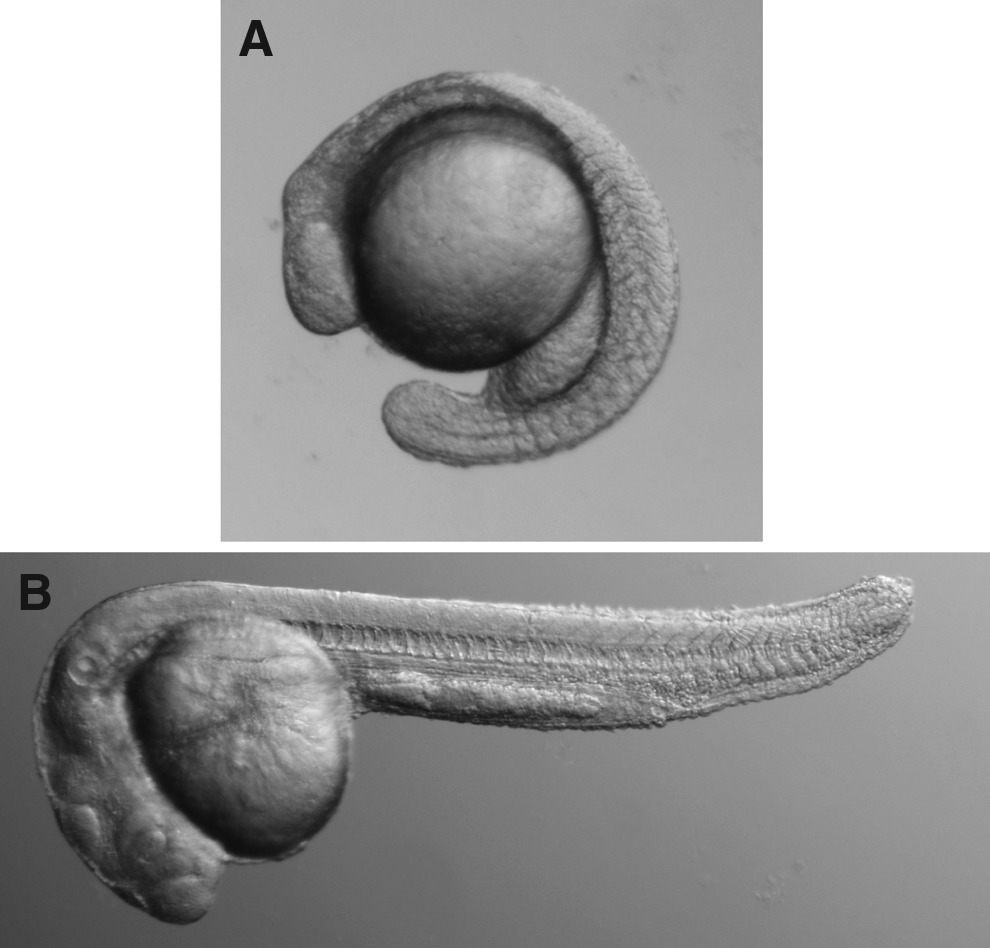
Light microscopic images of zebrafish embryos.
Immunohistochemistry
For all solution changes, embryos are allowed to settle in the tube, and solution is gently aspirated to prevent disturbing embryos. After fixation, embryos are washed at room temperature in PBS with TritonX-100 (1X PBS, 0.5% Triton X-100) (PBT) three times, 5 min each with gentle rocking. Next, embryos are blocked in 500 μL of 10% normal goat serum (NGS) (Sigma G6767-100) diluted in PBT for 1 h at room temperature while gently rocking. Embryos are incubated in anti-acetylated tubulin (Sigma T6793) (anti-AT) primary antibody diluted 1:1000 in 10% NGS-PBT for 4 h at room temperature or overnight at 4°C while gently rocking. Following primary antibody incubation, embryos are washed three times in PBT (15 min each) at room temperature with gentle rocking. Secondary antibodies such as Cy3 conjugated goat anti-mouse (GAM-Cy3) (Jackson ImmunoResearch #115-165-003) diluted 1:200 in 10% NGS-PBT are incubated for 4 h at room temperature or overnight at 4°C while gently rocking. Secondary antibodies are washed off as previously described for the primary antibody. Embryos are stored in 4°C until the next scheduled laboratory session.
Microdissection and mounting
Labeled embryos are transferred to a petri dish containing PBT. Using a dissecting microscope and Dumont #3 forceps, the yolk sac is removed from the embryos. To do this, the head of the embryo is pinned with one pair of forceps, while the yolk is separated from the body of the embryo. Since damage to the somite region will impede visualization, students are reminded to use caution while dissecting. Dissected embryos are pipetted onto a microscope slide and excess PBT is removed with a kimwipe. One drop of Slowfade gold antifade reagent (Fisher Scientific # S36936) is applied to the embryos. After embryos are aligned on the slide (5–10 embryos/slide), a cover slip that contains a small amount of Vaseline on the four corners is applied. Sufficient pressure is applied to the cover slip to seal the slip, while preventing embryo damage. Excess slowfade is removed from the edges of the cover slip with a kimwipe and the slide is sealed with nail polish. These prepared slides, if stored in the dark at 4°C, are stable for months.
Microscopy and statistical analysis
Both wide-field fluorescence microscopy and CLSM can be used on mounted embryos described above. While the resolution possible by CLSM far exceeds wide field microscopy (compare Fig. 2A and B), the expense of a CLSM makes this approach prohibitive, unless a CLSM is available at the home institution. Spinal cords are imaged using 20X and 40X objective lenses. For 40X imaging, a long working distance lens is required, as the overlying somite tissue is relatively thick. For confocal imaging of the spinal cord, 0.5 to 1.0 μm optical sections are sufficient for the purposes of the laboratory exercise (Fig. 2A and B). After image acquisition, ImageJ (free download from NIH; http://rsbweb.nih.gov/ij/) software is utilized to measure the dorsoventral axis of the dorsal longitudinal fasciculus (DLF). Graphical analysis using Microsoft Excel allows for statistical comparison between WT embryos and embryos treated with LiCl (Fig. 2C). Means of approximately 10 embryos per treatment are calculated, followed by SEM. p values are calculated by using an unpaired Student's t-test.
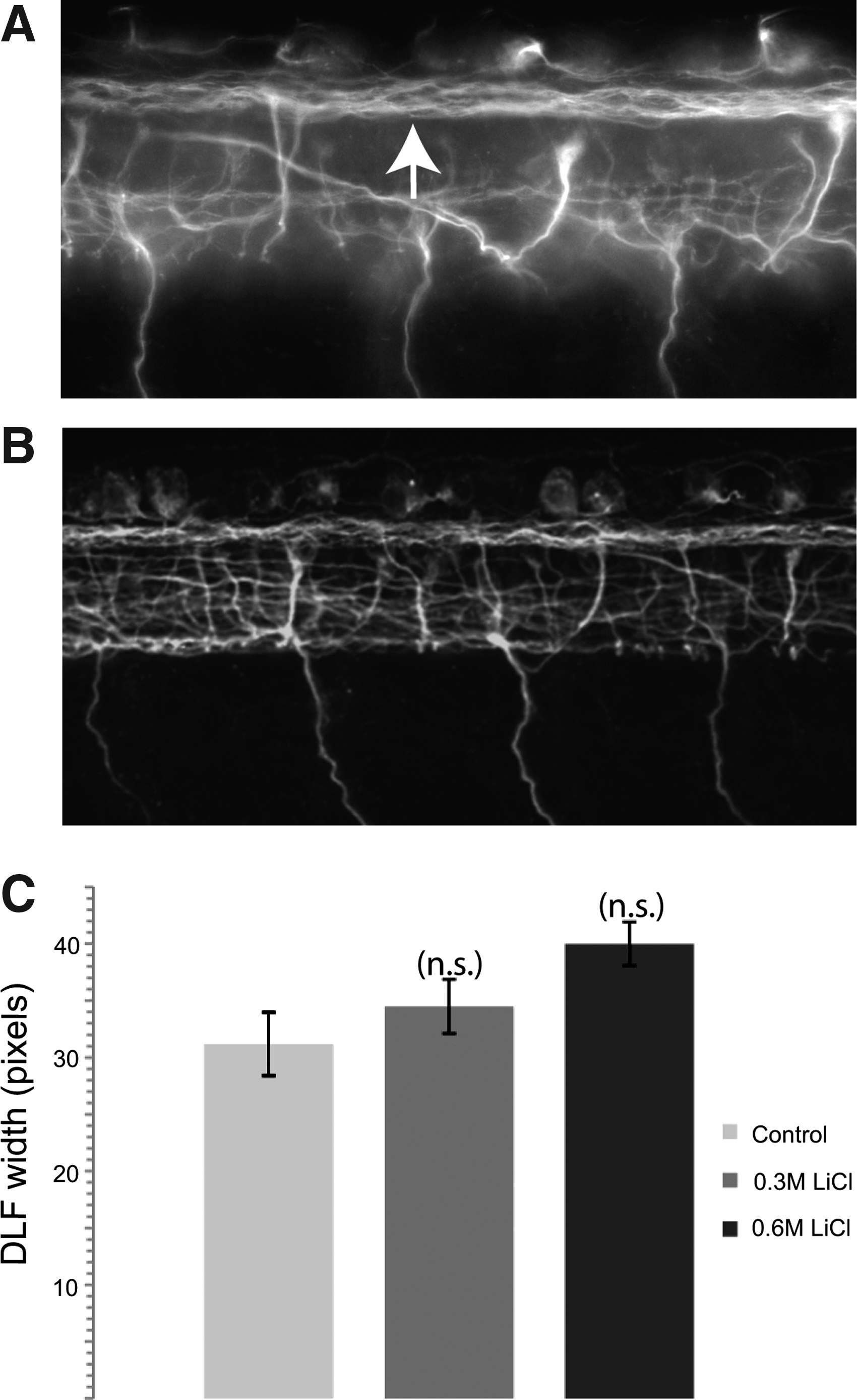
Examples of student generated images and data. Images of anti-acetylated tubulin immunofluorescence in the spinal cord of a 24 hpf embryo with
Lithium chloride treatment on live embryos
The following experiments were modified from LiCl laboratory exercises found at www.zfic.org. Using Kimmel et al. 12 as a guide, embryos are dechorionated with forceps at 14–16 hpf and divided into three groups in a 24-well plate. LiCl (Fisher #L121-100) is applied to embryos for either 10 min, or 6 h.9,13 A control group is raised in a solution of E3 and the other two groups in solutions of 0.3 M LiCl or 0.6 M LiCl diluted in E3. After incubation at 28.5°C embryos are washed three times with E3. Embryos are fixed and treated for anti-AT immunofluorescence as described above. If immunofluorescence cannot be carried out within a few days, embryos are cryopreserved first (see below).
Cryopreservation and rehydration of embryos
Cryopreservation can be used before the first block of laboratory exercises, but is optional. After fixing with PFA (see above), embryos are washed three times in PBT (5 min each) with gentle rocking at room temperature. Next, half of the PBT is replaced with 100% methanol (MeOH) and incubated at room temperature with rocking for 5 min. This step is repeated once. All of the liquid is then removed and replaced with 100% MeOH. Embryos are incubated for 15 min at room temperature with rocking. This step is repeated once. Now embryos can be stored −20°C. Before the immunohistochemistry protocol, embryos are rehydrated by repeating the dehydration steps but substituting MeOH for PBT. Briefly, half of the MeOH is aspirated, discarded, and replaced with PBT. Embryos are incubated for 5 min while gently rocking at room temperature. This is repeated once. Next, the remainder of the solution is removed and replaced with PBT. Embryos are incubated for 5 min while gently rocking at room temperature. This step is repeated once. Embryos are now ready for immunohistochemistry.
Results
The laboratory exercises outlined below constitute two blocks of laboratory sessions, 4 weeks each, for a total of 8 weeks. Refer to Appendix for detailed outline. Laboratories meet once per week for 3 hours. However, some exercises occur outside of laboratory time, due to limited resources (availability of microscopes) and procedural requirements (application of LiCl, followed by fixation later in the day). Since flexibility is required, students work in teams (typically pairs), which fosters collaboration, an integral component to the scientific process. The first 4-week block consists of immunofluorescence of wild-type zebrafish embryos. Embryos are visualized with both CLSM and wide field fluorescence microscopy (Fig. 2A and B). Concurrently, lecture material focuses on theory and methodologies of fluorescence microscopy. Additionally, students learn the basic theory of nervous system development, cell biological techniques, and cell signaling. During the second block in the laboratory, students apply knowledge gained previously in the course to address the role of Wnt activation on spinal tract formation. Students apply LiCl for varying amounts of time followed by fixation and anti-AT immunofluorescence. Embryos are mounted on slides and imaged using wide field fluorescence microscopy. Students assess overall spinal cord tract organization through careful comparison to stage-matched wild-type embryos. This process requires feedback from instructors, as students struggle, but ultimately master, qualitative changes in spinal tract organization. Once students are comfortable with this level of analysis, they calculate percentages of effected embryos. Students then employ quantitative analysis and measure the width of the DLF in the dorsoventral axis using ImageJ software. The DLF is measured in pixel length (as per ImageJ software analysis). Means +/− SEM are graphed for the formal laboratory report (Fig. 2C). A Student's unpaired t-test is performed to determine statistical significance.
Discussion
Student learning
Students are required to complete experiments outside of laboratory hours. To do this, they must collaborate with their partners or team members, delegate responsibility, and demonstrate accountability, all of which are done with minimal instructor guidance. This promotes independence and ownership of the learning experience, which is well received by the students.
All students produce both CLSM and wide field fluorescent images (see Fig. 2A and B for example of student work). Since these exercises take place over an 8-week interval and mounted embryos are stable for months, students have the opportunity to image embryos again if their initial attempts produce low quality images. The strengths and limitations of each type of microscopy (image quality versus cost of microscope and time required for imaging) are well described in their lab reports, and supported with student generated images. As the course proceeds, students become increasingly confortable with primary literature assignments, as evidenced by integration of background material in their laboratory reports. Results of LiCl treatment on DLF width vary within the student population. Regardless of outcome (larger DLF, smaller DLF, or no change), students are able to support their findings with known roles of Wnt or other morphogens. Furthermore, students are able to design future experiments that stem from their results, indicating that they understand the process of scientific inquiry.
Conceptual support for laboratory
During the 8 weeks of laboratory exercises, students receive theoretical background on cell biological methods, such as immunocytochemistry and pharmacology. In conjunction with this, students become familiar with zebrafish as a model system, including genetics, gene knock-down, transgenesis, and basic embryology. Students are introduced to developmental biology of the nervous system, incorporating the basics of proliferation, patterning, differentiation, axon guidance, and their molecular mechanisms. Various experimental approaches that examine these issues are taught, including BrdU labeling, in situ hybridization, and HuC immunofluorescence. For the laboratory exercises, students are aware that defects observed in axon tract formation may arise from earlier developmental defects such as patterning disruptions, which is pertinent for their interpretation of results.
Depending on the level of the course, a number of primary literature articles could serve as background material for the lab. Since canonical Wnt signaling is involved in many aspects of nervous system development, instructors can gear the background reading to be as focused or broad as desired. Megason and McMahon 14 demonstrate mitogenic activity of spinal cord Wnts, Bonner et al. 6 demonstrate distinct downstream functions and mechanisms of dorsal Wnt signaling in the zebrafish spinal cord. Muroyama et al. 15 establish that neuronal identity in the dorsal spinal cord relies on dorsal canonical Wnts, while Stachel et al. 9 and Klein and Melton 8 provide the initial evidence that LiCl dorsalizes zebrafish embryos and activates the Wnt pathway. Downes et al. 5 utilize transient expression of GFP to identify neuronal subtypes. Useful textbooks that support both lecture and laboratory include Molecular Biology of the Cell (5th edition, Alberts et al.) 16 and Development of the Nervous System (3rd edition, Sanes et al). 17 The Fourth Edition of Molecular Biology of the Cell is freely and readily available on NCBI PubMed.
These lab sessions succeed in teaching students basic cell biological methods, pharmacology, quantitative analysis, and microscopic imaging. As the experiments are performed on zebrafish, students are also exposed to the utility of this important vertebrate model organism. Performed on wild-type zebrafish, there is no need to maintain different mutant or transgenic lines of fish. Further, the labs utilize relatively simple and inexpensive methodologies (immunofluorescence, pharmacology). Thus, these labs represent effective transitional labs for instructors who are not classically trained in zebrafish.
Since the labs are modular, some aspects (such as CLSM) could be excluded. This would be prudent for institutions that lack a CLSM, as they are quite expensive. Though wide field fluorescence does not provide the same resolution or clarity compared to CLSM, it is quite sufficient to carry out the analysis of LiCl-treated embryos. In addition, other modules could be included such as application of LiCl at 2 hpf. 9 Since earlier treatments of LiCl dorsalize zebrafish embryos, 9 this module would address early developmental roles of Wnt signaling. Furthermore, transgenic zebrafish that express GFP in specific tissues can also be examined with anti-GFP immunofluorescence (Invitrogen A-11122) for double labeling experiments with anti-AT or another monoclonal antibody. Anti-GFP immunofluorescence also works on embryos that have been cryopreserved in MeOH. Over the course of 4 years, these laboratory modules have been taught to upper-level undergraduate students (both with and without the confocal module, early LiCl treatment, and transgenic GFP embryos).
These laboratory modules successfully train students in six critical areas of scientific investigation: (I) technical and (II) analytical skills; (III) critical reading of primary literature; (IV) collaboration, responsibility, and accountability; (V) hypothesis driven research; and (VI) accurately reporting scientific findings through written reports. Though these core concepts are mastered using zebrafish, they are not limited to this model organism. Furthermore, the six areas of scientific investigation are progressively learned, supporting retention of information. For example, early in the laboratory modules, students microdissect and learn basic zebrafish neuroanatomy. Students retain and utilize this during the entire course. Additionally, relevant primary literature is assigned throughout the laboratory modules, so that students are continually reinforcing and building on their knowledge. Finally, the collaborative nature of scientific research is fostered through working together in groups and discussing scientific findings with the entire class. Though all students are able to generate images, statistically evaluate data, and formulate written reports, there is no consistent LiCl result that can be reported from these student exercises. Instead, students are required to interpret their data and suggest alternative hypotheses and methodologies. For example, higher doses of LiCl may be used. Alternatively, LiCl may be applied earlier in development, since outcomes of earlier LiCl treatment are published. 9 Though students may leave the laboratory without a substantial experimental outcome, they leave the course with valuable insight of hypothesis-driven novel research.
By experiencing these labs, students are exposed early in their scientific careers to novel research using cutting-edge techniques in an important vertebrate model organism. In general, students receive these modules enthusiastically, and report intense interest in conducting experiments in which the outcome can be predicted based on published results, but has yet to be demonstrated. In evaluations, 50% of students favorably report that the laboratory was “like a graduate school experience.” Student comments included: “The lab was the best experience I have had in school so far. It pushed the boundaries of undergraduate science and had an almost graduate school feel,” and “we were also treated like graduate students. I appreciated this opportunity to work in an atmosphere similar to graduate school, so that I could get a taste of what I might want to pursue after college.” This inquiry-based approach to novel research in a classroom setting enhances critical thinking skills, helps students to retain relevant information, and is an early and appropriate motivator for students to continue scientific research in post-baccalaureate careers.
Footnotes
Disclosure Statement
No competing financial interests exist.