
Abstract
Significance:
Chronic wounds include, but are not limited, to diabetic foot ulcers, venous leg ulcers, and pressure ulcers. They are a challenge to wound care professionals and consume a great deal of healthcare resources around the globe. This review discusses the pathophysiology of complex chronic wounds and the means and modalities currently available to achieve healing in such patients.
Recent Advances:
Although often difficult to treat, an understanding of the underlying pathophysiology and specific attention toward managing these perturbations can often lead to successful healing.
Critical Issues:
Overcoming the factors that contribute to delayed healing are key components of a comprehensive approach to wound care and present the primary challenges to the treatment of chronic wounds. When wounds fail to achieve sufficient healing after 4 weeks of standard care, reassessment of underlying pathology and consideration of the need for advanced therapeutic agents should be undertaken. However, selection of an appropriate therapy is often not evidence based.
Future Directions:
Basic tenets of care need to be routinely followed, and a systematic evaluation of patients and their wounds will also facilitate appropriate care. Underlying pathologies, which result in the failure of these wounds to heal, differ among various types of chronic wounds. A better understanding of the differences between various types of chronic wounds at the molecular and cellular levels should improve our treatment approaches, leading to better healing rates, and facilitate the development of new more effective therapies. More evidence for the efficacy of current and future advanced wound therapies is required for their appropriate use.
Scope and Significance
C
Chronic wounds can be classified as vascular ulcers (e.g., venous and arterial ulcers), diabetic ulcers, and pressure ulcers (PUs). 6 Some common features shared by each of these wounds include prolonged or excessive inflammation, persistent infections, formation of drug-resistant microbial biofilms, and the inability of dermal and/or epidermal cells to respond to reparative stimuli. 7 –11 In aggregate, these pathophysiologic phenomena result in the failure of these wounds to heal. The underlying pathologies, however, differ among various types of chronic wounds.
This review provides a brief overview of the pathophysiology of chronic wound healing and discusses the established tenets and advanced treatment of chronic wounds, with an emphasis on diabetic foot ulcers (DFUs). Overcoming the factors that contribute to delayed healing is a part of the comprehensive approach to wound care and presents the primary challenges to the treatment of chronic wounds.
Translational Relevance
This review provides researchers with a comprehensive discussion of the pathophysiology and approaches for the evaluation and treatment of chronic wounds from the perspective of an experienced wound care physician. A better understanding of the challenges physicians face currently with chronic wounds should facilitate the development of new wound care products.
This review also demonstrates the lack of available evidence demonstrating efficacy for a majority of existing advanced wound care products. This fact should be taken into consideration by researchers during the development of clinical programs for wound care products.
Clinical Relevance
Understanding and addressing the challenges in the treatment of chronic wounds will lead to a better clinical outcome (faster and more durable wound closure), resulting in improved patient quality of life and reduced healthcare costs. This review provides a current expert opinion and can be used as a guideline for evaluation and appropriate treatment selection for nonhealing wounds.
Discussion
Physiology of wound healing
The physiological process of wound healing is achieved through four temporarily and spatially overlapping phases: hemostasis, inflammation, proliferation, and remodeling phases. 12,13 Immediately after injury, hemostasis occurs and is characterized by vasoconstriction and blood clotting, which prevents blood loss and provides the provisional matrix for cell migration. Platelets secrete growth factors and cytokines attract fibroblasts, endothelial cells, and immune cells to initiate the healing process. The subsequent inflammation phase lasts up to 7 days. The predominant cells at work in this phase are phagocytic cells, such as neutrophils and macrophages. Neutrophils release reactive oxygen species (ROS) and proteases that prevent bacterial contamination and cleanse the wound of cellular debris. Blood monocytes arrive at the wound site and differentiate into tissue macrophages. The latter not only remove bacteria and nonviable tissue by phagocytosis but also release various growth factors and cytokines recruiting fibroblasts, endothelial cells, and keratinocytes to repair the damaged blood vessels. As the inflammatory phase subsides accompanied by apoptosis of immune cells, the proliferation phase begins. This phase is primarily characterized by tissue granulation, formation of new blood vessels (angiogenesis), and epithelialization. The last phase occurs once the wound has closed and may last 1–2 years or longer. During this phase, the provisional matrix is remodeled into organized collagen bundles. 14,15
Pathophysiology of chronic wounds
Chronic wounds are defined as wounds that fail to proceed through the normal phases of wound healing in an orderly and timely manner. Often, chronic wounds stall in the inflammation phase of healing. Despite differences in etiology at the molecular level, chronic wounds share certain common features, including excessive levels of proinflammatory cytokines, proteases, ROS, and senescent cells, as well as the existence of persistent infection, and a deficiency of stem cells that are often also dysfunctional (Fig. 1).
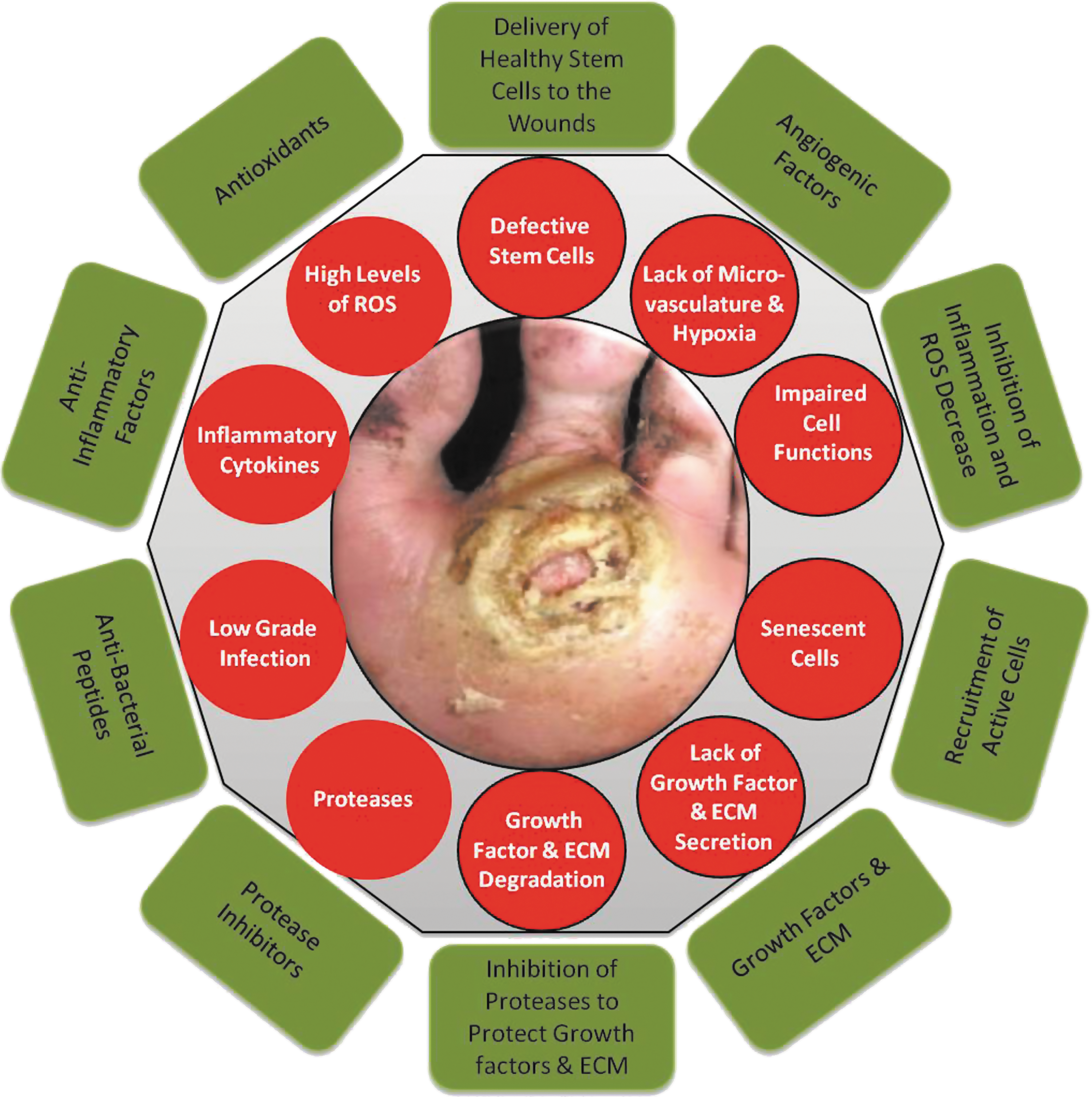
Molecular and cellular deficiencies in chronic wounds (red circles) and factors required to overcome them (green rectangles). Nonhealing ulcers and wounds represent a failure to achieve complete reepithelialization in the appropriate temporal sequence of tissue repair. Such wounds are characterized by excessive inflammation (including elevated levels of proteases, ROS, and inflammatory cytokines), by senescent cell populations with impaired proliferative and secretory capacities, and by defective MSCs. Excessive inflammation leads to degradation of newly synthesized growth factors and ECM. There is a need to restore the proper balance of cytokines, growth factors, and proteases, to recruit functional cells (epithelial cells, fibroblasts, and endothelial cells) to the wound area, and to deliver healthy functional MSCs directly to the wound to compensate for the patient's own dysfunctional stem cells. ECM, extracellular matrix; MSCs, mesenchymal stem cells; ROS, reactive oxygen species.
Due to repeated tissue injury, microorganisms and platelet-derived factors, such as transforming growth factor-β (TGF-β) or ECM fragment molecules, stimulate the constant influx of immune cells; the proinflammatory cytokine cascade therefore becomes amplified and persists for a prolonged time, leading to elevated levels of proteases. In acute wounds, proteases are tightly regulated by their inhibitors. In chronic wounds, protease levels exceed that of their respective inhibitors, leading to destruction of ECM and degradation of growth factors and their receptors. The proteolytic destruction of ECM not only prevents the wound from moving forward into the proliferative phase but also attracts more inflammatory cells, thus amplifying the inflammation cycle. 16
Immune cells produce ROS, which in low concentrations provides defense against microorganisms. In chronic wounds, however, the predominant hypoxic and inflammatory environment increases ROS production, which damages ECM proteins and causes cell damage. This sequence of events leads to an enhanced stimulation of proteases and inflammatory cytokines. 17 It has been suggested in an animal model that application of strong antioxidants reduces ROS to normal levels, which results in the reverse of the chronicity of wounds and improves healing. 18
Furthermore, chronic wounds are characterized by senescent cell populations with impaired proliferative and secretory capacities, rendering them unresponsive to typical wound healing signals. 15 It has been reported that fibroblasts from venous and PUs are senescent and have a diminished ability to proliferate. This diminished proliferative capacity is directly correlated with the failure of a wound to heal. 19 –21 Accumulated data also indicate that chronic wounds contain senescent keratinocytes, endothelial cells, fibroblasts, and macrophages. 22 –25 The senescent phenotype of cells in chronic wounds is attributed to oxidative stress that leads to DNA damage-related cell cycle arrest or to abnormal metabolic changes in diabetic patients, which results in defects in intracellular biochemical pathways such as the GSK-3β/Fyn/Nrf2 pathway. 24,26
In recent years, mesenchymal stem cells (MSCs) have been shown to play an important role in wound healing. 27 These cells can be recruited into the circulation in response to injury. Subsequently, they are found to engraft into the remodeling microvasculature. Nonetheless, it has also been shown that stem cells in animals and patients with diabetes or chronic wounds are both deficient and defective. 27 –29 Thus, these patients may require a direct delivery of healthy donor-derived functional MSCs to overcome this deficiency and achieve wound healing. 30
Nonhealing ulcers and wounds represent a failure to achieve complete reepithelialization in the appropriate temporal sequence of tissue repair. 31 Understanding the underlying molecular and physiologic perturbations of nonhealing wounds, one can appreciate the necessity to modify these wounds toward the characteristics of an acute healing wound. The need to restore the proper balance of cytokines, growth factors, proteases, and metabolically competent cells is illustrated in Fig. 1. 15
Patient and wound assessment
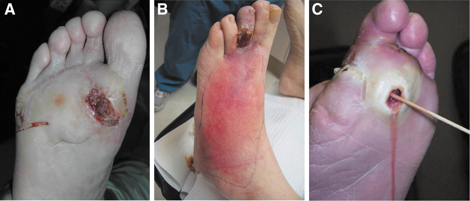
The care for chronic wounds therefore relies upon basic tenets that aim to not only remove or ameliorate the etiologic causes but also to address underlying systemic and metabolic perturbations such as infection or peripheral arterial disease. Proper care of the wound is facilitated initially by employing thorough patient and wound assessment. Factors contributing to the development or recalcitrance of the wounds are then addressed accordingly. Concurrent with the management of associated complications or etiologic factors, wound bed preparation plays a key role in encouraging the proper environment in which tissue repair can take place. Of course, appropriate diagnosis is mandatory to establish the etiology of the nonhealing wound. DFUs, venous leg ulcers (VLUs), PUs, post surgical wounds, and wound dehiscences are the most commonly seen wounds in wound care practice. Atypical wounds such as pyoderma gangrenosum can also be very challenging. Each has its own underlying etiopathogenesis that must be addressed upon initial presentation. Nonetheless, the clinician must be diligent in taking care to rule out the presence of malignancy in the wound either secondarily due to malignant degeneration as in the case of squamous cell carcinoma or as a primary lesion (Fig. 2A). Aside from neoplastic lesions, the basic tenets of wound assessment and care as presented in Table 1 serve as a guide to facilitate the management of most chronic wounds.
Basic tenets of wound care
ABI, ankle–brachial indices; BPG, bypass graft; DFU, diabetic foot ulcer; MRI, magnetic resonance imaging; PRN, as necessary; PU, pressure ulcer; VLU, venous leg ulcer.
Patient assessment
Patient assessment must start with a thorough patient history to determine medical comorbidities, contributing factors possibly leading to the chronic wound, prior trauma, prior history of wounds, current medications, and allergies. Obviously, the presence of diabetes mellitus with neuropathy will be important to note for those patients presenting with a DFU as will a history of deep venous thrombosis for those presenting with a suspected VLU. The importance of taking a medical history cannot be overemphasized. Laboratory studies at the time of presentation, including hematology, serum chemistries, and nutritional parameters, will also provide useful information to supplement initial impressions. 32 An assessment of the patients' living situation and their likely reliability in following prescribed treatments is important in the actual determination of which therapies should be employed to manage the wound(s).
Wound assessment
Assessment of the wound actually begins during the initial encounter with the patient, whereby his/her general appearance is noted as well as that of the wound itself. Morbid obesity or, conversely, a painfully thin patient body habitus is a clue to nutritional status that will have a bearing on treatment protocols as well as possibly on outcomes. A visual inspection of the wound will immediately identify very important attributes that will guide further evaluation and treatment. Obvious necrosis or gangrene portends a much more serious complexity, while a healthy granular appearance might preclude a wound complicated by significant ischemia. Significant erythema, on the other hand, can indicate cellulitis or infection requiring immediate hospitalization or might be an indicator of significant ischemia (dependent rubor) (Fig. 2B). Nonetheless, an initial visual impression of the wound provides important clues not only to the etiology of the lesion but also to its complexity.
Depth, extent (size), location, general appearance, odor, and notation of exudates are all essential components of wound evaluation and need to be recorded at baseline. 33 Probing the wound will determine its depth of penetration and any tracking along tendon sheaths or tissue planes. A positive probe-to-bone (PTB) test has a high predictive value for underlying osteomyelitis even in the absence of acute signs of deep infection (Fig. 2C). 34,35 A neurological evaluation should be performed to assess the presence and extent of neuropathy since this factor not only predisposes a patient to DFUs but is also a major risk factor for diabetes-related lower extremity amputation. 36 –38
A vascular examination is important for any type of chronic wound, but assessment of arterial perfusion is especially important for DFU patients since this parameter will drive all subsequent therapies and outcomes. Palpation of pulses commences initially, recognizing that this assessment can be highly variable among different clinicians. 39,40 Medial arterial calcification renders pulse palpation to be difficult even in the presence of adequate perfusion to the foot. Nonetheless, ankle–brachial indices (ABI) should be obtained at baseline for most patients for whom there is any question of a pulse deficit. Despite the falsely elevated ABI levels that medial arterial calcification produces, the American Diabetes Association has recommended that this diagnostic test be performed on diabetic patients over 50 years of age and whenever clinical circumstances (such as in the case of DFU) dictate this evaluation. 39 Regardless, when the ABI is below 0.9, the patient should be considered to have impaired arterial perfusion. To augment ankle–brachial studies, toe blood pressure readings, pulse volume recordings, transcutaneous oxygen measurements, and skin perfusion pressure measurements have been recommended as valid benchmarks for the prediction of wound healing. 41,42 Whenever peripheral arterial disease is suspected or confirmed by the noninvasive tests above, a referral to a vascular surgeon or interventionist is indicated for angiography and revascularization. Limb salvage rates have been reported as high as 80% for ischemic foot ulcers undergoing revascularization. 43 –46
Venous ulcers are obviously different from arterial wounds and require a different series of diagnostic testing to confirm superficial or deep venous reflux, perforator incompetence, and chronic (or acute) venous thrombosis. Venous duplex ultrasound, plethysmography, and other venous reflux tests are the mainstays of diagnosis. 47,48 Notwithstanding, mixed arterial and venous disease is always a concern for management that requires a combination of both arterial and venous noninvasive testing. 49
Ascertainment of infection and determination of its severity are also critical for appropriate wound management and classification. 33 As previously mentioned, a positive PTB test is a good indicator of underlying bone infection, especially in the presence of acute signs of infection. 35 When signs of infection at the wound are present, tissue cultures are indicated to guide specific antimicrobial treatment. In contrast, clinically uninfected wounds should not be cultured since such cultures will yield growth of contaminants that need not be treated. 50 Unfortunately, osteomyelitis can exist at the base of a wound without clinical signs of infection. Therefore, plain radiographs are necessary to rule out underlying bone changes or foreign bodies (or gas when clinical infection is present). However, the specificity (75–83%) of plain radiographs for the detection of osteomyelitis is higher than its sensitivity (43–75%); because of this, the use of other imaging methods are often required. 51 Serial plain radiography taken every 2–4 weeks is more useful than a single plain image due to delayed changes associated with infection progression. Serial plain radiography has greater sensitivity and specificity and is recommended when access to advanced imaging is limited. 50 Again, exposure of bone or a positive PTB test should prompt suspicion for bone infection and warrant further diagnostic testing. When X-rays are negative, but suspicion is high for bone infection, advanced imaging, such as nuclear medicine scans, magnetic resonance imaging, or computed tomography, is indicated to confirm the diagnosis and the extent of involvement. 33,50
Important for determining the etiology of foot wounds as well as for long-term management, structural deformities must be identified. Hammertoes, bunions, and especially Charcot deformities are important determinants of DFUs in association with neuropathy; these deformities are sites of increased pressure that must be ameliorated to provide for subsequent healing and prevention of recurrence. 33 Similarly, the etiology of the wounds must be ascertained and thereby removed to successfully administer treatment. Foot ulcers are often the result of shoe pressure, repetitive plantar stress, or injury, while VLUs are obviously the result of increased venous pressure and valvular incompetency.
Chronic wounds, following their systematic assessment, should be classified according to one of the published (and preferably validated) classification systems. For DFUs, the Wagner, University of Texas, and PEDIS classification systems each have their place and have been embraced by the wound care community. 52 –54 VLUs are classified by the CEAP classification system and PUs by the NPUAP classification schemes. 55,56 Classification can ultimately guide treatment by prompting systematic evaluation of important underlying components and, furthermore, can predict outcomes based on the severity of the disease. 57
Imaging is a valuable method for assessment of the wound healing process. Biopsy of wound tissue is considered the gold standard for measuring tissue morphological changes. The usual analysis involves tissue staining with hematoxylin and eosin, which is examined under light microscopy. However, it is an invasive and time-consuming method. The invasiveness of the method is the primary reason for the development and implementation of noninvasive optical imaging techniques. Currently, there are many different noninvasive methods, including laser Doppler imaging, indocyanine green videoangiography, near-infrared spectroscopy, in vivo capillary microscopy, orthogonal polarization spectral imaging, reflectance-mode confocal microscopy, hyperspectral imaging, optical coherence tomography, laser speckle imaging, photoacoustic microscopy, and others. Although the majority of these methods have more utility in basic research, several noninvasive imaging techniques are already in use for assessment of burns and chronic wounds. 58 –62 These published reports show the potential utility of noninvasive imaging as diagnostic and prognostic tools of the future to prevent the incidence of ulceration, diagnose infections, monitor complications and progress of treatment, and predict treatment outcomes.
Basic tenets of treatment of chronic wounds
Managing chronic wounds, although often times challenging, need not be considered a daunting task if basic principles of care are routinely followed. A thorough assessment of the patient and wound will guide subsequent treatment by elucidating underlying areas of concern that need to be specifically addressed. Therefore, a systematic approach to both assessment and treatment should most often lead to favorable outcomes. Due to the frequent complexity of patients and wounds, a multidisciplinary approach to management has been proven highly successful and is widely recommended. 33,63,64
Medical/holistic management of the patient must commence concurrently with wound management. Diabetic patients frequently need improved control of their hyperglycemia, renal insufficiency, nutrition, and other associated medical comorbidities that may adversely affect the healing of their wound(s). Patients with a VLU might often have hemodynamic perturbations requiring improved medical management, while PU patients, often bedridden from intercurrent illness, will have significant nutritional deficits that need to be corrected to optimize tissue repair. 65,66
The vascular examination performed during the wound assessment will have determined the need for necessary interventions. 40 Since many DFUs have a component of vascular insufficiency, referral to a vascular surgeon or vascular interventionist for arterial imaging (angiography, duplex scanning, etc.) and subsequent revascularization need to take place early in the course of treatment. The exact roles of endovascular and open bypass procedures are still evolving, but are primarily determined by arterial anatomy, wound severity, and patient comorbidities. 67,68 The ultimate goal is to restore a palpable pulse in the affected foot. In some limited cases where revascularization has failed or is not feasible, hyperbaric oxygen therapy (HBOT) might be indicated. 69,70 Topical oxygen therapy, long criticized as having no role in this regard, is emerging again as an adjunctive measure to improve tissue oxygenation. 71,72 In contrast, venous insufficiency must be addressed initially with adequate compression wrapping with or without intermittent pneumatic compression to counteract the detrimental effects of the venous hypertension causing associated VLUs. 49 Where compression therapy is ineffective or for recurrent VLUs due to significant venous disease, surgical intervention on the superficial, deep, and/or incompetent perforators is indicated. 47,49 Again, care must be taken to identify mixed arterial and venous disease in such circumstances since the associated arterial insufficiency complicates customary treatment protocols for venous ulcerations.
Infection is similarly an important risk factor for wound healing failure and, in the case of DFUs, for subsequent lower extremity amputation. 73,74 Even excessive bioburden can inhibit normal progression to wound healing. 75 While acutely infected wounds are easily diagnosed, neuropathy can mask the presence of deep infections or abscess. Hence, clinical suspicion must remain high when insensate patients complain of pain or flu-like symptoms. Once diagnosed, infection complicating chronic wounds must be treated aggressively. This includes thorough debridement, surgical drainage of abscesses, debridement of infected bone, and tissue culture-guided antimicrobial therapy. As previously mentioned, clinically noninfected ulcers should not be cultured nor treated with systemic antimicrobial therapy. 50 However, if osteomyelitis is suspected, bone culture, followed by specific antimicrobial therapy (and perhaps surgery), is warranted for this recalcitrant infection. 76
Specific wound care or wound bed preparation commences concurrently with the aforementioned interventions when feasible. 15,75,77 Revascularization, however, often follows control of infection and initiation of wound care procedures. The acronym TIME has been used over the last decade or so to facilitate an organized approach to wound bed preparation and has been summarized nicely by Leaper et al. 78 –80 This acronym refers to Tissue assessment and management, Infection/Inflammation management, Moisture imbalance management, and Edge of wound observation and management (Table 2). The TIME principles are an integral, although incomplete, part of this discussion and incorporate the basic tenets of wound care that are critical for managing chronic wounds.
TIME principles of wound bed preparation
Revised based on Leaper et al. 78
Debridement has long been recognized as a critical component for wound care and has been shown by several investigators to expedite healing. 81,82 Sharp debridement removes nonviable tissue and slough along with bacterial biofilms that prolong the inflammatory response in the chronic wound. 75 In effect, thorough debridement converts the chronic wound from one that is excessively inflamed, as previously described, to more of an acute profile that can jump-start the wound toward a healing trajectory. 15,77 While sharp debridement is considered to be the most efficient way to debride a wound (with scalpel, curette, tissue nippers, etc.), hydrosurgical or ultrasonic debridement can also be used in this regard. 77 The term ulcerectomy has been used to denote complete excision of ulcers down to healthy bleeding tissue, resulting in expedited healing of DFUs. 83 Maintenance debridement with enzymes (collagenase) is frequently used between clinic visits to gently remove slough or to enzymatically debride thick crusts (especially in neuroischemic wounds). 75,84 Biodebridement with maggots has been used for many years in patients not suitable for surgical debridement and has shown some promise in removing slough and necrotic tissue while promoting granulation tissue development. 85,86 Simple hydrogels or hydrocolloid dressings can provide for slow autolytic debridement of slough and dried crusts, especially in ischemic patients or those who cannot undergo surgical debridement. Regardless of the method used, effective debridement of chronic wounds is accepted as an essential component of care throughout the wound healing continuum. 15,33,64,87 Nonetheless, healing can be delayed if debridement is performed too frequently and/or extensively. Development of diagnostic tools, including biomarker analysis and noninvasive imaging, is necessary to better distinguish viable from nonviable tissues in the wound and guide debridement practices. 88
The importance of offloading the chronic wound cannot be overemphasized. 89 In fact, when this component of wound care is neglected, the chances of a successful outcome are extremely low. When one recognizes that most wounds, especially DFUs, have excessive pressure as their proximate cause, it is quite understandable that the high pressures must be ameliorated before healing can take place. For nonplantar wounds caused by tight shoes, it is imperative that the source of offending pressures be eliminated. A number of studies and reviews have confirmed the essential role of offloading in this regard. 90 –92 For DFUs, the total contact cast has long been considered as the gold standard for offloading by virtue of its pressure redistribution properties as well as irremovability. Numerous additional offloading modalities have been reported for DFUs, including braces, removable cast walkers, irremovable cast walkers (often referred to as instant total contact casts), half shoes, modified surgical shoes, foot casts, and various felt or foam dressings. 89,90,93 While each device has its own advantages for any given patient, almost any offloading modality is superior to no offloading for the management of DFUs.
Along the same lines, compression therapy for chronic VLUs is equally important. Since venous hypertension is at the source of these lesions, hydrostatic pressure into the skin and subcutaneous tissues underlying venous ulcers must be mitigated by external compression. 49 Different modalities, such as the classic Unna's boot, three- or four-layer compression bandages, and short stretch compression bandages, have long been used in this setting. 94 For ulcerated patients with significant venous insufficiency and associated chronic lymphedema, intermittent pneumatic compression pump therapy can also be recommended. While not supplanting the need for directly applied compression wraps, pump therapy will augment their effect and assist in maintaining long-term control of peripheral edema and lymphedema. 49,95
Surgical offloading is a term that (as its name implies) refers to the surgical management of foot deformities causing high pressures resulting in chronic ulcerations. 96 –98 While not generally considered a primary treatment for most neuropathic DFUs, those that prove to be recalcitrant to standard wound bed preparation and effective offloading should be considered for surgical internal decompression. Many such procedures have been reported as effective over the last several decades. 99 –102 Metatarsal head osteotomies or resections, sesamoidectomies, hammertoe repair, bunionectomies, first metatarsal phalangeal joint arthroplasties, plantar exostectomies, arthrodeses, and partial calcanectomies have all been described for the management of noninfected as well as infected chronic foot ulcers. Soft tissue tendon balancing procedures, such as tenotomies, tendon transfers, and lengthening procedures, including tendo-Achilles lengthening and gastrocnemius-soleus recession, can be done as isolated procedures or in concert with osseous procedures to reduce deformities and high forefoot plantar pressures. 103,104 Several authors have proposed a validated classification scheme for such operations based on the presence or absence of wounds as well as their acuity. 97,105 Elective procedures are done in the absence of neuropathy, while Prophylactic operations are performed in neuropathic individuals to prevent initial ulceration over deformities or to prevent their recurrence. Curative procedures are undertaken to internally decompress chronic wounds, reduce high plantar pressures, or to remove foci of bone infection/osteomyelitis to engender final healing. Emergent operations, often including amputations, must be performed to control acute infection as limb or life-threatening measures. As has been validated, this classification trends toward an increasing need for hospitalization and frequency of amputation in the progression from elective through emergent procedures. 105 Of course, residual postoperative wounds in these patients must be treated with the same tenets of wound care as discussed above until final healing occurs. 96
Topical wound therapies/dressings
While there are a myriad of topical therapies/antimicrobials and dressings available to the clinician, very few have prospective data to support their effectiveness in promoting wound repair. Nonetheless, many therapies can indeed be useful, despite reliance on anecdotal experience. Hence, clinicians tend to use what they are accustomed to or what seems to be effective based on personal experience. Topically applied agents for wounds run the gamut from sterile saline or hydrogel to povidone–iodine solutions, cadexomer iodine, hypochlorous acid, honey, and collagenase. One topical antimicrobial agent, superoxidized solution, has recently been formally studied for efficacy in healing DFUs and was found to be effective in this regard. 106 Similarly, the inexhaustible availability of dressings can make selection of the appropriate wound covering somewhat daunting. While standard cotton gauze dressings have long been considered standard of care (even in clinical trials), many other primary and secondary dressings are commercially available. Highly absorbent and moisture-retaining foam dressings, acrylics, alginates, hydrofibers, hydrocolloids, honey alginates, oxidized regenerated cellulose, micronized collagen, and many others can be considered as circumstances warrant. Many of the aforementioned products also are available with silver for control of bacterial overburden. While the use of silver-containing products is widespread, misuse of this antibacterial element for prolonged periods is also common. While its primary use is for the reduction of bacterial colonization, there are little data to support its efficacy as a wound healing agent. 107,108 It cannot be emphasized enough that standard dressings and topical therapies never supplant the need for debridement, effective offloading, and appropriate management of infection and ischemia.
Advanced therapies
All currently published guidelines and consensus reviews on the management of chronic DFUs, VLUs, and PUs support the belief that all such wounds should initially be treated with standard wound care principles as have been discussed. 33,49,56,64,109 In most cases, these basic tenets of wound care should be carried out before consideration of the use of more advanced therapies. Most wound care protocols now advocate the use of such standard measures for an initial period of 4 weeks, after which an assessment of wound area reduction should be made. While the 1999 American Diabetes Association publication was one of the first consensus documents on DFU assessment and treatment, it only mentioned that wounds failing to heal by 4 weeks were associated with worse outcomes, including amputation. 110 In 2003, however, Sheehan et al. published the often quoted article supporting the ability of the 4-week healing rate to predict complete healing by 12 weeks. 111 Using data from another large, multicenter randomized controlled trial (RCT), it was determined that the midpoint (median) percent wound area reduction from baseline at 4 weeks between those that healed and those that remained unhealed at 12 weeks was 53%. 112 Those who exceeded this midpoint healed in 58% of cases by 12 weeks. In contrast, those that did not achieve 53% wound area reduction by 4 weeks only healed in 9% of cases (p<0.001). Even more striking, the mean 4-week percent change in ulcer area was 82% in healers versus 25% in the nonhealing group regardless of the treatment arm. Subsequently, the 4-week 50% wound area reduction has been widely adopted and confirmed as a robust indicator for predicting healing at 3 months. 33,38,64,87,109,113 –115 Consistent with this premise, wounds failing to achieve a 50% area reduction at this time point need to be reassessed and subsequently considered for advanced therapies in the absence of underlying disease or nonadherence to prescribed basic treatment. 33,64 Figure 3 illustrates a fairly common algorithm for DFU treatment incorporating these principles. Figure 4 similarly illustrates an algorithm for VLU treatment.
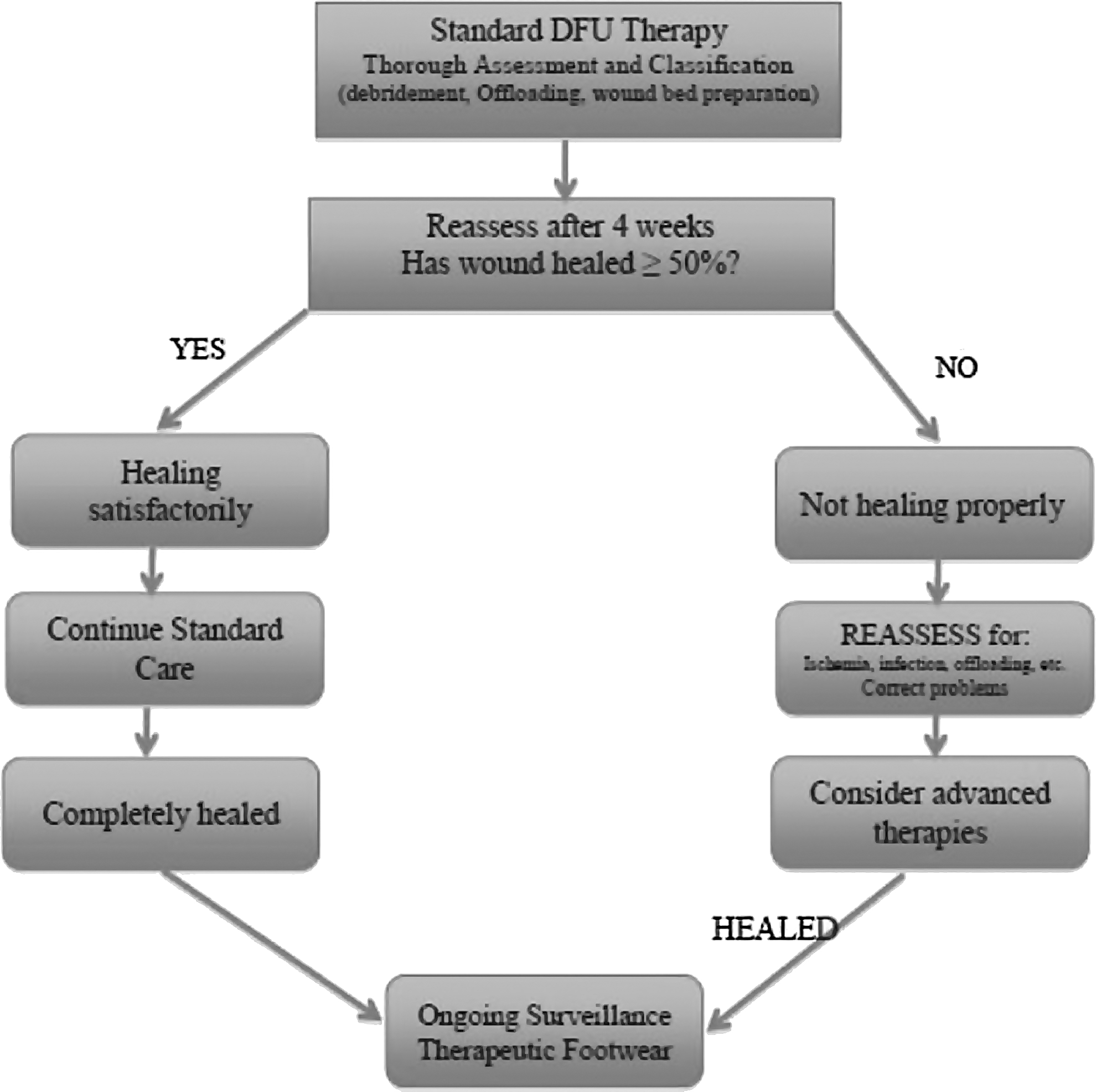
Simplified algorithm for diabetic foot ulcer (DFU) treatment.
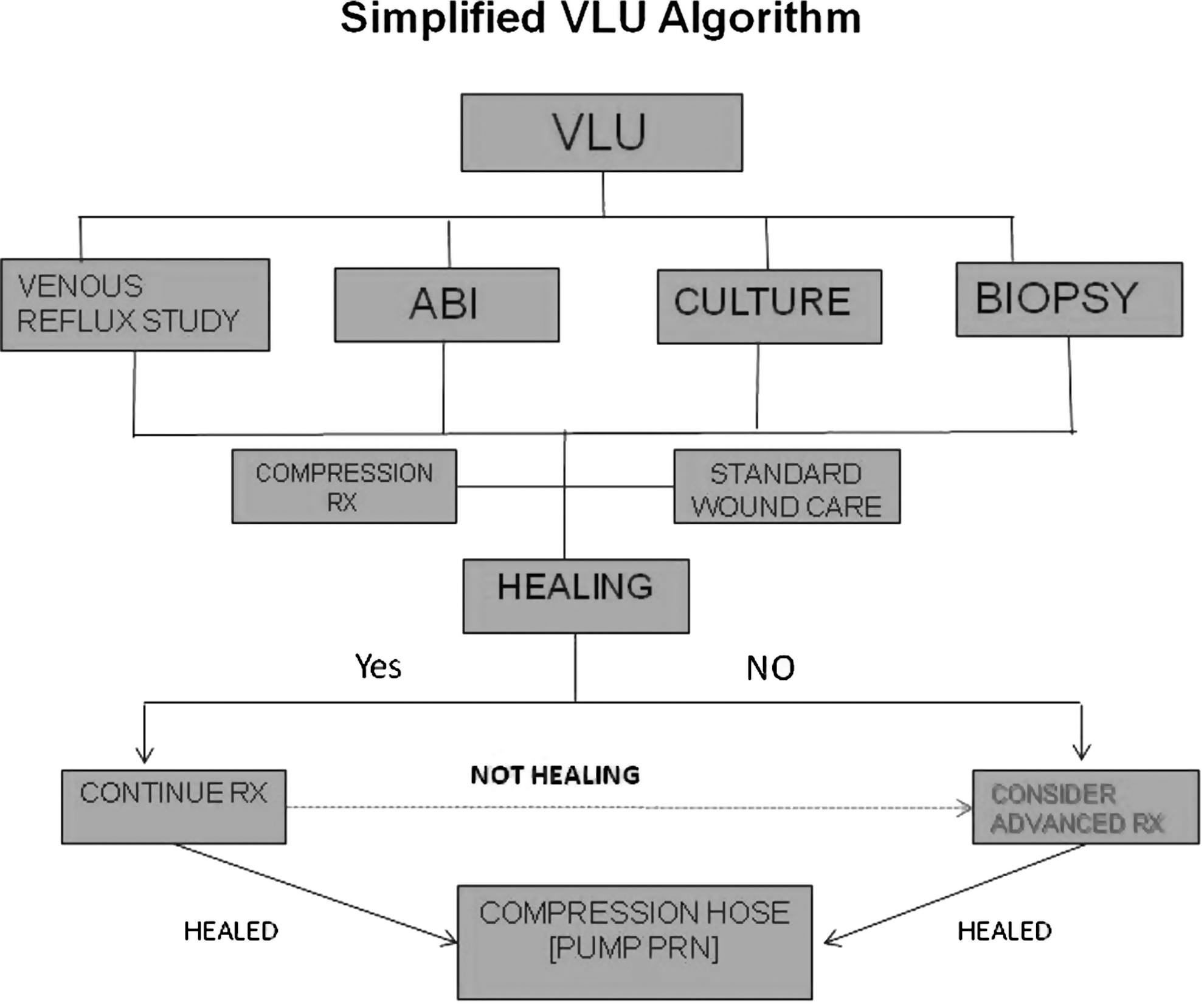
Simplified algorithm for venous leg ulcer (VLU) treatment. ABI, ankle–brachial indices.
Once it is determined that the patient might benefit from an advanced therapeutic agent, there are a number of options currently available. 64,116 –118 While there have been several key RCTs published on advanced wound care agents for chronic wounds, most such products do not have the benefit of high-level evidence or even nonrandomized prospective studies to attest to their efficacy. Advanced wound therapies can best be discussed by broadly categorizing them according to their specific technologies or engineering, tissue types, cell types, or protein content (i.e., growth factors). Even this scheme will not capture the myriad products, proteins, and molecules that have undergone trials and failed to demonstrate superiority over standard of care treatment. While not exhaustive, Table 3 provides a listing of the more common wound care technologies currently in use in the United States and abroad.
Wound care technologies
Negative pressure wound therapy
Since its introduction in the mid 1990s, negative pressure therapy has assumed a major role in the management of traumatic, acute, and chronic wounds, as well as for stabilizing skin grafts, flaps, and surgical incisions. 119 –123 Since the early studies of Morykwas et al. and Argenta and Morykwas that demonstrated the numerous attributes of NPWT, a very large body of evidence has been published supporting the clinical efficacy of this very important biophysical modality. 121,124 –129 Clinical trials have been conducted in the ensuing years that have proven the superiority of NPWT over standard therapy for managing open amputation wounds, DFUs, VLUs, and other wounds. 130 –132 As an adjunct to standard chronic wound care, NPWT very efficiently manages wound drainage and can provide expedited granulation tissue development, wound area contraction/reduction, preparation for delayed closure or grafting, or primary healing. 133,134 NPWT is also quite useful as a bolster to enhance the incorporation of skin grafts onto recipient wound beds. 135 Application techniques are important, however, since inappropriate placement of tubing can potentially cause skin pressure lesions. Therefore, a bridging method that provides for suction tubing placement away from plantar surfaces has become an important application technique. More recently, these electrically powered devices have added the ability to instill saline or other antimicrobial agents to assist in the cleansing of the wounds concurrent with providing the aforementioned benefits of NPWT. 128,136 –138 A mechanically powered, ultralight, and portable NPWT device has also been introduced in recent years. 135,139,140 Two articles comparing this device with the traditional electrically powered device in a prospective RCT have shown equivalent DFU healing outcomes with faster application times and a high degree of patient satisfaction. 141,142 Clinicians have also combined NPWT with other advanced therapies such as acellular and cellular matrices. 143 Such multimodal therapies can be administered concurrently or sequentially as wound characteristics change to provide the optimum therapy for any given wound.
Hyperbaric oxygen therapy
HBOT has been advocated as being beneficial for a wide variety of chronic wounds for over two decades. 144 –150 While there are numerous retrospective and prospective studies, case series, cohort studies, and several trials indicating the efficacy of HBOT for the treatment of DFUs and VLUs, the general quality of these studies is not robust since inclusion criteria and outcomes are highly variable. 69,144,145,150 –156 While the recent Cochrane review in 2012 indicated significant short-term improvement for healing DFUs over controls at 6 weeks (relative risk [RR] 5.20; p=0.02), this benefit was not evident at 1 year or longer. 157 While there was a suggestion that HBOT may decrease the major amputation rate in DFU patients, pooled analysis of the data did not yield a significant estimate for this association (RR 0.36, 95% confidence interval 0.11 to 1.18, p=0.08). The 2010 RCT by Londahl et al. reported favorable outcomes at 1 year for HBOT in those patients with Wagner grade 2, 3, or 4 DFUs failing prior standard interventions for >3 months (52% vs. 29%, p=0.03). 69 Unfortunately, the heterogeneity of the patients and time to reported outcomes (9 months or 1 year) make comparisons with other DFU trials difficult. A more recent review of HBOT in a large observational cohort study by Margolis found that the use of HBOT neither improved the likelihood that a wound would heal nor prevented amputation in patients with adequate lower limb arterial perfusion. 158 This review and the aforementioned Cochrane review both called for a reevaluation of HBOT with rigorously designed adequately powered trials to assess efficacy in healing chronic wounds.
Since the very poorly conducted 14-day study by Leslie et al. in 1988, the use of topical wound oxygen for DFUs and VLUs has been considered as highly controversial, especially by advocates of HBOT. 71 The lack of a formal study in this regard has been offset by several reviews, case series, and experimental studies on the effect of topically administered oxygen at the wound surface that seem to indicate a benefit in promoting wound repair. 154,159 –169 While several RCTs with different devices have been ongoing, there have been no publications to date comparing 12-week healing rates with those of standard care.
Biophysical modalities
Electrical stimulation has been the most studied biophysical device for healing chronic wounds to date, primarily utilized by physical therapists and physiatrists. 170 –173 An abundance of studies advocates the beneficial healing effect of electrical stimulation at various modes and frequencies for a variety of chronic wounds. 174 –177 Other nonthermal forms of electromagnetic energy have also been used for wound healing, including pulsed radiofrequency energy (PRFE), pulsed shortwave diathermy, and pulsed electromagnetic fields. 173,178,179 One in vitro study of PRFE effects on cultured dermal fibroblasts and keratinocytes found this electromagnetic field to upregulate expression of a variety of genes involved in modulating the inflammatory stage of wound healing. 180 In general, by interacting with endogenous bioelectric currents, electromagnetic fields indirectly upregulate the production of nitric oxide and multiple growth factors, resulting in cellular mobilization, angiogenesis, and expedited wound repair. 173,176
Ultrasound, most frequently used for diagnostic and musculoskeletal therapy purposes, has also assumed a role in wound management. Several lower frequency devices are currently available for debridement that use the delivery of sound waves to generate cavitation at the wound bed. 173,181 Wounds with thick fibrinous slough and necrosis can thereby be very aggressively debrided with low-frequency ultrasound (LFU) devices, although trials to show improved healing rates have not been conclusive. 181 –183 Noncontact LFU using a saline droplet carrier to the wound surface has been proposed as another LFU device that can improve healing rates of chronic ulcers, including those complicated by ischemia. 170,184 –188 While this noncontact LFU modality can assist with wound bed preparation, it cannot aggressively debride wounds as well as the aforementioned units. Several retrospective observational studies and one low quality RCT suggested improvements in healing rates over standard of care comparators, but confirmatory prospective studies have not been forthcoming. 184 –186,188
Extracorporeal shock wave therapy (ESWT) has been used for a number of years for a variety of musculoskeletal conditions and has recently been adapted for the treatment of cutaneous wounds. ESWT is defined as a series of high-energy acoustic pulses delivered to tissues by electrohydraulic, electromagnetic, and piezoelectric sources. 189 The pressure pulses generated promote a cascade of cytokine and growth factor upregulation leading to enhanced neovascularization, anti-inflammatory response, and tissue regeneration. 190,191 Several recent reviews and one study comparing ESWT with HBOT on DFU healing support its potential role in expediting wound repair. 189 –191 Nonetheless, RCTs are still required to evaluate the clinical efficacy of this modality in healing chronic wounds of different etiologies.
Biological and bioengineered therapies
Concurrent with the explosive growth of negative pressure therapies and HBOT for the management of chronic wounds in the last two decades, there has been an enormous amount of research and interest in advanced biological therapies. In this regard, biological therapies refer to tissue-based treatments (acellular and cellular), autologous platelet-rich plasma (PRP), as well as recombinant human growth factor therapies. While the latter can be considered as a category unto themselves due to the enormous amount of preclinical research on cytokine and growth factor-mediated wound repair, there are currently only several such therapies commercially available to clinicians—recombinant human platelet-derived growth factor (rhPDGF), fibroblast growth factor (rhFGF), and epidermal growth factor (rhEGF). Numerous other growth factors have been isolated and investigated, such as vascular endothelial growth factor (VEGF), keratinocyte growth factor-2, TGF-β, and granulocyte-macrophage colony-stimulating factor, but these currently are not approved for use in wound care. 192
Platelet-rich plasma and growth factors
The clinical interest in growth factor therapies over the last 25 years actually stemmed from the early work of Knighton et al. on autologous PRP and the cascade of growth factors released from activated platelets during the centrifugation of whole blood. 193 –195 Several small trials and retrospective reviews have affirmed the potential efficacy of topically applied, activated, autologous platelet supernatants for expedited healing of chronic lower extremity wounds. 196 –199 After recognizing the potential benefit of topically applied PDGF to chronic wounds, becaplermin gel (rhPDGF) was studied in the setting of chronic neuropathic DFUs and became the first commercially available advanced therapy for the management of these difficult wounds (and the only growth factor approved for use in the United States). 200 –202 In the 20-week phase III clinical trial by Wieman leading to its approval, topically applied rhPDGF gel was found to significantly increase the incidence of complete wound closure by 43% and decrease the time to healing by 32% over placebo-controlled standard wound care. 203 The efficacy of the single topically applied growth factor was corroborated as being beneficial to the healing of chronic nonischemic foot ulcers when combined with good standard wound care in several other studies pooling data from multiple sources. 200,202,204
rhEGF is perhaps the best studied growth factor for cutaneous ulcers and wounds, but is not available in the United States. 205 –212 Applied as a topical cream, or more commonly, by intralesional injections, EGF has been found also to expedite the healing of a variety of types of cutaneous wounds. Several small trials have been published from Asia and Cuba attesting to the healing benefits of EGF compared with controls. 207 –209,213 However, the heterogeneity of wounds, applications, patient management protocols, and outcomes has not lead to clinical trials in the United States nor to widespread global adoption of this therapy.
FGF has been studied primarily in Asia for a variety of chronic wounds, including DFUs, VLUs, and PUs, and is approved in Japan for this use. 214 –218 FGF, considered a potent angiogenic growth factor, also has an isoform (FGF-10) commonly known as keratinocyte growth factor-2 or repifermin. 192,217 This agent has been studied in the United States for chronic VLUs as a topical spray. Repifermin was shown to accelerate wound healing with significantly more patients achieving 75% wound closure with repifermin than with placebo after 12 weeks. The treatment effect appeared more marked for a subgroup of patients with initial wound areas ≤15 cm2 and wound ages of ≤18 months. 217 Unfortunately, there were no significant differences between the study and control groups for the primary outcome (healing at 12 weeks), likely due to methodological issues in the study protocol. Neither repifermin nor FGF has been approved in the United States for wound management.
Acellular therapies
The most common types of advanced biological therapies for chronic wounds can generally be classified as acellular therapies—those dermal, amniotic, or collagen-based tissues (human or animal) that have been decellularized during their processing. Often referred to as acellular and/or ECMs, these biological products serve as substrates into which cells can migrate and initiate angiogenesis, thereby promoting granulation tissue development and tissue regeneration. 116 Once considered only as inert structural collagen tissue providing a scaffold for cellular ingrowth, ECMs are now known to play an active part in tissue regeneration through a dynamic interaction with growth factors and host cells. 219 –221 ECMs contain not only structural collagen but also glycosaminoglycans (including hyaluronan), proteoglycans, and glycoproteins—all essential components to replace the defective ECM of injured tissues. 222,223 Currently available nonhuman ECM products with clinical data to support their efficacy include porcine-derived small intestinal submucosa, porcine urinary bladder matrix, bovine dermis, equine pericardium, and sheep (ovine) bladder. 224 –233 Another matrix dressing (Hyalomatrix®; Anika Therapeutics, Inc., Bedford, MA) comprising primarily hyaluronan, one of the main constituents of ECM, has also been studied in Europe as a dermal substitute for use in burns, VLUs, and DFUs. 234 –237 Integra® bilayer wound matrix (Integra LifeSciences, Plainsboro, NJ) is a dermal regeneration template in this category (bovine collagen, glycosaminoglycans, and silicone layer) that has primarily been used in burns for a number of years, but is being more frequently used in lower extremity chronic wounds and foot ulcers. 238,239 Although there are few published prospective studies to support their efficacy, most of these products are commercially available in the United States at relatively low cost and are approved for use in multiple chronic wound types.
Human dermal allografts have become increasingly popular in recent years for augmenting tissue regeneration in chronic lower extremity wounds and have been formally studied in DFUs and VLUs. These are defined by the Food and Drug Administration as human cellular and tissue-based products. The allografts are harvested from screened donors, and each is prepared with proprietary processes to decellularize and cryopreserve the dermis while maintaining the natural structure of the collagen and ECM. 240 –245 As with the aforementioned ECM products, the dermal matrices serve as scaffolds for cellular repopulation and angiogenesis, with varying degrees of incorporation into the recipient wound bed. 246,247 One of these cryopreserved dermal allografts, referred to as a human skin allograft, claims that both the extracellular and cellular components are preserved during the minimal processing. 242,243 Two of the available dermal regenerative matrices have successfully undergone small RCTs (Graftjacket®; KCI, San Antonio, TX and TheraSkin®; Soluble Systems, Newport News, VA), while a third is currently undergoing a clinical trial for DFUs (dCell®; Tissue Regenix, San Antonio, TX). 240,243 Available data suggest that acellular dermal matrices, in addition to basic wound care principles, may provide an effective technique for tissue regeneration in deep and cutaneous extremity wounds. 221,246
Amniotic membranes (AMs) and umbilical cord tissues have been used for many years for corneal ulcers and were actually the earliest reported biomaterials used for wound repair. In recent years, there has been a greatly expanded interest in these tissues for chronic wounds, likely due to the wide availability of placental tissues after cesarean deliveries. Due to their rich cellular content in the native state, AMs contain a number of cytokines and growth factors bound to the ECM after decellularization and preparation that remain available to augment angiogenesis and tissue repair when implanted into chronic wounds. 248 –251 AMs are available in the cryopreserved state or as dehydrated products for direct implantation. 250,252,253 Several products are also available in a micronized formulation that can be applied topically or hydrated for injection into wounds or other inflamed tissues (tendonitis, plantar fasciitis, etc.) to augment healing. 254 Amniotic fluid with granulized amniotic matrix is also available for the management of chronic wounds. 255 While several retrospective and prospective studies have supported the benefit of amniotic matrix products for chronic wound repair, the only published randomized and comparative studies to date have utilized the dehydrated human amnion/chorion membrane (dHACM). 255 –262 In the initial small RCT of just 25 DFU patients in a single center, Zelen et al. reported a 92% healing rate after 6 weeks in the dHACM (Epifix®; Mimedix Group, Marietta, GA) group compared with 8% in the standard of care group (p<0.001). 261 In a crossover study of unhealed patients in the control arm of the RCT, 91% healed by 12 weeks with biweekly dHCAM application. 259 Subsequent studies ascertained that weekly applications of this allograft provided more rapid healing of DFUs than biweekly application and that healing frequency with the amnion/chorion product was significantly higher than patients assigned to either a bilayered skin substitute or to standard of care treatment in another comparative trial. 260,262 Many of the aforementioned studies, however, were sponsored by a single company with a single product and the same investigative group. Since a number of other AM products are now commercially available, further studies should be forthcoming to determine if there is a distinction between different amniotic matrices pertaining to efficacy in wound repair.
Bioengineered cellular therapies
For more than a decade, two allogeneic bioengineered skin replacement therapies utilizing neonatal expanded cells have been available in the United States market to address chronic wounds. Both products underwent formal, controlled clinical trials before their approval for use. The first cellular product (Apligraf®; Organogenesis, Canton, MA) is a bilayered construct consisting of a bovine collagen matrix seeded with living human neonatal fibroblasts and a neonatal keratinocyte neoepidermis. It is approved for both chronic VLUs as well as DFUs. This living skin equivalent not only addresses the deficient ECM of chronic wounds by adding a collagen matrix but also introduces immune-privileged living cells that proliferate and actively synthesize growth factors, cytokines, and ECM products. 263 –265 In the pivotal, multicenter, 12-week clinical trial, patients randomized to the Apligraf group achieved complete healing in 56% of cases compared with 38% in the control group (p=0.0042). 266 With an average of four applications, the bilayered skin equivalent also healed the chronic DFUs significantly faster than those patients treated with standard care (65 vs. 90 days, p=0.0026). Other subsequent investigations found similar efficacy of this bioengineered product for healing chronic diabetic foot wounds as well as VLUs. 267,268
Dermagraft® (Organogenesis, Canton, MA) is a human fibroblast-derived dermal substitute (HFDS) comprising a cryopreserved, absorbable, three-dimensional polyglactin mesh substrate seeded with living neonatal dermal fibroblasts. 269 Similar to the bilayered skin replacement, these cells secrete a host of growth factors, cytokines, matrix proteins, and glycosaminoglycans that induce tissue regeneration through the development of granulation tissue and ingrowth of host fibroblasts and keratinocytes. 270,271 This dermal substitute was proven effective in healing chronic DFUs in the pivotal trial by Marston et al. in 2003. 272 In this multicenter clinical trial, those patients assigned to the HFDS group healed significantly faster after 12 weeks compared with the standard wound care group (30% vs. 18.3%, p=0.023). Despite the ostensibly low overall healing rate, the study group achieved a 64% increased healing compared with the standard care group. This was a greater margin of effect than reported in any previous trial. Despite the failure to achieve superiority in its primary outcome of complete healing in a large VLU trial, significant differences in complete healing were achieved for a subgroup of ulcers ≤12 months in duration (p=0.029). 273 Other authors have reported success with the dermal substitute in a variety of lower extremity wounds, often used in concert with other wound healing modalities. 274 –277 A recent study investigated the incidence of amputations and bone resections within the two arms of the DFU pivotal trial and found a decreased incidence of these complications in the HFDS group, likely related to a lower incidence of infection and faster healing in the investigational treatment group. 278
Stem cell therapies
Perhaps the most recent advancements for wound care therapies are that of stem cell therapies, primarily bone marrow derived, and most recently, placental-derived stem cells. Both sources are considered as adult stem cells, and the cell lineage of interest are the MSCs. Briefly, MSCs are multipotent progenitor cells that can directly differentiate into mesenchymal tissues, such as bone, tendon, and cartilage. Their ability to affect cutaneous repair, however, is through an indirect paracrine function (trophic activity), whereby they synthesize essential growth factors and cytokines that affect cell migration, proliferation, and metabolic activity of host cells and tissues. 30,279,280 In this manner, MSCs play an active role in the inflammatory, proliferative, and remodeling phases of wound repair. 281 Interestingly, MSCs can respond to the host environment by upregulating anti-inflammatory cytokines in the presence of inflammation and respond to hypoxic environments by upregulating the release of VEGF to induce angiogenesis. During the remodeling phase, MSCs produce growth factors, such as TGF-β3, to limit excessive scarring as well as modulate the balance between matrix metalloproteinases and tissue inhibitors of metalloproteinases while regulating collagen deposition. Important for allogeneic implantation, MSCs are characterized as being immune privileged since they lack cell surface antigens that would typically engender a foreign body reaction. 281
Bone marrow-derived stem cells (BMSCs) have been of interest for some time now and studies have indicated their ability to augment repair or regeneration of numerous tissues, including cardiac, bone, cartilage, blood vessels, and skin. 30,282,283 Although much interest has focused on orthopedic and critical limb ischemia applications, BMSCs are increasingly being studied for use in enhancing chronic wound and cutaneous repair. 118,284 –291 Several articles and clinical studies have specifically focused on DFU management. 292 –294 One of the earlier studies by Yamaguchi et al. took a novel approach by decorticating exposed bone at the base of the ulcer to locally release bone marrow cells directly into the wound. 290 This was followed several weeks later by application of autologous epidermal grafts, resulting in significantly improved healing compared with standard wound care (p<0.0001). In another small case series by Rogers et al., BMSCs were harvested from the ipsilateral distal tibial metaphysis, and the aspirate was applied topically or by peripheral injection under the debrided wounds with good results. 287 Large randomized studies have not yet been published comparing wounds treated with BMSCs with standard care. Another issue for consideration in such trials will be standardizing methods for obtaining and processing the marrow aspirates, as well as potential complications associated with this surgical procedure.
Placental tissues—including the umbilical cord, the amnion, and the chorion—are a rich source of MSCs and are readily available without the ethical concerns of embryonic stem cells. 281,292 Furthermore, the MSCs in these tissues do not suffer from the age-related effects nor decreased cell counts as found in MSCs harvested from adult patients with comorbid diseases. 279,293,295 Placental-derived MSCs show minimal differences from those obtained from different sites, and yet retain all of the metabolic, paracrine, and immunomodulative properties previously described while maintaining their immune-privileged status. 281 Traditionally, placental or amniotic tissues used for wound repair were prepared from fresh cesarean section donors. One small modern study investigated the efficacy of fresh AM on the healing of chronic VLUs and found a positive effect on pain reduction and a significant clinical response (20% completely healed) within the 3-month follow-up period. 257 Overcoming the difficulties of fresh transplantation, a new cryopreserved AM product (Grafix®; Osiris Therapeutics, Inc., Columbia, MD) has become commercially available in recent years. This human MSC-rich wound matrix contains viable cells, including fibroblasts and epithelial cells, in addition to MSCs and a natural ECM. 281 In a single-center retrospective study of 67 chronic lower extremity wounds, including VLUs and DFUs, this viable wound matrix in association with good standard wound care was found to close 76.1% of wounds at 12 weeks with a mean time to healing of 5.8 weeks. 296 A subsequent 12-week RCT comparing the efficacy of Grafix to standard of care for the healing of DFUs was published in 2014. 297 In this pivotal trial where the primary outcome was complete wound healing at 12 weeks, those patients assigned to weekly applications of the viable human matrix healed significantly more DFUs than those in the control group (62% vs. 21%, p=0.0001). The median time to healing was also significantly faster in the study group (42 vs. 69.5 days, p=0.019). Of particular interest, those unhealed patients in the control group were allowed to crossover to receive up to 12 weeks of viable stem cell matrix therapy. The probability for closure in these patients was 67.8% with a mean time to closure of 42 days. 297 The results of this study represent a margin of effect between active and control groups of 191%, higher than any other published study to date.
• Chronic wounds require a systematic approach, including both patient and wound assessment, no matter what the underlying etiology of the wound.
• The care of chronic wounds relies upon basic tenets that aim to remove or ameliorate the etiological causes and to address systemic and metabolic perturbations.
• Basic tenets include vascular examination, followed by appropriate interventions, management of wound-related infection, wound debridement, and offloading.
• Wound bed preparation plays a key role in providing the proper environment in which tissue repair can take place.
• In addition to offloading with the purpose of eliminating excessive pressure, the term surgical offloading has been introduced, meaning the surgical management of foot deformities underlying chronic ulcerations.
• Most wound care protocols advocate the use of advanced therapies after 4 weeks for nonhealed wounds.
• There are a number of advanced therapies for chronic wounds. However, most do not have a high level of evidence or even nonrandomized prospective studies assessing their efficacy. Selection of advanced therapies should be based on the evidence available.
Summary
Complex wounds represent a major challenge for the clinician and wound care specialist. Despite all the recent advances in wound care therapies and our understanding of the pathophysiology underlying chronic wounds, nothing can supplant the need for adhering to the basic tenets of wound care: thorough assessment with medical and nutritional optimization, debridement (including surgery), offloading (or compression), management of ischemia, management of infection, and appropriate wound bed preparation. Following these principles and established wound care guidelines should often lead to satisfactory outcomes. In addition, the rational use of advanced wound care therapies is encouraged when wounds do not respond sufficiently to good standard care after 4 weeks or sooner as circumstances dictate. In most cases, selection of advanced therapies should be based on the evidence available to help facilitate their most appropriate use.
Footnotes
Acknowledgment and Funding Sources
The authors thank Alla Danilkovitch, PhD, for her review and thoughtful suggestions. No competing financial interests exist.
Author Disclosure and Ghostwriting
The opinions expressed are those of the authors and do not represent those of the Department of Veterans Affairs or the United States Government. No ghostwriters were used to write this article.
About the Authors